
TL;DR
This study combines a mathematical model with CT scans to understand how blood flow forces relate to heart disease plaque locations.
Contribution
The novel integration of shear stress modeling with CT imaging reveals new insights into arterial architecture's role in heart disease.
Findings
Shear stress patterns were mapped using CT scans and mathematical modeling.
Arterial architecture was found to influence the location of lipid plaques.
The method provides a new way to study heart disease mechanisms.
Abstract
Shear stress in arteries, which is a measure of the force exerted by blood flow on the arterial wall, is associated with the location of lipid plaques that cause heart disease. In this study, a mathematical model of shear stress was combined with cross-sectional x-ray images of an artery taken using Computed Tomography (CT) scanning, allowing the authors to explore patterns of shearing stress and shed light on the role of arterial architecture in heart disease.
Genes, proteins, chemicals, diseases, species, mutations and cell lines named across the full text — each resolved to its canonical identifier and authoritative record.
Click any figure to enlarge with its caption.
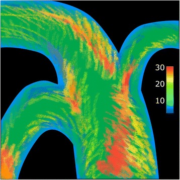 Figure 1
Peer Reviews
No public reviews on file for this paper yet. If you reviewed it on a platform where reviews are public (OpenReview, ICLR, NeurIPS, ICML), you can paste yours below so the community can read it here.
Videos
No videos yet. Explain this paper in a talk, walkthrough, or lecture? Add one.
Taxonomy
TopicsCoronary Interventions and Diagnostics · Cardiovascular Health and Disease Prevention