Developmental disruptions underlying brain abnormalities in ciliopathies
Jiami Guo, Holden Higginbotham, Jingjun Li, Jackie Nichols, Josua Hirt, Vladimir Ghukasyan, E. S. Anton

TL;DR
This paper explores how disruptions in primary cilia function during brain development lead to brain abnormalities in ciliopathies.
Contribution
The study identifies specific roles of ciliopathy genes in mouse cortical development and links these to brain abnormalities in ciliopathies.
Findings
Ciliopathy genes affect key stages of mouse cortical development, including progenitor development and neuronal migration.
Disrupted developmental events are directly linked to brain abnormalities observed in ciliopathies.
The study provides insights into the functions of ciliopathy genes in corticogenesis.
Abstract
Primary cilia are essential conveyors of signals underlying major cell functions. Cerebral cortical progenitors and neurons have a primary cilium. The significance of cilia function for brain development and function is evident in the plethora of developmental brain disorders associated with human ciliopathies. Nevertheless, the role of primary cilia function in corticogenesis remains largely unknown. Here we delineate the functions of primary cilia in the construction of cerebral cortex and their relevance to ciliopathies, using a shRNA library targeting ciliopathy genes known to cause brain disorders, but whose roles in brain development are unclear. We used the library to query how ciliopathy genes affect distinct stages of mouse cortical development, in particular neural progenitor development, neuronal migration, neuronal differentiation, and early neuronal connectivity. Our…
Genes, proteins, chemicals, diseases, species, mutations and cell lines named across the full text — each resolved to its canonical identifier and authoritative record.
Click any figure to enlarge with its caption.
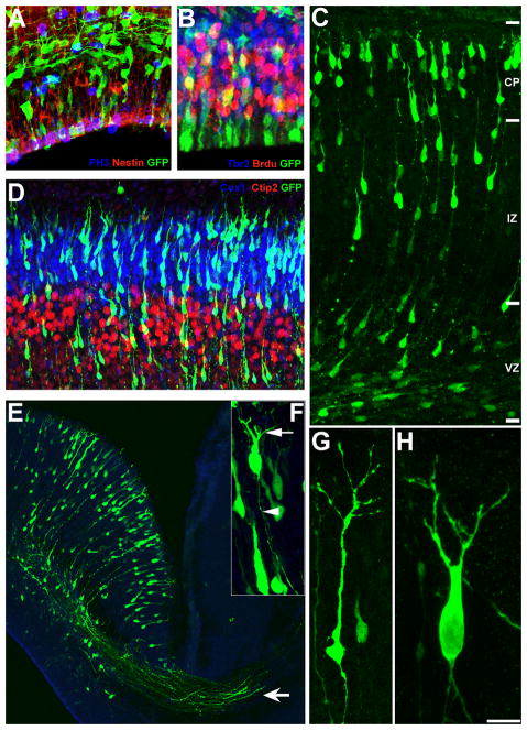 Figure 1
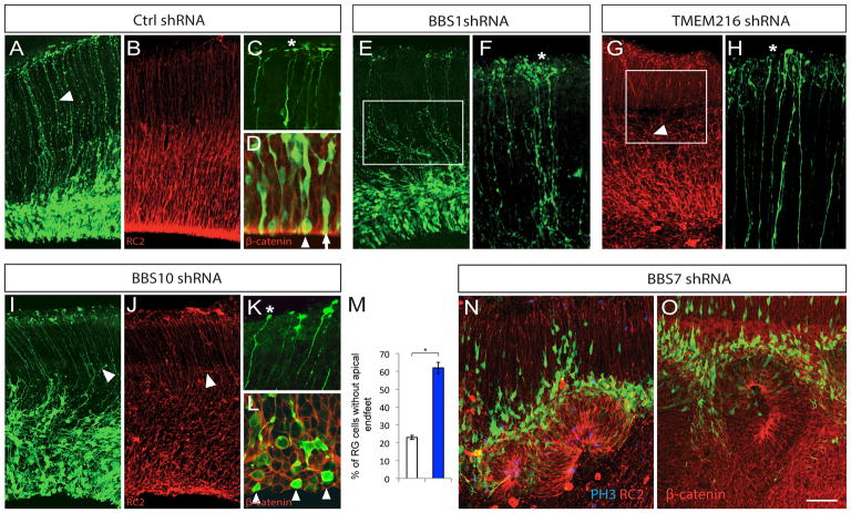 Figure 2
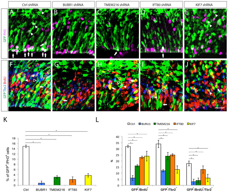 Figure 3
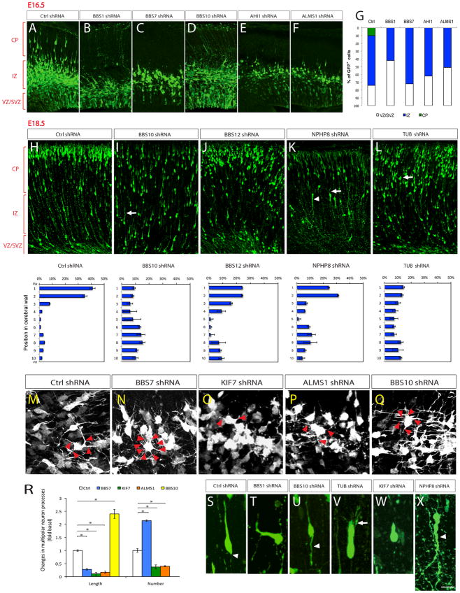 Figure 4
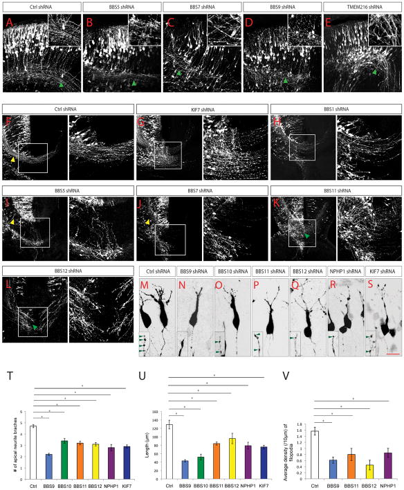 Figure 5
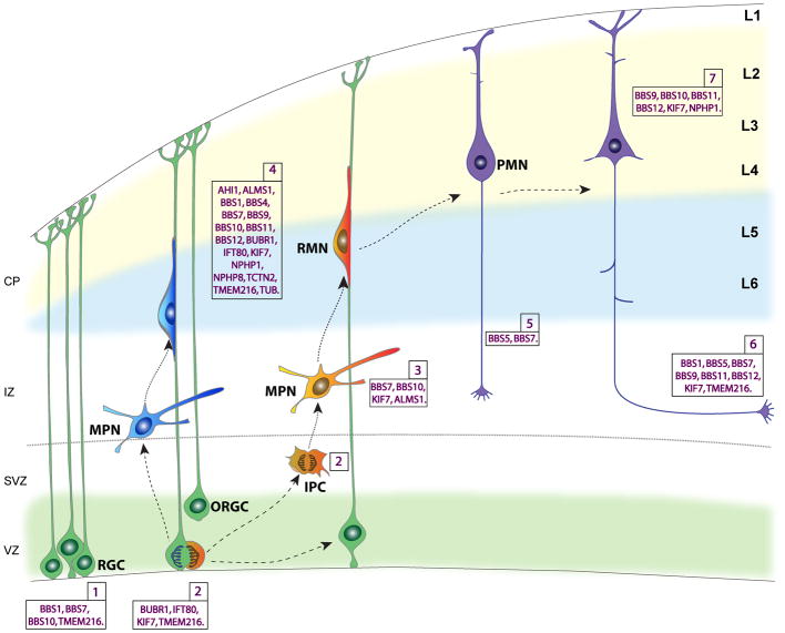 Figure 6
Peer Reviews
No public reviews on file for this paper yet. If you reviewed it on a platform where reviews are public (OpenReview, ICLR, NeurIPS, ICML), you can paste yours below so the community can read it here.
Videos
No videos yet. Explain this paper in a talk, walkthrough, or lecture? Add one.
Taxonomy
TopicsEmployment, Labor, and Gender Studies
Introduction
Cerebral cortex forms as a result of a coordinated sequence of events: radial progenitor formation, neurogenesis, neuronal migration, post-migratory neuronal differentiation, and connectivity. An efficient intracellular response to extracellular signaling cues is fundamental for coordinating the various cellular events that underlie the formation of cerebral cortex. Primary cilia, the microtubule-based, slender, antenna-like projections from cells, are essential integrators and conveyors of signal transduction. Cortical neuronal progenitors and developing neurons have a primary cilium. The significance of cilia function for cortical development and function ^1^ is evident in developmental brain disorders such as Joubert, Meckel-Gruber, orofaciodigital, and Bardet-Biedl syndromes (commonly referred to as ciliopathies), where disrupted cilia function and the resultant changes in cortical formation underlie cognitive deficits and intellectual disabilities ^2–4^. Moreover, neuropsychiatric disorders such as autism spectrum disorders and schizophrenia are also associated with human ciliopathies ^2–5^. These observations suggest that impaired cilia function can hinder the development of neural circuitry and activity, leading to significant functional deficits. Therefore, delineating specific functions of ciliopathy-associated genes in distinct stages of cortical development can help decipher the biological basis of brain function abnormalities in ciliopathies.
Functional genomic analysis of cilium has led to the identification of a diverse group of cilia and centrosome specific proteins ^1–4,6–8^. Among them, mutations in 87 of these genes have been associated with human ciliopathies (Supp. Tables 1, 2); mutations in 77 of these genes thus far are known to result in neurobehavioral or neurodevelopmental deficits in humans (Supp. Tables 1, 2, 3). A necessary first step in our ability to devise effective therapeutic interventions for these cilia-related neurodevelopmental diseases is an understanding of their biological basis. Disrupted neural circuitry due to genetic mutations in cilia-specific genes in ciliopathy patients could arise as a result of deficits in the generation of appropriate number and types of neurons, disrupted neuronal migration, placement, or connectivity in the developing brain. Pinpointing which of these processes is disrupted as a result of a genetic mutation can be a complex and time-consuming process. However, a shRNA library-based gene knockdown approach coupled with routinized assays for distinct stages of cortical development allow for rapid and efficient assessment of the brain developmental roles of ciliopathy genes. With this goal in mind, we compiled a library of shRNA targeting 30 genes known to be linked to human ciliopathies with neurological deficits (Supp. Table 4). We used this library to query how ciliopathy-related genes affect distinct stages of brain development, in particular neural progenitor development, neuronal migration, neuronal differentiation, early neuronal connectivity, following shRNA-mediated gene knockdown or control shRNA expression in embryonic brains. These studies give us a comprehensive insight into how human ciliopathy-related genes affect distinct stages of cerebral cortical formation and reveal hitherto undefined neurodevelopmental pathways whose disruption are likely to be integrally related to the development of neurological deficits in human ciliopathies.
Results
shRNA library of ciliopathy genes
Mutations in 87 cilia related genes have thus far been associated with human ciliopathies (Supp. Tables 1, 2, 3). Mutations in 77 of these genes are known to be associated with neurobehavioral or neurodevelopmental deficits in humans (Supp. Tables 1, 2, 3). The developmental and neurological consequences of mutations associated with the majority of these genes are yet to be clarified. We compiled a library of shRNAs targeting 30 of the ciliopathy genes with defined links to neurodeficits in humans to systematically evaluate their function in cerebral cortical development. Previously well-studied ciliopathy related genes with known developmental functions (for example, ARL13B^9,10^, INPP5E ^11,12^, IFT88 ^13^) were excluded. This library was assembled using the resources available at the UNC Gene Therapy Center (Supp. Table 4). Three to six different shRNAs for each gene were combined into two pools to achieve knockdown efficiency. Effects were confirmed with duplicated pools. As control for off-target disruptions, a vector containing scrambled shRNA was used. Target gene knockdown efficiency of shRNAs was validated using quantitative reverse transcription-polymerase chain reaction (qRT-PCR) (Supp. Table 5). Validity of specific shRNAs was further checked by randomly analyzing off target effects on other ciliopathy genes in the list. No such off target knockdown was detected. Further, several shRNAs were found not to induce expected changes in specific gene expression. They were used as additional controls and did not produce any changes in our cortical development assays.
shRNA library screen for ciliopathy gene functions in brain
To understand how ciliopathy-related genes affect early brain development, different ciliopathy gene-specific shRNA or control shRNA vectors were electroporated into the developing cerebral cortex at E14.5. For some of the genes (BBS7, BUBR1, KIF7), rescue of shRNA effects was tested using co-electroporation of shRNA resistant human cDNAs (UNC Gene Therapy Center). Electroporated embryos were allowed to survive for two or four days in vivo, and were analyzed for progenitor proliferation/ organization, neuronal migration, laminar organization, and differentiation of post-migratory cortical neurons and their projections. The effect of shRNAs on distinct aspects of cortical development were assayed as follows.
Cortical progenitors electroporated with shRNAs at E14.5 were labeled with radial glial (RG)-specific anti-RC2 or anti-Nestin antibodies or intermediate progenitor-specific anti-Tbr2 antibodies at E16.5. E16.5 embryos were pulse labeled with BrdU 1 hour prior to removal. Radial glial morphology and the apical and basal endfeet of GFP^+^/RC2^+^ RG were examined for changes in RG polarity. The apical molecular polarity of RG was evaluated by the characteristic apical β-catenin enrichment at the ventricular surface. The radial glial progenitor division was evaluated using PH3 and BrdU immunolabeling. The number and position of GFP^+^/BrdU^+^ or PH3^+^ nuclei were used to examine changes in the rate of cell division and the position of progenitor nuclei during cell cycle. Actively proliferating intermediate progenitors were co-labeled with anti-Tbr2 and anti-BrdU antibodies to monitor the changes in their proliferation patterns (Figure 1).
The extent of migration of newly generated GFP^+^ neurons was assessed by measuring the distance between ventricular surface and the leading front of GFP^+^ migratory neurons, and by binning the distribution of GFP^+^ neurons across the developing cortical wall. Directionality (polarity) of neuronal migration was categorized as “oriented” or “misoriented” based on the angle of orientation of the leading process of migrating neurons relative to the pial surface (oriented: 75°–90°, misoriented: < 75°). The final positioning of cortical neurons within the developing cortical layers was investigated by co-labeling GFP^+^ neurons with antibodies to layer specific markers Tbr1 (layer VI), Ctip2 (layer V), and Cux1 (layer II–III). Distribution of co-labeled neurons were quantified and compared between control and shRNA-expressing groups (Figure 1).
Post-migratory differentiation of GFP^+^, shRNA expressing neurons in the cortical plate (CP) was evaluated by determining whether these neurons display an axon, and if the axon projects first apically and then laterally towards contralateral cortical or subcortical regions. The elaboration of apical and basal neurites was evaluated by measuring the length and number of the primary neurite branches and the density of filopodia on primary apical neurites (Figure 1).
The effect of ciliopathy-specific genes on cortical progenitors
The generation and maintenance of apico-basally polarized radial glial scaffold is an essential first step in cerebral cortical formation. Of the 30 ciliopathy genes tested, knockdown of 4 genes (BBS1, BBS7, BBS10, and TMEM216) resulted in disruption of the apical-basal polarity of RG cells (Supp. Table 6). Control RG progenitors display characteristic polarized morphology, with cell soma in the VZ, an apical endfoot, an elongated polarized basal process oriented towards the pial surface, and branched basal endfeet attached to the pial basement membrane (BM) (Figure 2A–D). Knockdown of BBS1 (Bardet-Biedl syndrome 1) resulted in wavy RG processes (Figure 2E) and excessively branched basal endfeet (Figure 2F, asterisk). In contrast, TMEM216 (transmembrane protein 216) or BBS10 (Bardet-Biedl syndrome 10) deficiency led to short or retracted basal RG processes with aberrant branching (Figure 2G, I, J) and clubby endfeet structures (Figure 2H, K; asterisk). Further, BBS10 knockdown also led to loss of apical enrichment of β-catenin and significantly increased percentage of RG cells without apical endfeet (Figure 2D, L, M). In sharp contrast to the effects of the above 3 genes, BBS7 knockdown caused the ventricular epithelium to invaginate towards the pial surface, resulting in rosette-like structures with actively dividing progenitors localized in the center of the rosette lumen and migrating neurons radiating outwards (Figure 2N, O; Supp. Figure 1). Taken together, these results indicate that different ciliopathy-specific genes (BBS1, BBS7, BBS10, and TMEM216) modulate distinct aspects of apical-basal polarity of RG scaffold and the integrity of the proliferative niche organization.
As cerebral cortex forms, precise control of radial progenitor cell cycle dynamics is required for appropriate precursor pool expansion and neurogenesis. Symmetrical divisions of RG produce daughter radial progenitors and help expand the progenitor pool. As neurogenesis begins, radial progenitors divide asymmetrically and produce daughter neurons and intermediate precursor (IP) cells. IPs then undergo symmetric neurogenic divisions and predominantly give rise to the neurons of upper layer identities. Knockdown of BUBR1 (Bub1-related kinase; BUB1B), IFT80 (intraflagellar transport 80 homolog), KIF7 (kinesin family member 7), and TMEM216 led to decreased cell divisions of both apical radial progenitors (Figure 3A–E, arrows; Supp. Figure 1) and basal IPs as indicated by the reduced GFP^+^/PH3^+^ cells (Figure 3A–E, arrowheads) and basally localized GFP^+^/Tbr2^+^, or GFP^+^/Tbr2^+^/BrdU^+^ cells (Figure 3F–J; Supp. Figure 1; Supp. Table 6). These findings suggest that distinct ciliopathy genes may selectively modulate progenitor cell division and thus, cortical neurogenesis.
Ciliopathy genes and neuronal migration
Newborn neurons migrate radially through the intermediate zone (IZ) to reach their specific cortical layers in the developing cortical plate (CP). During the initial phase of radial migration, newborn neurons within the lower intermediate zone transiently assume a characteristic “multipolar” morphology to explore their microenvironment for directional cues, and then transform into a bipolar morphology as they migrate radially (Figure 4A). The bipolar neurons subsequently migrate towards CP by adhering to the basal radial glial process, with their leading process oriented towards the pial surface (Figure 4A). Knockdown of BBS1, BBS7, BBS10, AHI1 and ALMS1 resulted in a significantly higher percentage of newborn GFP^+^ neurons remaining as Tuj-1^+^ (and Tbr2^−^) multipolar neurons in the lower IZ at E16.5 (Figure 4B–G), suggesting a defect in the appropriate multipolar-to-bipolar transition. Notably, knockdown of these genes only delayed but not permanently stalled this transition, as indicated by the presence of bipolar migratory neurons at E18.5 in these cortices (Figure 4 and Supp. Figure 1). Compared to control, knockdown of BBS7, ALMS1 (Alstrom syndrome 1), and KIF7 resulted in thinner and shorter processes of the multipolar neurons, whereas knockdown of BBS10 resulted in longer processes (Figure 4M–R; Supp. Figure 2; Supp. Table 7). BBS7 knockdown also led to an increased number of processes, whereas knockdown of ALMS1 and KIF7 led to decreased number of processes in multipolar neurons (Figure 4M–P, R; Supp. Figure 2; Supp. Table 7).
In total, knockdown of 17 ciliopathy genes including AHI1, ALMS1, BBS1, BBS4, BBS7, BBS9, BBS10, BBS11 [TRIM32], BBS12, BUBR1 [BUB1B], IFT80, KIF7, NPHP1 (nephronophthisis 1), NPHP8 [RPGRIP1L], TCTN2, TMEM216, and TUB (Tubby protein homolog) resulted in delayed migration (Figure 4A–L; Supp. Figures 1, 2; Supp. Table 7). The migration delay is associated with increased leading process branching in BBS1, BBS4, BBS10, BUBR1 [BUB1B], IFT80, NPHP8 [RPGRIP1L], and TUB deficient neurons (Figure 4I, K, L, V [arrows]; Supp. Figure 2 D, E, H, I [arrows]), and misorientation in BBS1, NPHP8 [RPGRIP1L] and TCTN2 deficient neurons (Figure 4K, T, X; Supp. Figure 2 L [arrowhead]; Supp. Table 7). Moreover, knockdown of BBS4, BBS12, BUBR1 [BUB1B], IFT80, KIF7, NPHP1, NPHP8 [RPGRIP1L], and TUB resulted in shortened leading process (Figure 4; Supp. Figure 2; Supp. Table 7), whereas knockdown of BBS10 and NPHP8 [RPGRIP1L] led to aberrantly elongated trailing processes (Figure 4U, X [arrowheads]). Taken together, these results demonstrate that ciliopathy genes can regulate distinct aspects of cortical neuron migration, including the transient multipolar stage, multipolar-to-bipolar transition and glial-guided radial migration. Although control shRNA expression consistently did not reveal altered array of migration patterns, a recent study on doublecortin and neuronal migration highlights the importance of further validation of neuronal migration effects observed with shRNAs using additional in vivo mouse genetic tools ^14^.
Ciliopathy genes that required for neuronal identity and laminar organization
Upon reaching the CP, newly generated neurons migrate past older neurons to occupy more superficial regions of the CP, where they terminate their migration and acquire distinct laminar and neuronal subtype identities. Compared to control, knockdown of AHI1, ALMS1, BBS1, BBS4, BBS7, BBS9, BBS10, BBS11 [TRIM32], BBS12, BUBR1 [BUB1B], IFT80, KIF7, NPHP1, NPHP8 [RPGRIP1L], TCTN2, TMEM216, and TUB resulted in significantly increased percentage of GFP^+^/Cux1^+^ neurons localized at deeper CP positions where Ctip2^+^ or Tbr1^+^ neurons are normally localized (Supp. Table 8; Supp. Figure 3). Therefore, consistent with their role in neuronal migration, knockdown of these genes impair the laminar placement of neurons with upper layer identities. Quantification of the percentage of GFP^+^/layer marker^+^ (Cux1, Ctip2, or Tbr1) neurons indicate no significant differences between control and shRNA groups, suggesting that neuronal layer identity was not altered by shRNA expression (Supp. Table 8). Together, these data demonstrate a critical role for groups of ciliopathy genes including AHI1, ALMS1, BBS1, BBS4, BBS7, BBS9, BBS10, BBS11 [TRIM32], BBS12, BUBR1 [BUB1B], IFT80, KIF7, NPHP1, NPHP8 [RPGRIP1L], TCTN2, TMEM216, and TUB in the coordination of migration and placement of neurons in the developing cerebral cortex.
Effect of Ciliopathy genes in post-migratory differentiation of neurons
After arriving at their laminar destination within the developing cortical plate, projection neurons extend axons and dendrites as they assemble into functional neural circuits. During this process, neurons initially extend an axon, through the corpus callosum to the contralateral cortex or through the internal capsule to sub-cerebral targets. In addition, neurons also extend apical dendrites towards the pial surface and extensive basal dendrites from the cell soma. The dendrites of cortical neurons also develop small protrusions called spines, which are the major sites of synaptic contacts. We found that knockdown of BBS5, BBS7 resulted in significantly reduced axon outgrowth (Supp. Table 9; Figure 5A, F, I, J). The axons of BBS5, BBS7, BBS9, BBS11 [TRIM32], BBS12, and TMEM216 deficient neurons also display aberrant trajectory and fasciculation (Figure 5A–E, K, L). Knockdown of BBS1, BBS5, BBS7, BBS11 [TRIM32], BBS12, and KIF7 caused a substantial reduction in axons that project across the midline to contralateral cortex (Figure 5F–L; Supp. Figure 1; Supp. Table 9). This defect in midline crossing in BBS5, BBS7 deficient axons is caused not only by reduced axon outgrowth but also by disrupted axon guidance. These axons reached midline, but instead of crossing, project aberrantly towards subcortical targets (Figure 5I–J). Although the dendrites and dendritic spines are not fully formed at E18, deficiency in BBS9, BBS10, BBS11 [TRIM32], BBS12, NPHP1, or KIF7 resulted in reduced apical neurite branching and total length (Figure 5M–U; Figure S1; Supp. Table 9). Additionally, BBS9, BBS11, BBS12, and NPHP1 knockdown also resulted in lower density of filopodia (Supp. Table 9), many of which eventually form dendritic spines (Tada and Sheng, 2006). In summary, these results suggest that ciliopathy related genes BBS1, BBS5, BBS7, BBS9, BBS10, BBS11 [TRIM32], BBS12, KIF7, NPHP1, and TMEM216, differentially modulate appropriate patterns of post-migratory neuronal differentiation in cerebral cortex, including axonal growth, axonal guidance, neurite extension and arborization.
Discussion
Structural and functional neurological deficits are a common feature of ciliopathies in humans. However, the role of primary cilia-related genes linked to ciliopathies in brain development and function remains largely unknown. In this study, we carried out a systematic in vivo characterization of the role of 30 ciliopathy genes in major steps of cerebral cortical formation, ranging from progenitor development, neuronal migration, neuronal differentiation, and early neuronal connectivity. Importantly, this electroporation based approach enables the exploration of cilia-related gene functions in cerebral cortical development while avoiding hydrocephalus, noted often in cilia mutants. Our observations provide a comprehensive survey of ciliopathy gene functions in the context of cerebral cortical development. The developmental expression patterns of ciliopathy genes often correlate with their distinct functions during cerebral cortical development ^15,16,17^ (Supp. Figure 4). Our results link deficiency in 17 of ciliopathy genes to a variety of neurodevelopmental defects that may help us understand the diverse clinical features of ciliopathies, including cortical hypoplasia, ectopias, and axonal fiber tract defects, and the resultant functional outcomes such as intellectual disabilities ^1,4,18^. Currently, the known ciliopathy- related gene mutations are thought to lead to loss of function of respective proteins. The loss-of function approach used in our screen is consistent with this, but is limited in its ability to model gain of function mutations. However, further examination of these gene functions with shRNA resistant gene constructs (Supp. Figure 1), gene specific shmiRNA, mouse genetic tools, and related human gene mutations will be necessary to fully understand the role of these genes in brain developmental abnormalities in ciliopathies ^14^. Use of human mutant alleles will be particularly helpful in uncovering potential gain of function mutations. Gene manipulations at developmental stages other than what were described in this study may help confirm or reveal additional stage specific effects of ciliopathy genes, especially for the ones without any detectable phenotypes in our assays. Cell biological exploration of these gene functions in cilia compartments and associated centrosome is also necessary. Nevertheless, this study provides a template for further exploration of the biological basis of the brain structural and functional deficits associated with ciliopathies. These new insights into the functions of ciliopathy genes help link human brain malformations seen in ciliopathies to disruptions in specific cortical developmental events (Figure 6).
The 4 ciliopathy genes (BUBR1 [BUB1B], IFT80, KIF7, and TMEM216) we identified as modifiers of progenitor proliferation encode proteins localized to distinct ciliary compartments (Supp. Table 1). BUBR1 [BUB1B], localized in centrosome/basal body, is a key spindle assembly checkpoint protein, whose insufficiency leads to cell cycle misregulation and ciliogenesis defects ^19^. Importantly, mutations in BUBR1 [BUB1B] cause a rare human disorder mosaic-variegated aneuploidy (MVA) or premature chromatid separation syndrome (PCS), a novel ciliopathy syndrome characterized by microcephaly and mental retardation ^19,20^. TMEM216 is a component of transition zone and is required for centrosome/basal body docking to initiate ciliogenesis ^21–23^. TMEM216 is a causative gene for ciliopathy syndromes JSRDs and MKSs, with associated mental retardation ^21–23^. Our observation of disrupted neural progenitor proliferation in BUBR1 [BUB1B] and TMEM216 deficient brains is consistent with the clinical symptoms, such as microcephaly, found in human patients. On the other hand, IFT80 and KIF7 are members of IFT complexes and deficiency of IFT80 or KIF7 results in impaired ciliary signaling transduction without loss of cilia ^24–26^. Mutations of IFT80 cause Jeune asphyxiating thoracic dystrophy (JATD) and short rib polydactyly type III (SRP type III) ^24^. Mutations in KIF7 cause hydrolethalus (HLS) and acrocallosal syndromes (ACLS) ^25^. The 4 ciliopathy genes we identified as regulators of progenitor proliferation could represent two distinct molecular pathways: centrosome-dependent and ciliary signaling-dependent, underlying some of the cortical deficits (e.g., microcephaly) associated with these gene mutations.
Knockdown of BBS1, BBS10, BBS7, and TMEM216, lead to disruption of distinct aspects of the polarized RG scaffold. BBS1, BBS10 and TMEM216 are required for the maintenance of RG scaffold; BBS10 is required for the polarized organization of RG scaffold, whereas knockdown of BBS7 resulted in the formation of rosette-like heterotopias caused by invagination of the progenitor niche. These observations suggest their converging yet differential influence of cilia-related genes on the RG polarity. Further understanding of the functions of these proteins localized to different ciliary compartments in the regulation of distinct aspects of RG polarity could help dissect the molecular mechanisms underlying RG organization and function. Cortical heterotopias is an abnormal brain patterning defect commonly seen in Bardet-Biedl syndrome (BBS), Joubert syndrome (JBTS), Meckel–Gruber syndrome (MKS) ^9,13,27^, caused by defects in the formation and maintenance of polarized RG progenitors and RG-guided neuronal migration. Importantly, mutations in BBS1, BBS7, and BBS10 cause BBS, whereas mutations in TMEM216 cause JBTS and MKS ^18,23,28^. The presence of cortical heterotopia in these ciliopathies is associated with epilepsy and mental retardation ^2,4,7,29,30^. Taken together, these data demonstrate the converging role of multiple ciliopathy genes in the development and function of RG progenitors. Disruption of these processes may underlie cortical malformations such as microcephaly and heterotopias seen in ciliopathies related to mutations in these genes.
Defects in neuronal migration and differentiation of cortical neurons can impair the organization and activity of the cortical circuitry, and are thought to underlie a broad spectrum of neurodevelopmental and psychiatric disorders ^31,32^. Neuronal migration defects such as cortical periventricular heterotopia and polymicrogyria are common features of various ciliopathies (Supp. Table 1). Surprisingly, our results show that 17 different ciliopathy genes modulate distinct phases of cortical neuron migration, highlighting the critical nature of primary cilia in neuronal migration during cortical development. These results also provide molecular bases and genotype-phenotype correlations for the brain abnormalities caused by aberrant neuronal migration in ciliopathies. For example, ciliopathy patients carry mutations in AHI1 or BBS genes show cortical heterotopia and polymicrogyria ^28,33^. Consistently, our results suggest that neurons expressing AHI1 or BBS (BBS1, BBS4, BBS5, BBS7, BBS9, BBS10, BBS11 [TRIM32], BBS12) shRNA show migration defects. We also observed that knockdown of ciliopathy genes ALMS1, BUBR1 [BUB1B], IFT80, NPHP1, NPHP8 [RPGRIP1L], TCTN2, TMEM216, and TUB retarded neuronal migration. Neurons deficient in these genes display excessive branching or mis-orientated leading processes. Since knockdown of BBS1, BBS10, and TMEM216 also led to disrupted RG morphology, the migration defects seen in these 3 ciliopathy gene knockdowns may in part result from compromised RG scaffold, whereas the migration defects with the other genes could be due to their specific neuronal functions. Knockdown of 5 genes (BBS7, BBS10, ALMS1, KIF7, and AHI1) led to defects in mutipolar-bipolar transition stage, suggesting a selective requirement for primary cilia in the early stages of neuronal migration. Though the molecular mechanisms involved in the transition from multipolar to radial migration are yet to be fully understood, multiple cues such as adhesion molecules (e.g. TAG1, N-cadherin, Connexin 43), cytoskeletal regulators (e.g. CDK5 and ARX), intracellular signaling molecules (e.g. Rap1, RhoA, Mark2, LKB1), transcription factors (e.g. FoxG1, Scratch 1, 2, KLF4) and extracellular signals such as Neurog2, Semaphorin 3A, Netrin, and Reelin are involved in this process ^34–42^. Our results suggest that primary cilium and its associated signaling proteins could help sense, integrate and convey signaling cues necessary for this transition.
Consistent with the migration defects, we found aberrant laminar placement of neurons following disruption of 17 ciliopathy genes (AHI1, ALMS1, BBS1, BBS4, BBS7, BBS9, BBS10, BBS11 [TRIM32], BBS12, BUBR1 [BUB1B], IFT80, KIF7, NPHP1, NPHP8 [RPGRIP1L], TCTN2, TMEM216, and TUB). Recent studies also showed that genetic deficiency of BBS1 or BBS4 in mice results in perturbed migration and lamination defects ^31,43^. The identification of multiple ciliopathy gene with previously unknown roles in neuronal migration and placement suggest that this stage of cortical development is highly vulnerable in ciliopathies and the resultant changes in the formation of cortical circuits may underlie cognitive deficits associated with ciliopathies.
Postmigratory cortical neurons undergo extensive neurite outgrowth to generate the appropriate axon-dendritic architecture of the neuronal circuits. A steady and polarized delivery of membrane proteins, such as guidance cue receptors, and cytoskeletal components to the growing neurites is required to enable the neurites to extend in appropriate directional patterns as they contact and form synapses with appropriate partners. Consistently, primary cilia mediated signaling cascades (e.g. GPCR and Wnt signaling) and cilia-associated centrosome mechanisms have both been shown to regulate neuronal dendritic and axonal outgrowth and refinement ^44–47^. We found that 2 BBS genes (BBS5, BBS7) are required for axonal outgrowth, whereas 1 transition zone gene (NPHP1), 4 BBS genes (BBS9, BBS10, BBS11 [TRIM32], and BBS12), and an IFT regulator, KIF7, modulate neurite outgrowth and filopodial formation, indicating the differential and specific influence of ciliopathy genes over post-migratory neuronal differentiation.
Defects in axonal tract development, including callosal agenesis, disrupted pyramidal decussation and superior cerebellar peduncle (SCP) decussation, are often seen in ciliopathy patients, and these axonal tract defects are thought to underlie some of the motor and cognitive deficits in ciliopathies ^2,4,17,25,29,48^. It is known that midline glial structures (e.g. glial wedge) and secreted guidance molecules (e.g. WNT, Slit2, Shh, Netrin1) released from these structures are crucial for guiding axonal midline crossing and corpus callosum formation ^48–52^. For example, Shh functions to guide commissural axons by acting directly as a chemoattractant for the axon growth cones. Consistently, mutations in KIF7, known to disrupt Shh pathway, cause acallosal syndromes in humans and lead to dysgenesis or agenesis of corpus callosum in mice ^25,26,53^. Our observations indicated that in addition to KIF7, knockdown of 5 of the BBS genes, BBS1, BBS5, BBS7, BBS11 [TRIM32], and BBS12, also led to axonal guidance and midline crossing defects. How these cilia-related proteins, localized primarily in cilium near the cell soma, modulate of dynamics of the axonal growth cone remains unclear. The answer might lie in non-ciliary expression of ciliopathy genes at other sub-cellular compartments such as growth cone. To date, several ciliopathy genes have been shown to have non-ciliary expression (e.g. KIF7 in growth cone, KIF3A, BBS4, BBS8 in synapse) or cilia-independent functions (e.g. KIF7 in axonal guidance, BBS4 in synaptic transmission), suggesting additional key roles for some of the ciliopathy genes in protein trafficking and membrane transport ^53–58^. Future effort aimed at dissecting the role of ciliary vs. non-ciliary function of ciliopathy genes during corticogenesis will be essential to delineate how they contribute to the etiology of constellation of symptoms associated with ciliopathies.
Many of the causative genes of ciliopathy syndromes display genetic and/or direct physical interactions, leading possibly to perturbations in common cellular processes ^6,59^. For example, it has been shown that KIF7 genetically interact with BBS loci ^25^. Deficiency of KIF7 and several BBS genes led to similar defects in progenitor proliferation and neuronal migration, indicating convergent biological influences of genetic interaction between KIF7 and BBS genes. On the other contrary, some ciliopathy related genes can exert effects on multiple aspects of human brain development (Supp. Table 10). For example, mutations in AH1 can disrupt neurogenesis, neuronal migration, and axon growth, leading to hypoplasia, heterotopia, and failed axonal decussation in humans (Supp. Table 10). If and how AH1 convergently interact with other ciliopathy genes to modulate these diverse cellular processes remains unclear.
Based on localization in cilia and functional interactions, ciliopathy genes have also been classified into functional networks ^6,60^. BBSome is the best studied of the ciliopathy protein complexes. BBSome contains 7 highly conserved BBS proteins (BBS1, BBS2, BBS4, BBS5, BBS7, BBS8 [TTC8], BBS9) that localize to basal body and functions as a unit that mediates vesicle trafficking to the ciliary membrane ^61^. Three chaperonin-like BBS proteins (BBS6 [MKKS], BBS10, and BBS12) form a higher-order complex, mediate BBSome subunit association, and are required for BBSome assembly ^61,62^. Deficiency of BBS genes cause Bardet–Biedl syndrome, one of the well-characterized ciliopathies ^28^. Our results show that deficiency in 8 BBS protein family members (BBS1, BBS4, BBS5, BBS7, BBS9, BBS10, BBS11 [TRIM32], BBS12) led to significant cortical defects including disrupted RG scaffold, perturbed neuronal migration, and impaired neuronal differentiation, thus highlighting the essential roles of BBS genes in brain development. Importantly, the results of our functional assays further clarify some of the known effects of BBSome ciliopathy in humans: It has been suggested that deficiency in any member of BBSome or chaperonin-like BBS complex disrupts the functionality of the BBSome and cause similar phenotypes in human patients and BBS animal models ^59,62–66^. Consistent with this, our results show that deficiency of BBSome members and chaperonin-like BBS proteins often led to defects in common neurodevelopmental events. We found that BBS1, BBS7, and BBS10 are involved in RG progenitor development, knockdown of BBS1, BBS4, BBS7, BBS9, BBS10, BBS11 [TRIM32], or BBS12 resulted in neuronal migration defects, whereas loss of BBS1, BBS5, BBS7, BBS9, BBS10, BBS11 or BBS12 led to post-migratory, neuronal differentiation defects. However, our analysis also revealed other previously unknown distinctive requirements for individual BBSome members and chaperonin-like BBS proteins during corticogenesis. For example, knockdown of BBS1 and BBS10 resulted in excessive branching, and reduced branching of RG basal endfeet, respectively (Figure 2). Also, knockdown of BBS7 and BBS10 led to shorter, and longer multipolar neuronal processes, respectively (Figure 4). Thus, it is likely that even though BBSome and chaperonin-like BBS proteins function to synergize and converge on common developmental pathways, they still exert different regulatory roles during corticogenesis. The same also appears to be the case for other ciliopathy networks such as NPHP-JBTS-MKS complex.
Ciliopathies display a broad range of clinical features, high degree of pathological variability, genetic heterogeneity, and phenotypic overlaps, thus complicating their diagnosis and treatment ^4^. Elucidation of multifaceted functions of cilia and associated protein complexes is fundamental to our effort to understand the biological bases of ciliopathies and develop efficient therapeutic interventions. This comprehensive in vivo functional investigation of the causative genes of ciliopathies in the context of cortical development not only delineates varied functions of cilia in the construction of the cerebral cortex, but also revealed potential developmental bases of brain abnormalities in ciliopathies. In the future, the use of causative human mutant alleles with defined links to cortical abnormalities in this type of screen or analysis of genotyped neural cells generated from ciliopathy patient-derived induced pluripotent stem cells (iPSCs) can help further define the biological basis of ciliopathy mutations and enhance our understanding of cilia-related gene functions in brain development. Establishing a causal link between genotype and phenotype of ciliopathies with such approaches will be vital to develop rational therapeutic interventions for ciliopathies.
Methods
Mice
Mice were cared for according to guidelines approved by the University of North Carolina. Light/dark cycle in the vivarium is 7/7 hours. Animals were housed in groups of 3 adults per cage. C57/BL6 pregnant female mice were used for in utero electroporation. The day of vaginal plug detection was considered E0.5.
shRNA library of ciliopathy genes
A library of shRNAs specific to the 30 human ciliopathy genes was assembled using the resources available at the UNC Gene Therapy Center. Lists of shRNA constructs and sources are provided in the supplemental information (Supp. Table 4). Plasmids used for in utero electroporation were prepared using the EndoFree Plasmid kit (Qiagen). For each gene, two pools of shRNAs containing two to three individual shRNAs were randomly combined and used for electroporation. Non-silencing scrambled shRNA (sc-108080, Santa Cruz), GIPZ or pLKO.1 shRNA vectors (GE Dharmacon and UNC Gene Therapy Center) were used as control. Knockdown efficiency (KE) of shRNAs (fold change compared to control) was measured using quantitative real time-PCR and are indicated for each shRNA group. Knockdown efficiency of 70% was used as a threshold for shRNA pools. Each pool consisted of equivalent amount of different shRNAs (2 or 3/ pool) listed. For BBS8, only the effective pool of ShRNA containing three different shRNAs was used. shRNAs from Santa Cruz are prepooled (4shRNAs/gene) by the vendor.
Tissue Culture, transfection, RNA isolation and purification
Mouse kidney inner medullary collecting duct (IMCD3) epithelial cells were grown in Dulbecco’s Modification of Eagle’s Medium (DMEM)/ F12 media supplemented with 10% fetal bovine serum (FBS) and 1% penicillin streptomycin (P/S) antibiotics at 37°C with 5% CO_2_. The cells were allowed to reach 80% confluency and ciliogenesis was induced by serum starvation of the cells for 24 hours. IMCD3 cells were then transfected using Invitrogen Lipofectamine 2000 Reagent according to manufacture’s instructions (Sigma). For each well of cells in a 6-well plate, 1.5 μg of shRNAs and pCIG2 was used. The cells were imaged three days post-transfection to measure transfection efficiency (percentage of GFP^+^ cells). Transfected IMCD3 cells were then lysed for RNA purification using a Qiagen RNeasy mini kit according to manufacture’s instructions.
RT-PCR and qPCR
Reverse Transcription-PCR was performed using the Invitrogen SuperScript III First-Strand Synthesis System. Real Time PCR was performed using Applied Biosystem Power SYBR Green PCR master mix and the appropriate forward and reverse primers (Supp. Table 5). The real time PCR reactions were performed in triplicates. Peptidylprolyl isomerase A (Ppia) was used as an endogenous reference. The comparative (ΔΔC_T_) method of relative quantification was used to determine the level of gene knockdown.
In utero electroporation
In utero electroporations were performed as described previously^67,68^. Briefly, 1–2 μl of pool of plasmid DNA (1.5 μg/μl) were injected into the lateral ventricles of E14.5 brains and electroporated using 5 pulses at 30 V for 50 ms at 950 ms intervals through the uterine wall using a BTX ElectroSquarePorator (ECM 830). In shRNA+cDNA rescue experiments, human cDNAs were used at 1μg/μl concentration. Each pool of shRNAs or control vectors was electroporated into 4 different embryos. Embryos were then allowed to develop for 2 or 4 days prior to analyses.
Immunohistochemistry
The primary antibodies used were anti-RC2 (1:1, Iowa Hybridoma), anti-β-catenin (rabbit polyclonal, 1:1000;), anti-GFP (chicken, 1:1000; Abcam), anti-BrdU (mouse monoclonal, 1:50; BD Biosciences), anti-phospho-histone H3 (PH3) (rabbit polyclonal, 1:200; Millipore), anti-Tbr2 (1:500, AB23345, Abcam), anti-Ctip2 (1:500, AB18645, Abcam), anti-Cux1 (1:100, SC13024, Santa Cruz Biotechnology), anti-Tbr1 (1:500, AB31940, Abcam), anti-Brn1 (1:1,000, gift from A. Ryan, McGill University)^10^. Secondary antibodies were AlexaFluor 488 or Cy3-conjugated (Invitrogen and Jackson ImmunoResearch). Nuclei were counterstained with DAPI (Sigma). Electroporated mouse brains were removed and fixed with 4% paraformaldehyde overnight at 4°C. Brains were then embedded in 3% agarose and sectioned at 50 μm with a Vibratome (VT1000S; Leica Microsystems). Sections were blocked with PBS/10% goat serum/0.2% Triton X-100 for 1 h, and incubated in primary antibodies (see above) overnight at 4°C. After three washes in 1xPBS, sections were incubated with secondary antibodies (1:1000) at room temperature for 2 hours, washed, and mounted with Citifluor anti-fading solution (Agar). For BrdU labeling, BrdU (50 mg/kg) was injected intraperitoneally into pregnant mice one hour prior to brain tissue processing.
Quantitative analysis of progenitor proliferation, polarity, and niche organization
GFP^+^ radial glial progenitors were co- immunolabeled with RC2 or anti-nestin antibodies. The characteristic apical end feet attached to pial surface and branched basal endfeet were imaged and quantified per section in control and shRNA brains. Lack of apical endfeet, increased or decreased branching of basal en feet were measured. The apical molecular polarity of RG was detected by apical β-catenin enrichment at the ventricular surface of GFP^+^ radial progenitors. Changes in radial glial progenitor proliferation were detected using PH3 and BrdU immunolabeling. The number and position of GFP^+^/BrdU^+^ or PH3^+^ nuclei in the VZ was measured (25×10^3^ μm^2^ area) to examine changes in the rate of cell division and the position of progenitor nuclei during cell cycle. Actively proliferating intermediate progenitors in control and shRNA brains were co-labeled with anti-Tbr2 and anti-BrdU antibodies and quantified (25×10^3^ μm^2^ area) to monitor the changes in their proliferation patterns.
Quantitative analysis of neuronal migration and placement
Cerebral wall was divided into ten equal bins and the percentage of GFP^+^ neurons in each bin was counted^67^. Number of leading processes per neuron was counted in migrating neurons in the intermediate zone. The percentage of migrating neurons with leading processes oriented at an angle <75° to the pial surface was also measured. Percentage of GFP^+^/Layer marker^+^(Tbr1, Ctip2, or Cux1) neurons located below cortical plate, away from their normal laminar locations was quantified to detect changes in the laminar placement of shRNA expressing neurons. Images were acquired using Zeiss 710 and Zeiss 780 confocal systems. Imaging analysis was done using Zeiss LSM Image Browser and ImageJ software (NIH).
Quantitative analysis of post-migratory differentiation of neurons
Number of GFP^+^, shRNA expressing neurons in the cortical plate (CP) displaying an axon and the projection of the axon apically and then laterally towards contralateral cortical or subcortical regions was quantified per section. The length of apical and basal neurites and the number of the primary neurite branches were counted per GFP^+^ neuron. The density of immature filopodia on primary apical neurites was quantified per 10μm length.
Statistical analysis
GraphPad or Excel was used for data analysis. Two-tailed Student’s t test and two-way ANOVA with Tukey-Kramer multiple comparison test were performed using GraphPad. Data were collected and processed blindly. All data are expressed as means ± standard error of the mean (SEM). Statistical comparisons between different knockdown groups were not performed as there are differences in the efficacy of the knockdown of different genes.
Supplementary Material
1
The reference list from the paper itself. Each links out to its DOI / PubMed record.
- 1Guemez-Gamboa A Coufal NG Gleeson JG Primary Cilia in the Developing and Mature Brain Neuron 8251152110.1016/j.neuron.2014.04.024201424811376 PMC 4104280 · doi ↗ · pubmed ↗
- 2Lancaster MA Gleeson JG The primary cilium as a cellular signaling center: lessons from disease Current Opinion in Genetics & Development 1922022910.1016/j.gde.2009.04.008200919477114 PMC 2953615 · doi ↗ · pubmed ↗
- 3Louvi A Grove EA Cilia in the CNS: the quiet organelle claims center stage Neuron 691046106010.1016/j.neuron.2011.03.002201121435552 PMC 3070490 · doi ↗ · pubmed ↗
- 4Hildebrandt F Benzing T Katsanis N Ciliopathies The New England journal of medicine 3641533154310.1056/NEJ Mra 1010172201121506742 PMC 3640822 · doi ↗ · pubmed ↗
- 5Marley A Von Zastrow MA simple cell-based assay reveals that diverse neuropsychiatric risk genes converge on primary cilia P Lo S ONE 7e 4664710.1371/journal.pone.0046647201223056384 PMC 3463515 · doi ↗ · pubmed ↗
- 6Sang L Mapping the NPHP-JBTS-MKS protein network reveals ciliopathy disease genes and pathways Cell 14551352810.1016/j.cell.2011.04.019201121565611 PMC 3383065 · doi ↗ · pubmed ↗
- 7Novarino G Akizu N Gleeson JG Modeling human disease in humans: the ciliopathies Cell 147707910.1016/j.cell.2011.09.014201121962508 PMC 3202432 · doi ↗ · pubmed ↗
- 8Eggenschwiler JT Anderson KV Cilia and developmental signaling Annual review of cell and developmental biology 2334537310.1146/annurev.cellbio.23.090506.1232492007 PMC 209404217506691 · doi ↗ · pubmed ↗