B-mode and contrast-enhanced ultrasound versus magnetic resonance imaging in hepatic angiomyolipoma: a case report
Andrea Balbinot, Sofia Maria Bakken, Nicola Venturoli, Deborah Malvi, Elisa Albertini, Maria Giulia Pirini, Antonietta D’Errico, Carla Serra

TL;DR
This case report compares ultrasound and MRI in diagnosing a rare liver tumor in a patient with a genetic condition.
Contribution
The study highlights the diagnostic value of B-mode and contrast-enhanced ultrasound in hepatic angiomyolipoma.
Findings
B-mode and contrast-enhanced ultrasound correctly identified a benign hepatic lesion.
MRI initially suggested hepatocellular carcinoma, but ultrasound findings were confirmed by histology.
HAML is rare and can be challenging to diagnose, but ultrasound may aid in accurate assessment.
Abstract
Angiomyolipoma is a solid mesenchymal tumour that usually affects the kidney. Hepatic localization of angiomyolipoma (HAML) is rare and usually asymptomatic however it presents a challenging differential diagnosis. We present the case of a 45-year-old man affected by tuberous sclerosis complex type 2 (TSC2) and an hepatic lesion suspected to be hepatocellular carcinoma on magnetic resonance but whose Bmode ultrasound and contrast-enhanced ultrasound (CEUS) findings were consistent with benignity, as confirmed by histology.
Genes, proteins, chemicals, diseases, species, mutations and cell lines named across the full text — each resolved to its canonical identifier and authoritative record.
Click any figure to enlarge with its caption.
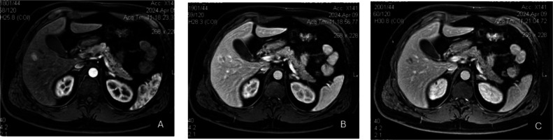 Figure 1
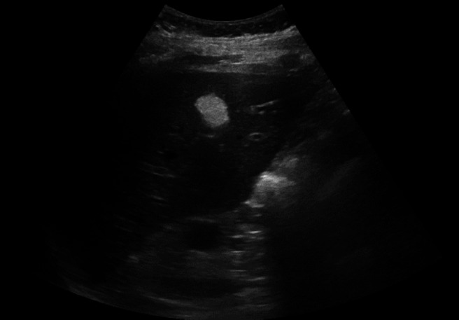 Figure 2
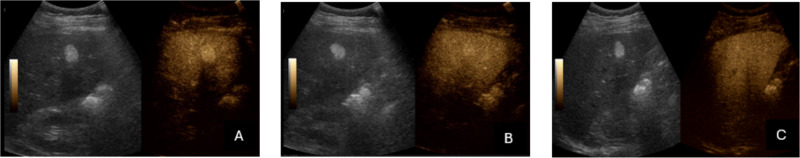 Figure 3
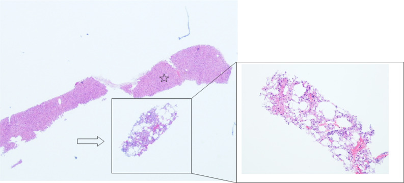 Figure 4
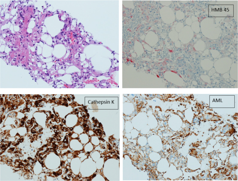 Figure 5
Peer Reviews
No public reviews on file for this paper yet. If you reviewed it on a platform where reviews are public (OpenReview, ICLR, NeurIPS, ICML), you can paste yours below so the community can read it here.
Videos
No videos yet. Explain this paper in a talk, walkthrough, or lecture? Add one.
Taxonomy
TopicsTuberous Sclerosis Complex Research