Sudden Death of a 17‐Month‐Old Chimpanzee ( Pan troglodytes ) due to a Congenital Undiagnosed Ventricular Septal Defect
Ivanela Kondova, Merel Langelaar, Arno A. W. Roest, Peter J. Heidt

TL;DR
A 17-month-old chimpanzee suddenly died from an undiagnosed heart defect similar to those seen in humans.
Contribution
This case highlights the occurrence of undiagnosed VSD in non-human primates leading to sudden death.
Findings
The chimpanzee had a congenital ventricular septal defect (VSD).
The heart showed left ventricular dilatation and right ventricular hypertrophy.
The VSD was not diagnosed before the sudden death of the infant.
Abstract
Ventricular septal defect (VSD) is a congenital defect that is frequently observed in humans. We report the sudden death of an infant chimpanzee due to a congenital undiagnosed VSD. During necropsy, the examination of the heart revealed a VSD, dilatation of the left ventricle and myocardial hypertrophy of the right ventricle.
Genes, proteins, chemicals, diseases, species, mutations and cell lines named across the full text — each resolved to its canonical identifier and authoritative record.
Click any figure to enlarge with its caption.
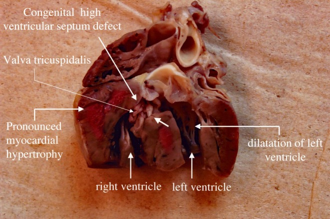 FIGURE 1
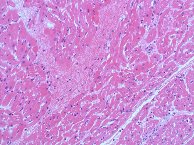 FIGURE 2
Peer Reviews
No public reviews on file for this paper yet. If you reviewed it on a platform where reviews are public (OpenReview, ICLR, NeurIPS, ICML), you can paste yours below so the community can read it here.
Videos
No videos yet. Explain this paper in a talk, walkthrough, or lecture? Add one.
Taxonomy
TopicsCongenital heart defects research · Congenital Heart Disease Studies · Cardiovascular Conditions and Treatments
Introduction
1
Chimpanzees ( Pan troglodytes ) are genetically and physiologically close to humans, and documentation of naturally occurring congenital anomalies is rare. In humans, congenital heart defects (CHDs) are the most frequent congenital abnormalities at live birth. Structural abnormalities of the heart present at birth are well documented in human medicine but reports of such conditions in non‐human primates, and especially great apes, are rare. Ventricular septal defect (VSD) is one of the most commonly observed congenital conditions in human newborns, resulting in abnormal communication between the left and right ventricles. It allows oxygen‐rich blood to move back into the lungs instead of being pumped to the rest of the body. The oxygen‐rich blood mixes with oxygen‐poor blood. These changes may increase blood pressure in the lungs and require the heart to work harder to pump the blood around. Small VSDs cause no problems and usually close on their own. Babies with medium or larger VSD may need surgery early in life to prevent complications.
This case report is selected from the archives of the Biomedical Primate Research Centre (BPRC) in the Netherlands. The research on chimpanzees at the BPRC ended in 2003, and in 2006, all the chimpanzees were donated to different zooparks in Europe.
We describe a sudden death due to a VSD with a diameter of ±1 cm in a young chimpanzee from the BPRC colony in the Year 2002. This report aims to contribute to the limited literature on CHDs in great apes and support comparative pathology and clinical management in zoological settings.
Case Report
2
A 17‐month‐old male chimpanzee was found dead in his group‐cage on 10‐06‐2002. The animal was in good body condition with unremarkable clinical history. In an attempt to reanimate the animal, heart massage was performed by the veterinarian without result. Necropsy was performed immediately after the death of the animal.
A multiplicity of subcutaneous haemorrhages was observed during the necropsy. However, the quantity of the haemorrhages was not considered to be related to the death of the animal, but considered to be related to the reanimation attempts. Histopathological examination verified the acute character of the haemorrhages.
The necropsy showed that the death of the animal was caused by a high‐positioned VSD of ±1 cm diameter, leading to failure of the circulatory system (Figure 1). Furthermore, dilatation of the left ventricle and myocardial hypertrophy of the right ventricle were evident. No other lesions that could be related to heart failure were found during the necropsy.
Gross appearance of the heart showing the ventricular septal defect.
Histological examination revealed multifocal segmental fibre degenerations with reactive resorptive inflammatory cell infiltration accompanied by multifocal intramural haemorrhages in the myocardium (Figure 2).
Microscopic findings of the heart (H&E staining, ×200). Segmental fibre‐degeneration with reactive resorptive inflammatory infiltrates and intramural extravasated erythrocytes (haemorrhage).
Discussion
3
A congenital VSD in a chimpanzee is mainly observed during necropsy. In the case that we present, the infant chimpanzee showed no signs indicating a VSD, such as poor eating, failure to thrive, fast breathing or breathlessness and easy tiring. The finding of a hypertrophied right ventricle and the sudden death without any clinical signs suggest that pulmonary hypertension due to increased pulmonary flow through the VSD played an important role in this unexpected death.
We came across only one published case of a VSD in a chimpanzee. This heart abnormality was found during necropsy in a prematurely born chimpanzee in which after birth heart murmur, tachypnoea, dyspnoea and disturbances of blood flow were observed [1]. Cases of a VSD in other non‐human primates are a case in a Bonnet macaque ( Macaca radiata ) [2], a case in a 15‐year‐old Sumatran orangutan ( Pongo abelii ) [3] and a case in a 3‐year‐old gorilla ( Gorilla gorilla gorilla ) [4]. A VSD was also observed in other animal species: pika [5], alpaca [6], horse [7] and calves [8]. The incidence in different animal species is unknown. However, a congenital VSD is regularly observed in human children because of the healthcare in developed countries, but it can exist undiagnosed, leading to the sudden death of a child [9]. The rarity of sudden death in affected children was demonstrated in a multicentre study from 10 countries, in which only two cases (1%) of lethal VSD were found out of a total of 186 cases of sudden death [10]. In a population‐based cohort study, including more than 25 000 newborns born in the greater Copenhagen area, a total of 850 newborns (3.3% of 25.556) with a VSD were identified. The newborns underwent echocardiography within 60 days after birth. After 1 year, 83.5% of all VSDs had closed spontaneously, resulting in a decrease of prevalence from 3.3% at birth to 0.5% in 1‐year‐old children [11].
Since the incidence of congenital VSDs in human primates is around 3% it can be expected that this percentage is also occurring in non‐human primates, for example chimpanzees. The numbers of congenital VSDs observed in non‐human primates in the literature are low. This may be due to the fact that they are often not noticed. They are mostly reported in case studies rather than large‐scale prevalence studies, which is the reason that the correct percentages are missing. It is unlikely that the incidence in non‐human primates is different from that in humans. The chance of a young chimpanzee dying from this defect, based on the human percentages, is therefore small. That we diagnosed such a case in the BPRC chimpanzee colony with a limited number of births (67 births between 1976 and 2000) was therefore unexpected, based on the fact that only in 0.5% of 1‐year‐old human children this defect is (still) present.
Conflicts of Interest
The authors declare no conflicts of interest.
The reference list from the paper itself. Each links out to its DOI / PubMed record.
- 1A. A. Binhazim , P. Alford , and M. Miller , “Ventricular Septal Defect in an Infant Chimpanzee ( Pan troglodytes ),” Journal of Medical Primatology 23, no. 6 (1994): 362–364, 10.1111/j.1600-0684.1994.tb 00299.x.7897643 · doi ↗ · pubmed ↗
- 2T. V. Anilkumar and S. Sandhyamani , “Atrial Septal Defect in a Bonnet Macaque ( Macaca radiata ),” Journal of Medical Primatology 19, no. 8 (1990): 749–752.2084263 · pubmed ↗
- 3R. A. Cook , P. Sheppardson , M. Mc Ginn , M. L. Roskop , and B. Y. Wong , “Evaluation of a Ventricular Septal Defect in an Orangutan: A Case Report,” Journal of Medical Primatology 15, no. 4 (1986): 303–308.3746889 · pubmed ↗
- 4C. Machado , F. G. Mihm , and C. Noe , “Diagnosis of a Ventricular Septal Defect in a Gorilla Using In Vivo Oximetry,” Journal of Zoo and Wildlife Medicine 20, no. 2 (1989): 199–202, http://www.jstor.org/stable/20094940.
- 5H. Shinohara and H. Nishimura , “Spontaneous Closure of Isolated Ventricular Septal Defect in the Pika (Ochotona rufescens rufescens),” Experimental Animals 35, no. 2 (1986): 169–173, 10.1538/expanim 1978.35.2_169.3732408 · doi ↗ · pubmed ↗
- 6H. Poser , A. Dalla Pria , G. M. De Benedictis , C. Stelletta , M. Berlanda , and C. Guglielmini , “Ventricular Septal Defect and Double‐Chambered Right Ventricle in an Alpaca,” Journal of Veterinary Cardiology 17, no. 1 (2015): 71–76, 10.1016/j.jvc.2014.10.005.25595611 · doi ↗ · pubmed ↗
- 7C. W. Lombard , W. K. Scarratt , and C. D. Buergelt , “Ventricular Septal Defects in the Horse,” Journal of the American Veterinary Medical Association 183, no. 5 (1983): 562–565.6618989 · pubmed ↗
- 8K. J. Mitchell and C. C. Schwarzwald , “Echocardiography for the Assessment of Congenital Heart Defects in Calves,” Veterinary Clinics of North America: Food Animal Practice 32, no. 1 (2016): 37–54, 10.1016/j.cvfa.2015.09.002.26922111 · doi ↗ · pubmed ↗