Case of a genodermatosis presenting with verrucous lesions mimicking treatment-refractory warts
Raj P. Fadadu, Charisse Orme

Abstract
Genes, proteins, chemicals, diseases, species, mutations and cell lines named across the full text — each resolved to its canonical identifier and authoritative record.
Click any figure to enlarge with its caption.
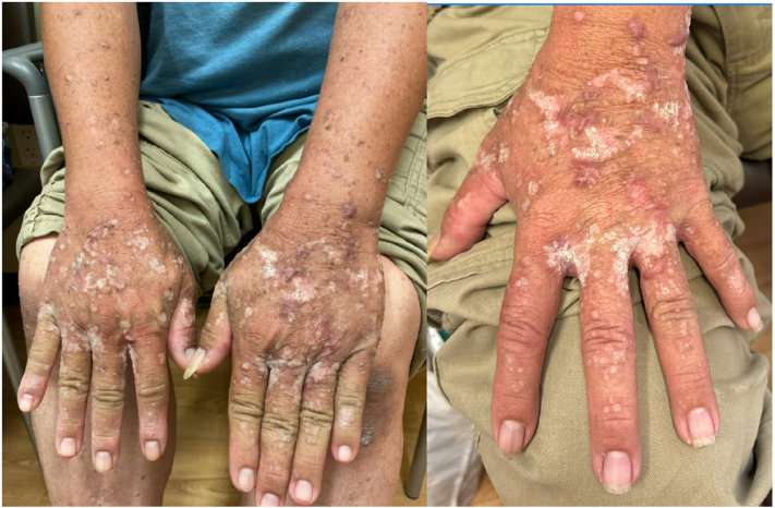 Figure 1
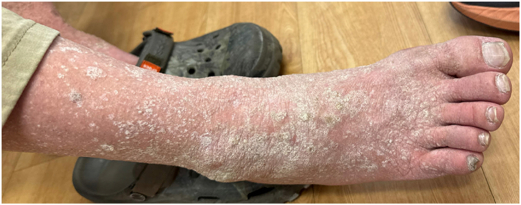 Figure 2
Peer Reviews
No public reviews on file for this paper yet. If you reviewed it on a platform where reviews are public (OpenReview, ICLR, NeurIPS, ICML), you can paste yours below so the community can read it here.
Videos
No videos yet. Explain this paper in a talk, walkthrough, or lecture? Add one.
Taxonomy
TopicsGenetic and rare skin diseases. · Skin and Cellular Biology Research · Oral Health Pathology and Treatment
Case description
A 52-year-old man presented to dermatology clinic with numerous, chronic verrucous lesions on the hands and feet. The lesions started on the hands and feet around 30 years ago and were becoming more numerous and spreading proximally. They were refractory to many topical and destructive wart therapies, including salicylic acid, 5-flourouracil, imiquimod, intralesional Candida antigen injections, cryotherapy, and shave removal. In addition, he had colon cancer at age 45 years, and his family history was notable for a brother and aunt with colon cancer.
On examination, he had confluent verrucous papules and plaques on the bilateral hands and feet and distal arms and legs, with pink keloids on the hands (Figs 1 and 2). No intraoral pathologies were identified. Prior dermatopathology results from shave biopsies demonstrated verruca vulgaris, acral keratosis, and verrucous keratosis.Fig 1. White verrucous papules and plaques and keloids on the hands and arms.Fig 2. Confluent white and hyperkeratotic verrucous papules and plaques of the right foot and distal leg.
Labs initially ordered included complete blood cell count, complete metabolic panel, T-cell subset panel, HIV screening, immunoglobulin panel, and serum protein electrophoresis. Results were notable for CD8+ cell count of 149 (normal range: 180-1170 cells/μL), CD3+ cell count of 466 (840-3060 cells/μL), and CD4+ cell count of 303 (490-1740 cells/μL).
Question 1: What is the most likely diagnosis?
- **A.**Warts, hypogammaglobulinemia, infections, and myelokathexis (WHIM) syndrome.
- **B.**Epidermodysplasia verruciformis.
- **C.**Multiple hamartoma tumor syndrome (Cowden syndrome).
- **D.**Acquired natural killer-cell immunodeficiency.
- **E.**Arsenic keratoses.
Correct answer: C. Multiple hamartoma tumor syndrome (Cowden syndrome).
Discussion
After receiving the result of T-cell lymphopenia, we were concerned about immunodeficiency and genetic testing was ordered, which revealed a heterozygous pathogenic variant (c.1003C>T; p.Arg335∗) in the PTEN (phosphatase and tensin homolog) gene on chromosome 10, supporting a diagnosing of Cowden syndrome.
Cowden syndrome, or multiple hamartoma tumor syndrome, is a genodermatosis associated with mutations in the PTEN gene inherited in an autosomal dominant manner.1 It belongs to the PTEN hamartoma tumor syndrome spectrum, which also includes genodermatoses like Bannayan-Riley-Ruvalcaba syndrome. Physiologically, PTEN is a tumor suppressor gene that downregulates the phosphatidylinositol 3-kinase/AKT/mammalian target of rapamycin (mTOR) pathway.1 Loss of PTEN function leads to excessive mTOR signaling in certain tissues, causing high cellular proliferation. The gene also plays a role in immune development, though there are only few cases of Cowden syndrome presenting with immune dysregulation, such as T-cell lymphopenia in this patient.2
The mucocutaneous symptoms of Cowden syndrome encompass a broad spectrum, and most patients develop skin involvement early by age 30 years. The most common mucosal sites of involvement are the tongue and lips, which can develop cobblestone-appearing or papillomatosis lesions.1^,^3 Keratotic and verrucous papules on palmoplantar areas are often seen. Other skin findings include sclerotic fibromas, trichilemmomas of the central face, acrochordons, vascular malformations, and soft tissue tumors.3 Patients have a higher risk for melanoma (around 5% lifetime risk) compared to the general population. Regarding extra-mucocutaneous symptoms, there is increased risk for skeletal abnormalities, macrocephaly, neurocognitive delay, and numerous solid-organ malignancies, including colon, endometrial, breast, kidney, and thyroid.4
Management of mucocutaneous symptoms is primarily supportive, though there is limited evidence that topical or oral sirolimus, which inhibits mTOR, may be beneficial.5 Genetic counseling is important for patients and their families. Multidisciplinary management is essential for cancer surveillance given high risk for multiple solid-organ malignancies.
Declaration of generative AI and AI-assisted technologies in the writing process
There was no use of artificial intelligence related to this article.
Conflicts of interest
None disclosed.
The reference list from the paper itself. Each links out to its DOI / PubMed record.
- 1Farooq A.Walker L.J.Bowling J.Audisio R.A.Cowden syndrome Cancer Treat Rev 368201057758310.1016/j.ctrv.2010.04.00220580873 · doi ↗ · pubmed ↗
- 2Browning M.J.Chandra A.Carbonaro V.Okkenhaug K.Barwell J.Cowden's syndrome with immunodeficiency J Med Genet 5212201585685910.1136/jmedgenet-2015-10326626246517 PMC 4661225 · doi ↗ · pubmed ↗
- 3Lim A.Ngeow J.The skin in Cowden syndrome Front Med 8202165884210.3389/fmed.2021.658842 PMC 822253634179044 · doi ↗ · pubmed ↗
- 4Pilarski R.Burt R.Kohlman W.Pho L.Shannon K.M.Swisher E.Cowden syndrome and the PTEN hamartoma tumor syndrome: systematic review and revised diagnostic criteria J Natl Cancer Inst 1052120131607161610.1093/jnci/djt 27724136893 · doi ↗ · pubmed ↗
- 5Bhanot A.Harrell K.Levin J.Treatment of trichilemmomas with topical sirolimus JAMA Dermatol 1593202334034210.1001/jamadermatol.2022.570136598777 · doi ↗ · pubmed ↗