Retrospective analysis of possible associations between Pneumocystis spp. and five immunosuppressive viral pathogens in three wild carnivore species
Branislav Kureljušić, Vesna Milićević, Dimitrije Glišić, Ana Vasić, Božidar Savić, Nemanja Jezdimirović, Nicolle Gobbo Oliveira Erünlü, Barbara Blasi, Christiane Weissenbacher-Lang

TL;DR
This study examined the link between Pneumocystis fungi and viruses in wild carnivores, finding limited evidence of viral impact on fungal infections.
Contribution
The study provides new insights into the potential interactions between Pneumocystis spp. and immunosuppressive viruses in wild carnivores.
Findings
Pneumocystis spp. were detected in 40.7% of sampled animals, with no significant association with most viral pathogens.
Co-infections of Pneumocystis spp. and PRV were found in three animals, but no clinical Pneumocystis pneumonia was observed.
Viral loads were high in co-infected samples, but Pneumocystis loads were linked only to subclinical infections.
Abstract
Pneumocystis spp. proliferate under immunosuppressive conditions in mammalian lungs, and several pathogens have been discussed as potential contributors to fungal proliferation. This study aimed to investigate the possible associations between Pneumocystis spp. and immunosuppressive viruses in Serbian wild mammals. A total of 108 wild carnivores – including golden jackals (Canis aureus), Eurasian badgers (Meles meles), and red foxes (Vulpes vulpes) – were collected from Veliko Gradište, Stara Pazova, and Ugrinovci during the 2022/2023 hunting season. The presence of Pneumocystis spp., canine parvovirus 2 (CPV-2), pseudorabies virus (PRV), canine distemper virus, canine coronavirus, and canine herpesvirus was assessed using conventional PCR and real-time PCR. Pneumocystis spp. were detected in 40.7% of all sampled animals (20/60 golden jackals, 4/9 Eurasian badgers, and 20/39 red foxes).…
Genes, proteins, chemicals, diseases, species, mutations and cell lines named across the full text — each resolved to its canonical identifier and authoritative record.
Click any figure to enlarge with its caption.
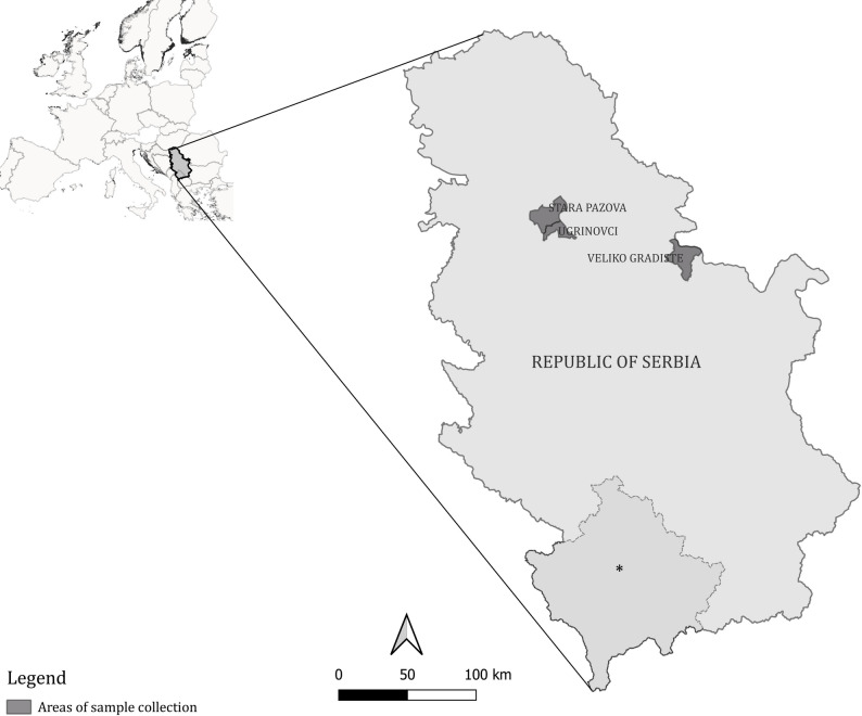 Figure 1
- —Ministry of Science, Technological Development and Innovation of the Republic of Serbia
- —OeAD-GmbH – Austria´s Agency for Education and Internationalisation
Peer Reviews
No public reviews on file for this paper yet. If you reviewed it on a platform where reviews are public (OpenReview, ICLR, NeurIPS, ICML), you can paste yours below so the community can read it here.
Videos
No videos yet. Explain this paper in a talk, walkthrough, or lecture? Add one.
Taxonomy
TopicsPneumocystis jirovecii pneumonia detection and treatment · Toxoplasma gondii Research Studies · Pneumonia and Respiratory Infections
Background
The golden jackal (Canis aureus), Eurasian badger (Meles meles) and red fox (Vulpes vulpes) are widely distributed across Europe [1–4]. Typically, the golden jackal was confined to southeastern Eurasia, but since the mid-20th century, its range has expanded from the Balkans along the Danube basin into Central Europe. In Serbia, populations of golden jackals are well-established and increasing [1, 4]. The Eurasian badger inhabits nearly all European countries west of the River Volga [2]. As a highly adaptive generalist, it thrives in diverse habitats [5]. However, concerns persist due to population fluctuations, leading to protected status in some European countries for several decades [6]. The red fox is the most widespread wild carnivore globally, with a natural range covering much of the Northern Hemisphere [3]. Over time, it has adapted to various habitats [7], and introduced populations are now found in Australia and North America [8]. These three carnivore species are frequently targeted by surveillance programs (e.g., rabies and bovine tuberculosis monitoring), due to their ability to transmit a wide range of pathogens. Their epidemiological significance is largely attributed to their abundance and extensive distribution [9–11].
Pneumocystis spp. are highly diversified opportunistic fungi that attach to type-1 alveolar epithelial cells [12]. Severe, often fatal pneumonia caused by Pneumocystis spp. proliferation has been primarily documented in immunosuppressed humans [13]. In contrast, infections in immunocompetent hosts are typically asymptomatic and referred to as “colonization” or “subclinical infection” [14]. Pneumocystis spp. have been detected in the lungs of various mammals [15], and it is believed that all mammal species are susceptible to infection [16]. However, studies on wild carnivores are limited. While prevalence may be high, these animals rarely develop severe infections [15, 17–19]. The proliferation of Pneumocystis spp. is closely linked to immunosuppressive conditions [12]. In addition to congenital immunosuppression [20, 21], acquired immunomodulatory factors, such as co-infections, have been proposed as contributors [15]. However, the impact of highly immunosuppressive pathogens on Pneumocystis spp. proliferation remains poorly understood and requires further investigation.
Several viruses – including canine parvovirus 2 (CPV-2), pseudorabies virus (PRV), canine distemper virus (CDV), and canine coronavirus (CCoV) – are known to interfere with the host’s immunity to facilitate their replication. CPV-2 induces significant lymphopenia in dogs [22]. Although interspecies transmission has been confirmed [23], little is known about its clinical effects in wild carnivores, particularly regarding immunosuppression. PRV employs strategies to evade host immune responses, such as inducing autophagy or suppressing apoptosis to enhance its own replication and dissemination [24]. This pathogen affects various feline and canine species, including red foxes, wolves, and minks [25]. While interspecies transmission has been extensively studied [25], its immunomodulatory effects in these species remain unclear. CDV infection has been associated with lymphocyte depletion in Italian red foxes. However, due to advanced autolysis, carcasses of Eurasian badgers and other animals were unsuitable for necropsy and pathohistological analysis, and CDV was confirmed only via PCR in Eurasian badger samples [26]. CCoV infection in carnivores is characterized by immunosuppression, including lymphocyte depletion and intestinal wall breakdown [27]. Moderate lymphocyte depletion and lymphoid follicles necrosis have been observed in dogs [27], but similar effects in wild carnivores have not been documented. Canine herpesvirus (CHV), endemic in domestic dogs worldwide, can reactivate under immunosuppressive conditions [28]. In wildlife, stressors such as mating, lactation, food or water shortage, or opportunistic infectious diseases are discussed as possible inducers of immunosuppressive conditions [29]. Although CHV is not considered a direct immunosuppressive agent, it was included in this study because comparable immunosuppressive conditions could also exacerbate Pneumocystis spp. proliferation. Synergistic interactions with Pneumocystis spp. cannot be ruled out due to a lack of studies.
Reports on potential associations between Pneumocystis spp. and infectious agents such as CDV or CPV-2 in wild carnivores are sporadic [15, 19, 30, 31]. Additionally, the occurrence of these pathogens in wild carnivores has been insufficiently studied, and it remains unclear whether they cause symptoms and lesions comparable to those observed in domesticated carnivores.
While detailed studies on these viruses are lacking, recent research has reported Protoparvovirus carnivoran 1 infection in Serbian golden jackals [32]. German red foxes and Eurasian badgers have tested positive for CPV-2 and CDV, with statistical evaluations conducted based on sex, age, and sampling season [33]. However, all samples were negative for PRV and CHV. Molecular detection of these viruses has been complemented by pathohistological analysis of the central nervous system, which revealed non-suppurative encephalitis [33]. Pseudorabies is one of the better described diseases in wild animals. A review of Sehl and Teifke [34] describes clinical signs (such as neurological signs, pruritus and skin abrasions, fever, anorexia, salivation, vomiting, diarrhea, dyspnea, vocalization, and apathy), pathogenesis, epidemiology, and postmortem findings in various animal species, including wild red foxes. Similar details are available in reviews on CDV in foxes and other mammals [26, 35]. CHV has been studied experimentally in red foxes, where clinical signs such as fever, lethargy, and jaundiced mucosal surfaces were observed [36]. Protoparvovirus carnivoran 1 has been phylogenetically analyzed in Serbian golden jackals [32], CDV in Serbian golden jackals [37] and Croatian red foxes [38], and CCoV in Chinese red foxes [39].
The aim of this study was to determine the prevalence of Pneumocystis spp., CPV-2, PRV, CDV, CCoV, and CHV in golden jackals, Eurasian badgers, and red foxes and to statistically evaluate possible correlations between these pathogens and the animals’ sex, age classes, and geographic origin. Pneumocystis spp., CPV-2, and PRV were semiquantified using real-time PCR.
Methods
Animal species and sample collection
The study investigated 108 wild carnivores, comprising 60 golden jackals (Canis aureus), 9 Eurasian badgers (Meles meles), and 39 red foxes (Vulpes vulpes). Species identification was performed based on morphological characteristics, including body measurements, coat coloration and, for golden jackals, the presence of distinctive features such as connate (partially fused) pads on the central digits of their front feet [40].
All animals were legally harvested during the regular hunting season, which spanned from late October 2022 to mid-January 2023, by local hunting organizations in Serbia. The studied species are legally classified as game animals under Serbian wildlife management regulations and are hunted primarily for population control and to mitigate potential human-wild life conflicts arising from overabundance, particularly in the case of golden jackals and red foxes. Eurasian badgers are hunted less frequently and only under general hunting quotas; their inclusion in this study was based on the availability of specimens during the sampling period rather than targeted removal. The animals were obtained as part of routine hunting activities conducted within the framework of a long-term wildlife health monitoring program coordinated by the Institute of Veterinary Medicine of Serbia in collaboration with the Ministry of Environmental Protection and the Ministry of Science, Technological Development and Innovation. This framework ensured that the animals represented a random subset of legally harvested individuals rather than those selectively removed due to specific health or behavioral concerns.
The animals originated from three different counties: 66 animals were collected from Braničevo (Veliko Gradište), 19 from Srem (Stara Pazova), and 3 from the City of Belgrade (Ugrinovci). Veliko Gradište is a rural area near the Serbian-Romanian border, while Stara Pazova and Ugrinovci are located in or near the City of Belgrade (Fig. 1). Details regarding sex, age, and origin of the investigated animals are summarized in Table 1. Age classification was based on body size and tooth wear (juveniles < 0.5 years; subadults 0.5-1 year; adults > 1 year).
Fig. 1. Map of Europe and Serbia with marked areas of sample collection. The map was created using the QGIS 3.28.6 software, Firenze edition *This designation is without prejudice to positions on status and is in accordance with United Nations Security Council Resolution 1244 (1999) and the advisory opinion of the International Court of Justice on the Kosovo Declaration of Independence
The animals were submitted to the Institute of Veterinary Medicine of Serbia, Department of Pathology, for sample collection. No detailed pathomorphological investigation was conducted as the primary focus was sample collection. During the section, no significant pathomorphological lesions were observed, except for those caused by ectoparasites, endoparasites, and traumas related to the shooting. No pathohistological investigations were performed.
Table 1. Sex, age, and geographic origin of the investigated animalsGolden jackal(n = 60)Eurasian badger(n = 9)Red fox(n = 39) Sex Female32623Male28316 Age Juvenile11014Subadult100Adult48925 Origin Veliko Gradište36822Stara Pazova6013Ugrinovci102Unknown1712
Extraction of DNA and RNA
All samples were collected under sterile conditions, stored at -80 °C until processing, and subjected to PCR analysis for pathogen detection. DNA from Pneumocystis spp. was extracted from lung tissue using the Nexttec DNA Isolation from Tissue & Cells Kit (Nexttec, Hilgertshausen, Germany) following the manufacturer’s protocol.
For virus detection, samples were mechanically homogenized using a mortar and pestle, diluted 1:10 in Dulbecco’s Modified Eagle Medium (DMEM; Thermo Fisher, Massachusetts, USA), and centrifuged at 1500 × g for 10 min. The supernatant was collected for further analysis and stored at -80 °C. For viral pathogen detection, brain tissue was used for CDV and PRV, while spleen tissue was analyzed for CPV-2. For CHV, a pooled sample of lung, kidney, liver, and spleen tissues was examined to account for the systemic distribution of the virus. CCoV was investigated using a pool sample of lung tissue and lymph nodes, as these are primary target sites of infection. RNA and DNA were extracted using the IndiSpin Pathogen Kit (Indical Bioscience GmbH, Leipzig, Germany) according to the manufacturer’s instructions.
Polymerase chain reaction
Pneumocystis spp.
Prior to this study, Pneumocystis spp.-positive lung tissue samples from golden jackals, Eurasian badgers, and red foxes were sequenced to obtain sequencing data on the mitochondrial large subunit ribosomal RNA (mtLSU rRNA) gene for real-time PCR primer/probe design. The PCR mixture contained 12.5 µL Kapa 2G Fast HotStart ReadyMix (Merck, Vienna, Austria), 0.4 mM of each primer (fw: 5’-CTCATGTCAGCATTCTCTCTTTA-3’, rv: TAGGATATAGCTGGTTTTCTGCGA; [41]), 1 µL MgCl_2_ (Peqlab, VWR, Vienna, Austria), 1 µL template DNA, and distilled water to a total volume of 25 µL per reaction. The cycling conditions included an initial denaturation at 95 °C for 3 min, followed by 40 cycles at 95 °C for 15 s, 55 °C for 15 s, and 72 °C for 25 s, with a final extension at 72 °C for 1 min. Gel electrophoresis was performed as described previously [19], and the gel image is provided in Supplementary Fig. 1.
DNA extracted from Pneumocystis spp.-positive pig lungs served as positive control, while nuclease-free water was used as negative control. Positive PCR products (amplicon size 500 bp) were purified using the MinElute PCR Purification kit (Qiagen, Vienna, Austria) and submitted for Sanger sequencing (Microsynth, Vienna, Austria). Sequences were assembled using BioEdit Sequence Alignment Editor 7.7.1 [42] and analyzed with NCBI Basic Local Alignment Search Tool (BLAST) to confirm their genetic affiliation to the genus Pneumocystis. To give additional information about the genetic relationships between the three investigated (NCBI accession numbers (acc. nos.) PX703107-PX703109) and published Pneumocystis spp., phylogenetic analysis was performed using MEGA 11 with the Neighbour Joining method, and bootstrap values (100 replicates) were displayed next to the branches. The evolutionary distances were computed using the Maximum Composite Likelihood method and are in the units of the number of base substitutions per site. The mtLSU rRNA gene analyses included 16 sequences with 317 positions (Supplementary Fig. 2).
Based on the Pneumocystis spp. sequences of golden jackals, Eurasian badgers, and red foxes, primers and probes were designed for the respective mtLSU rRNA genes using the Sci Ed Central software package, following published guidelines [43]. BLAST searches were conducted to ensure no cross-reactivity with other organisms. The real-time PCR reaction master mix contained 10 µL Luna Universal Probe qPCR Mix (1X; Merck KG, Vienna, Austria), 0.4 µM of each primer (golden jackal and Eurasian badger: fw: 5’-TGGGACTTTTTGCGATAAGG-3’, rv: 5’-CTGTCACAGAGAAACCACTTC-3’ [amplicon size 110 bp], red fox: fw: 5’-TCTCGGATATTTAATCTCAG-3’, rv: 5’-AGAGAAATCACTTCCTTTAT-3’ [amplicon size 148 bp]), 0.2 µM of the probe (golden jackal and Eurasian badger: 5’-FAM-TCAAGAGGGAAACAGCCCAGAACA-TAMRA-3’, red fox: 5’-FAM-TGCGATAAGGTGGAAAGTCG-TAMRA-3’), 2 µL template DNA and distilled water to a total volume of 20 µL per reaction. Cycling conditions included an initial denaturation at 95 °C for 60 s, followed by 40 cycles at 95 °C for 15 s and 55 °C (Canis aureus, Meles meles)/50°C (Vulpes vulpes) for 30 s. Samples were classified as positive if the threshold cycle (Ct) value was < 40; samples without a Ct value were classified as “undetermined”.
The real-time PCR primers were initially tested with the aforementioned protocol (annealing temperatures: 55 °C for golden jackal and Eurasian badger primers, 50 °C for red fox primers). PCR products were sequenced to confirm their genetic affiliation to the genus Pneumocystis. Gel images and BLAST results are provided in Supplementary Fig. 1. Supplementary Fig. 3 shows an alignment of published mtLSU rRNA P. canis (NCBI acc. no. MT726217), Pneumocystis sp. host Canis aureus (NCBI acc. no. OL677878), and Pneumocystis sp. host Vulpes vulpes (NCBI acc. no. MH500780) sequences, along with sequences from this study (NCBI acc. nos. PX703107-PX703109), primers used for Sanger sequencing, and real-time PCR primers.
Viral pathogens
CPV-2 [44] and PRV [45] were analyzed by real-time PCR, with protocols modified to use Luna^®^ Universal Probe qPCR Master Mix (New England Biolabs, Ipswich, Massachusetts, USA). CDV [46] was investigated using real-time RT-PCR, employing the Luna^®^ Universal Probe One-Step RT-qPCR Kit (New England Biolabs, Ipswich, Massachusetts, USA). CHV detection was performed using PCR as described by Burr et al. [47], with the HotStarTaq Master Mix Kit (Qiagen, Les Ulis, France).
CCoV detection involved a two-step RT-PCR. The first round used a 20 µL reaction containing 4 µL of 5× Qiagen OneStep RT-PCR Buffer, 4 µL of QSolution, 0.8 µL dNTP mix, Qiagen enzyme mix (Qiagen, Les Ulis, France), 1.2 µL of each 10 mM primer (CCV1 fw: 5′-GGCGTAACTGATGGACCACG-3′, CCV2 rv: 5′-CTTGTACGGGCGGCAACATC-3′), 2 µL RNA template, and nuclease-free water. Cycling conditions were 94° C for 2 min, followed by 40 cycles of 94° C for 1 min, 50° C for 1 min, and 72° C for 1 min. The second round used 10 µL HotStarTaq Master Mix (Qiagen, Les Ulis, France), 0.6 µL of each 10 mM primer, 2 µL template, and water to 20 µL. Annealing was performed at 60 °C for 1 min, using the outer forward primer and the inner antisense primer CCV3 (5′-GCTCCACTAGCACCAGTGG-3′) [48].
For all real-time PCR and RT-PCR assays, positive controls consisted of previously confirmed viral DNA or RNA samples, while nuclease-free water served as the negative control. All reactions included both positive and negative controls alongside test samples.
Statistical analysis
Statistical analyses were performed using IBM SPSS Statistics version 29 (IBM Corporation, Armonk, NY, USA). Correlations between the pathogens were evaluated using Spearman’s rank correlation coefficient ρ. Differences between species, sexes, age classes, and origins were analyzed using the χ^2^ test. The statistical unit was the individual sample. The significance level was set at α = 0.05.
Results
A total of 44 out of 108 specimens (40.7%) tested positive for Pneumocystis spp. DNA. At the species level, Pneumocystis spp. DNA was detected in 20/60 (33.3%) golden jackals, 4/9 (44.4%) Eurasian badgers, and 20/39 (51.3%) red foxes. Geographically, 27 Pneumocystis spp.-positive samples were collected in Veliko Gradište, 9 in Stara Pazova, and 1 in Ugrinovci, while the origin of 7/44 Pneumocystis spp.-positive samples was undocumented. Details on the distribution of Pneumocystis spp. results by species and location are summarized in Table 2.
Ct values for Pneumocystis spp. ranged from 28.1 to 37.1 with an arithmetic mean of 33.6 (95% CI: 32.9–34.3). The mean Ct values for each host species were as follows: Pneumocystis sp. host Canis aureus was 32.8 (95% CI: 31.7–34.0), Pneumocystis sp. host Meles meles 33.5 (95% CI: 30.9–36.1), and Pneumocystis sp. host Vulpes vulpes 34.4 (95% CI: 33.4–35.4). No statistically significant differences were observed between species (p = 0.201), age groups (p = 0.158), sexes (p = 0.650), and geographic origins (p = 0.876).
Table 2. Distribution of Pneumocystis spp. and viral pathogen results on species and geographic origin levelOriginSpeciesPneumocystis spp.CPV-2PRVVeliko GradišteGolden jackal (n = 36)12 (33.3%)2 (5.6%)1 (2.8%)Eurasian badger (n = 8)4 (50.0%)00Red fox (n = 22)11 (50.0%)01 (4.6%)Stara PazovaGolden jackal (n = 6)2 (33.3%)00Eurasian badger (n = 0)000Red fox (n = 13)7 (53.9%)01 (7.7%)UgrinovciGolden jackal (n = 1)000Eurasian badger (n = 0)000Red fox (n = 2)1 (50.0%)00Unknown originGolden jackal (n = 17)6 (35.3%)1 (5.9%)1 (5.9%)Eurasian badger (n = 1)000Red fox (n = 2)1 (50.0%)01 (50.0%)
Phylogenetic analysis of the mtLSU rRNA sequences revealed a close relationship between Pneumocystis sp. host Canis aureus and Pneumocystis sp. host Vulpes vulpes with P. canis CK1. In contrast, Pneumocystis sp. host Meles meles formed a clade with P. canis CK2 and Pneumocystis sp. host Potos flavus, a Pneumocystis species derived from kinkajous. Pneumocystis sp. mustelae, another carnivore-derived Pneumocystis species, was more distantly related, but still clustered with the carnivore-associated clade (Supplementary Fig. 2).
Regarding viral pathogens, 3/108 (2.8%) samples tested positive for CPV-2, and 5/108 (4.6%) for PRV (Table 2). The corresponding Ct values are listed in Table 3. Phylogenetic results for CPV-2 have been previously published as part of a broader study [32]. Co-infections of Pneumocystis spp. and PRV were detected in one golden jackal and two red foxes. Three golden jackals were positive only for CPV-2, while one golden jackal and one red fox were positive only for PRV. No animals were co-infected with Pneumocystis spp., CPV-2, and PRV, or with CPV-2 and PRV. CDV, CCoV, and CHV were not detected in any sample. Detailed information on CPV-2- and PRV-positive samples is provided in Table 3.
No significant correlations between any of the investigated pathogens (Pneumocystis spp./CPV-2: ρ = -0.140, p = 0.148; Pneumocystis spp./PRV: ρ = 0.087, p = 0.380; CPV-2/PRV: ρ = -0.038, p = 0.698) were observed. CPV-2 Ct values in golden jackals ranged from 21.0 to 26.7, with an arithmetic mean of 24.0 (95% CI: 23.3–24.7). PRV Ct values in golden jackals ranged from 26.3 to 34.0, while in red foxes, PRV Ct values were higher, ranging from 29.5 to 34.7. The overall arithmetic mean of PRV Ct values was 31.3 (95% CI: 30.4–32.2).
All data generated during this study are provided in Supplementary File 1.
Table 3. Detailed information about samples positive for CPV-2 or PRVIDSpeciesVirus (Ct)Pneumocystis spp. CtSexAgeOrigin49Golden jackalCPV-2 (26.7)UndeterminedMaleAdultVeliko Gradište56Golden jackalCPV-2 (21.0)UndeterminedMaleAdultVeliko Gradište101Golden jackalCPV-2 (24.4)UndeterminedFemaleAdultUnknown48Golden jackalPRV (34.0)UndeterminedMaleAdultVeliko Gradište106Golden jackalPRV (26.3)37.0FemaleAdultUnknown32Red foxPRV (31.8)36.3FemaleJuvenileStara Pazova47Red foxPRV (34.7)31.0MaleAdultVeliko Gradište88Red foxPRV (29.5)UndeterminedFemaleAdultUnknown
Discussion
This study represents the first investigation of Pneumocystis spp. in wild carnivores from Serbia using molecular methods. Additionally, it explored co-infection, prevalence, and potential associations of Pneumocystis spp. with five viral pathogens. Previous studies on Pneumocystis spp. in golden jackals [49], Eurasian badgers [50] and red foxes [18, 50] have been limited to phylogenetic analysis. To our knowledge, these are the first results for Pneumocystis spp. in Eurasian badger and red fox populations from Serbia.
The overall prevalence of Pneumocystis spp. in wild carnivores was 40.7%, with high prevalence rates observed at the species level: 33.3% in golden jackals, 44.4% in Eurasian badgers, and 51.3% in red foxes. Previous studies reported a prevalence of 13.0% in 46 hunted golden jackals [49]. In contrast, Italian samples from Eurasian badgers (n = 16) and red foxes (n = 40), collected for rabies control, were negative [50]. However, a German study found a prevalence of 46.8% among 62 red foxes also collected for rabies control [18]. These differences in prevalence may be attributed to geographic and ecological variations, differences in host population density and behavior, as well as discrepancies in sampling strategies, diagnostic protocols, and detection sensitivity. Notably, the studies by Riebold et al. [18] and Kureljušić et al. [49] used recently designed pan-Pneumocystis spp. primers targeting the mtLSU rRNA gene, while Danesi et al. [50] employed a nested PCR with primers designed in the 1990s [51, 52]. The mtLSU rRNA gene is widely used for genotyping due to its high variability and multiple copies per cell, which enhance PCR sensitivity [53]. However, this variability can complicate primer design, as older primers may not detect all Pneumocystis spp. due to limited genetic information available at the time of their development. Our findings confirm the closer genetic relationship of carnivore-derived Pneumocystis spp. compared to those from other mammalian species, though single nucleotide divergencies can still impair primer binding and reduce PCR sensitivity.
Statistical analysis revealed no significant differences in Pneumocystis spp. load between sexes, age classes, and geographic origins. Two sampling locations, Stara Pazova and Ugrinovci, are suburban areas, while Veliko Gradište is predominantly rural. No significant differences were observed between suburban and rural areas, though the smaller sample sizes from Stara Pazova and Ugrinovci may have influenced these results. Similarly, Riebold et al. [18] did not find significant effects of trapping location on Pneumocystis spp. infection in red foxes, though sex and age were not statistically evaluated in their study.
Among the viral pathogens analyzed, only CPV-2 and PRV were detected, with low prevalence rates of 2.8% and 4.6%, respectively, likely due to the snapshot nature of the study. All three parvovirus strains detected in golden jackals were identified as Protoparvovirus carnivoran 1, genogroup Feline panleukopenia virus (FPV), marking only the second report of FPV infection in this species [32]. Although cross-species transmission could not be confirmed, continued monitoring of golden jackal health is essential to prevent virus exchange with domestic and native species and to assess the impact of infections on their populations.
The health status of the animals at the time of death was unknown. In clinically affected Serbian domestic dogs, a CPV-2 prevalence of 28% has been reported [54]. However, this figure is not directly comparable to our findings, as the domestic dog study focused on symptomatic animals, while our study sampled a random subset of free-ranging carnivores, independently from the clinical status. This comparison serves only as a general epidemiological reference, indicating that CPV-2 circulates in both domestic and wild canid populations in the region. CPV-2 has been minimally studied in wild animals, but a Namibian study reported one CPV-2-positive black-backed jackal (Lupulella mesomelas) out of 32 tested, with a Ct value of 26.45 [55]. In our study, golden jackal samples had Ct values ranging from 21.0 to 26.7, confirming the presence of viral nucleic acid. However, these values were not used to infer viral load or disease severity, as no quantitative comparison with clinically affected or healthy animals was conducted. Given that all CPV-2–positive golden jackals were adults and their health status was unknown, it is possible that these infections were subclinical, as previously reported in unvaccinated adult dogs [56]. This hypothesis requires confirmation through future studies integrating clinical, pathological, and virological data.
PRV has been documented in Serbia in wild boars [57] and one domestic dog [58]. While PRV has been detected in a wide range of animals [59], reports in wild carnivores are rare, including red foxes [60, 61]. To our knowledge, PRV has not been previously described in golden jackals or Eurasian badgers. In red foxes, PRV infections have been associated with clinical signs comparable to those of dogs such as motor incoordination, head-scratching, lunging, and biting branches and scrubs, with death occurring shortly after symptom onset [60, 61]. In this study, PRV was detected in two golden jackals and three red foxes, with Ct values between 26.3 and 34.7. These values are comparable to those reported in four Slovenian dogs (29.33–33.91) [62] and American hunting dogs infected after close contact with feral swine (up to 40.8) [63]. Sequencing data from a previous study suggested that a red fox was infected by a domestic rather than a wild source [60]. None of the Eurasian badger samples tested positive for viral pathogens, though the smaller sample size may have influenced this result. Eurasian badgers are a known reservoir of bovine tuberculosis and culling has been used to control the disease, often disrupting social structures and increasing badger movement and disease spread [10]. The impact of other diseases on badger populations remains under-researched.
The absence of other viral pathogens in this study may be attributed to its retrospective design, short sampling period, limited geographic scope, or the age and seasonality of the sampled animals [33]. The primary aim of this project was pathogen identification, and while samples were collected during autopsies, no detailed pathomorphological and pathohistological examination were performed. Future studies should include thorough pathological investigations to better interpret PCR results. Additionally, incorporating passive disease surveillance – focusing on animals found dead or showing clinical signs – would provide a more comprehensive understanding of pathogen circulation and disease impact.
The overall prevalence of Pneumocystis spp. in wild carnivores was high, while CPV-2 and PRV were only detected at low rates. Asymptomatic Pneumocystis spp. colonization appears to be the most common manifestation in wild carnivores, although severe Pneumocystis pneumonia may develop under conditions of immunosuppression [19, 21, 64–69]. Previous studies in species such as American minks, dogs, pigs, llamas, and goats have suggested that co-infections may trigger fungal proliferation [31, 70–74]. However, in the present study, only eight animals showed infections with high viral loads, and three of these animals were co-infected with PRV and Pneumocystis sp. host Canis aureus or Pneumocystis sp. host Vulpes vulpes. Despite the co-infections, the fungal load in these animals remained low. In humans, different herpesviruses were detected with similar frequencies in patients with clinically apparent Pneumocystis pneumonia and in those with lower fungal burdens. However, specific viral patterns were linked to worse outcomes: mono-infection with Epstein–Barr virus (EBV), as well as co-infections involving EBV with cytomegalovirus or EBV with herpes simplex virus type 1, were significantly associated with higher mortality in patients with Pneumocystis pneumonia compared with those without herpesvirus infection [75]. Hence, the low viral prevalence and lack of histopathology in this study preclude excluding a possible contribution of viral immunomodulation.
Danesi et al. [64] proposed a threshold Ct value of 26 to distinguish clinical Pneumocystis spp. infections from colonization in dogs. In this study, no animal exhibited a Ct value lower than this threshold, suggesting that none of the investigated wild carnivores had clinically apparent Pneumocystis pneumonia. The low proportion of positive viral results further supports the hypothesis that the investigated pathogens did not trigger immunosuppression-associated Pneumocystis spp. proliferation, despite their known potential to modulate or impair immune function.
CPV-2, a pathogen with recognized immunosuppressive properties [22], was not detected in any animal co-infected with Pneumocystis spp. This lack of co-infection might be attributed to the low number of CPV-2-positive samples in the study, limiting the ability to draw definitive conclusions. Similarly, PRV, which has immunosuppressive potential [76], was detected in two golden jackals and three red foxes. However, PRV’s relevance as an immunosuppressive factor in carnivores is limited due to its peracute and lethal course, with infected animals typically dying within 24–48 h [77]. In end hosts, PRV remains confined to the peripheral nervous system, causing a systemic inflammatory response, which leads to death. The low prevalence of PRV observed in this study is likely due to this rapid disease progression, as brain samples are not consistently positive in end hosts [77].
Although the role of immunosuppressive pathogens in Pneumocystis spp. proliferation has been widely considered, studies have not consistently established this connection. For example, porcine circovirus type 2 [78] and Betaarterivirus suid 2 [79], both of which have strong immunomodulatory effects in pigs, appear to play a minor role in Pneumocystis spp. proliferation. Instead, combinations of various bacterial infections, including those without direct immunosuppressive effects, may be a prerequisite for fungal proliferation [72, 80]. In cats, no association has been found between clinical Pneumocystis pneumonia and infections with feline leukemia virus or feline immunodeficiency virus [81, 82]. Conversely, demodicosis has been identified as risk factor for Pneumocystis spp. proliferation in dogs [83, 84].
Non-infectious factors, such as stress related to food shortage or environmental conditions, may also influence Pneumocystis spp. proliferation. However, these factors have been poorly studied in wild animals. Limited available data suggest a potential role for such stressors, but further research is needed to confirm these assumptions [85, 86]. Future studies should aim to investigate the interplay between infectious and non-infectious factors, as well as their combined impact on the health and disease dynamics of wild carnivore populations.
Conclusions
Investigating wild animals is generally challenging due to the low availability of samples, legal restrictions, and the labor-intensive nature of sampling. As a result, the true prevalence and epidemiology of pathogens affecting these populations often remain unclear. Study designs are frequently constrained by the availability of samples from specific species, sexes, age classes, and geographic origins. Furthermore, the evaluation of clinical signs in wild animals is typically not feasible, leaving postmortem assessment and pathogen detection as the primary diagnostic tools. This study provides valuable initial insights into the prevalence and potential impact of Pneumocystis spp. and viral infections in wild carnivores from Serbia. Our findings confirm the presence of Pneumocystis spp. and viral pathogens. However, the role of co-infections and other immunosuppressive factors in influencing fungal proliferation and disease progression remains poorly understood.
Supplementary Information
Supplementary Material 1. Collection date, species, sex, age, origin, and PCR/real-time PCR results. This file contains the data of species, sex, age, origin, and PCR/real-time PCR results of Pneumocystis spp., canine parvovirus 2 (CPV-2), pseudorabies virus (PRV), canine distemper virus (CDV), canine coronavirus (CCoV), and canine herpesvirus (CHV) generated during this study.
Supplementary Material 2. Gel images of the mtLSU rRNA sequencing PCR, of the testing of the mtLSU rRNA real-time PCR primers by conventional PCR, and NCBI BLAST results.
Supplementary Material 3. Neighbour Joining tree of the mtLSU rRNA nucleotide gene sequences including 3 Pneumocystis sequences derived from Canis aureus (NCBI acc. no. PX703107), Meles meles (NCBI acc. no. PX703109), and Vulpes vulpes (NCBI acc. no. PX703108) generated in this study and 12 published Pneumocystis sequences. Schizosaccharomyces pombe served as an outgroup. The percentage of replicate trees in which the associated taxa clustered together (100 replicates) is shown next to the branches.
Supplementary Material 4. Alignment of published sequences of the mtLSU rRNA gene (P. canis, NCBI acc. no. MT726217, Pneumocystis sp. host Canis aureus, NCBI acc. no. OL677878, and Pneumocystis sp. host Vulpes vulpes, NCBI acc. no. MH500780), sequences of this study (Pneumocystis sp. host Canis aureus [NCBI acc. no. PX703107]), Pneumocystis sp. host Meles meles [NCBI acc. no. PX703109], and Pneumocystis sp. host Vulpes vulpes [NCBI acc. no. PX703108]), primers used for Sanger sequencing, and real-time PCR primers.
The reference list from the paper itself. Each links out to its DOI / PubMed record.
- 1Wakefield AE. DNA sequences identical to Pneumocystis carinii f. sp. carinii and Pneumocystis carinii f. sp. hominis in samples of air spora. J Clin Microbiol. 1996;34(7):1754–9. 10.1128/jcm.34.7.1754-1759.199610.1128/jcm.34.7.1754-1759.1996 PMC 2291088784583 · doi ↗ · pubmed ↗
- 2Weissenbacher-Lang C, Kureljušić B, Nedorost N, Matula B, Schießl W, Stixenberger D et al. Retrospective analysis of bacterial and viral co-infections in Pneumocystis spp. positive lung samples of Austrian pigs with pneumonia. Browning GF, editor. P Lo S One. 2016;11(7):e 0158479. 10.1371/journal.pone.015847910.1371/journal.pone.0158479 PMC 494876927428002 · doi ↗ · pubmed ↗