Prolyl tRNA Synthetase Is Required for Mammarenavirus Multiplication
Haydar Witwit, Pablo Ibanez, Ruifeng Zhou, Nathaniel Jackson, Ruby Escobedo, Beatrice Cubitt, Roaa Khafaji, Rachel Y. Sattler, Luis Martinez-Sobrido, Juan Carlos de la Torre

TL;DR
This study shows that halofuginone inhibits mammarenavirus multiplication by targeting prolyl-tRNA synthetase, offering a potential new antiviral treatment.
Contribution
The study identifies prolyl-tRNA synthetase as a critical target for halofuginone's antiviral activity against mammarenaviruses.
Findings
Halofuginone inhibits prolyl-tRNA synthetase activity, leading to amino acid starvation and translation inhibition.
Halofuginone strongly inhibits Z budding activity in mammarenaviruses.
Exogenous proline prevents halofuginone's anti-LCMV activity, confirming the role of prolyl-tRNA synthetase inhibition.
Abstract
Several mammarenaviruses (MaAv), chiefly Lassa virus (LASV) in Western Africa and Junin virus (JUNV) in the Argentinean Pampas, cause severe disease in humans and pose important public health problems in their endemic regions. In addition, the globally distributed MaAv lymphocytic choriomeningitis virus (LCMV) is an underrecognized human pathogen of clinical significance, especially in congenital infections, and LCMV poses a serious risk for immunocompromised individuals. There are no FDA-approved MaAv vaccines or antivirals, and current anti-MaAv therapy is limited to an off-label use of ribavirin, whose efficacy remains controversial. This highlights an urgent unmet need for developing antivirals against human pathogenic MaAv. Halofuginone (HF), a derivative of the natural alkaloid febrifugine, has been shown to exhibit antiviral activity against several RNA viruses. Here, we present…
Genes, proteins, chemicals, diseases, species, mutations and cell lines named across the full text — each resolved to its canonical identifier and authoritative record.
Click any figure to enlarge with its caption.
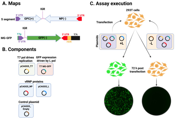 Figure 1
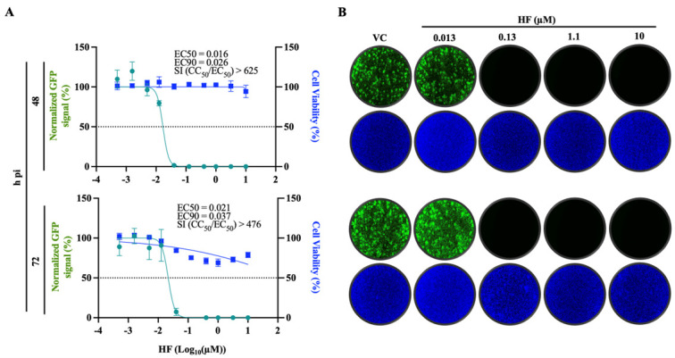 Figure 2
 Figure 3
 Figure 4
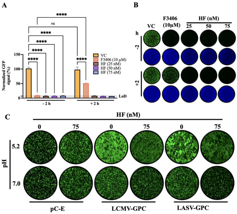 Figure 5
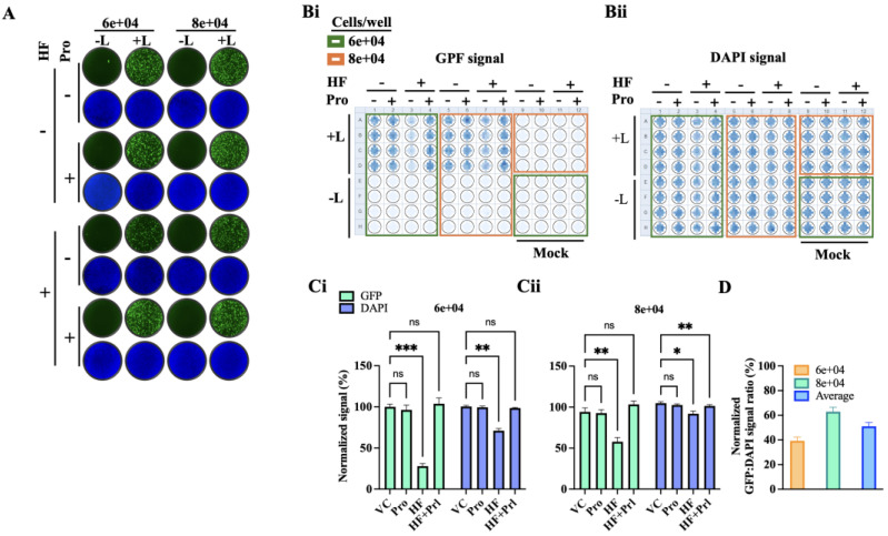 Figure 6
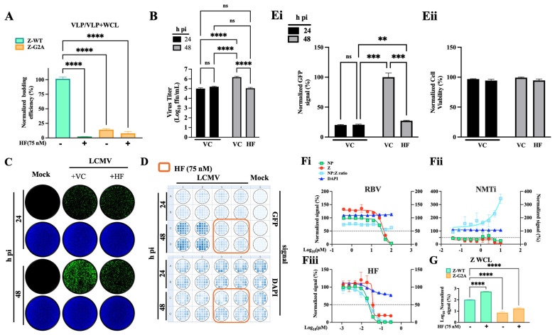 Figure 7
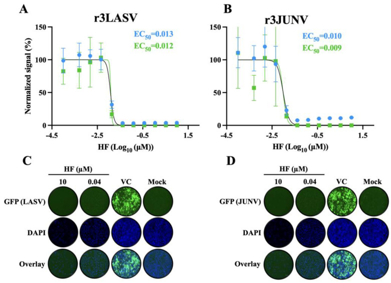 Figure 8
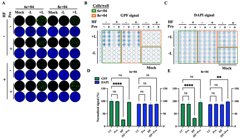 Figure 9
- —NIH/NIAID
- —Texas Biomedical Research Institute Forum Foundation
Peer Reviews
No public reviews on file for this paper yet. If you reviewed it on a platform where reviews are public (OpenReview, ICLR, NeurIPS, ICML), you can paste yours below so the community can read it here.
Videos
No videos yet. Explain this paper in a talk, walkthrough, or lecture? Add one.
Taxonomy
TopicsViral Infections and Outbreaks Research · Viral Infections and Vectors · Respiratory viral infections research
1. Introduction
Mammarenaviruses (MaAv) cause persistent infections in multiple rodent species worldwide, and human infections occur primarily through mucosal exposure to aerosols or direct contact of abraded skin with infectious materials. Several MaAv cause viral hemorrhagic fever disease (VHFD) in humans and represent important public health challenges in their endemic regions. Specifically, the MaAv Lassa virus (LASV) is highly prevalent in Western Africa where it is estimated to infect over 500,000 people annually [1], resulting in a high number of Lassa fever (LF) cases, a VHFD associated with high morbidity, and case fatality rates as high as 69% among hospitalized confirmed LF cases [2,3,4]. LASV endemic regions are expanding [5], and increased global travel has led to the importation of LF cases into non-endemic metropolitan areas [6,7]. Notably, LASV ranks very high among zoonotic viruses with potential for spillover and spread in humans [8]. Likewise, the MaAv Junin virus (JUNV) causes Argentine hemorrhagic fever, with case fatality rates exceeding 15%, and several other New World MaAv cause VHFD throughout South America [9,10]. Moreover, several MaAv, including LASV and JUNV, are credible biodefense threats and are classified as Category A agents [11]. In addition, mounting evidence indicates that the worldwide-distributed MaAv lymphocytic choriomeningitis virus (LCMV) is a neglected human pathogen of clinical significance in congenital infections and transplantation medicine [12,13,14]. Although most acquired LCMV infections in the immunocompetent are asymptomatic or result in self-limited mild illness [15], in some cases, LCMV-infected immunocompetent individuals can develop severe disease [16].
There are currently no FDA-approved MaAv vaccines or antivirals, and current anti-MaAv therapeutic options are limited to an off-label use of ribavirin, whose efficacy remains controversial [17]. A monoclonal antibody cocktail has shown protection in preclinical models of LF [18], but high cost, cold chain distribution and storage requirements, and parenteral delivery may limit their use to specialized treatment centers. The broad-spectrum polymerase inhibitor nucleoside analogs favipiravir [19,20,21,22] and 4’-fluorouridine (4’-FIU) [23,24], as well as cell entry inhibitors LHF-535 [25] and ARN-75039 [26], which target the fusion activity of the virus surface glycoprotein (GP), have shown promising results in preclinical models of LF, and LHF-535 is in phase I clinical trials. However, favipiravir did not show superior efficacy over ribavirin in a phase II clinical trial for the treatment of LF [27], whereas the linkage of 4’-FIU to mitochondrial dysfunction and cardiotoxicity may complicate its clinical development. Likewise, the high genetic diversity of LASV [28] can compromise cell entry inhibitors, as single mutations in GP can confer cross-resistance to these type of antivirals [29]. Thus, the development of broad-spectrum antivirals against human pathogenic MaAv represents a significant unmet biomedical need.
Halofuginone (HF), an FDA-approved feed additive derived from the natural alkaloid febrifugine, is a potent competitive inhibitor of the prolyl tRNA synthetase (PRS) activity of the bifunctional enzyme glutamyl-prolyl-tRNA synthetase 1 (EPRS1) that is currently under investigation as a therapeutic for the treatment of fibrotic disorders, cancer, and autoimmune diseases [30,31,32]. Notably, HF has also been shown to exhibit potent antiviral activity against different viruses, including CHIKV, DENV, and SARS-CoV-2 [33,34]. Here, we present evidence that HF exhibits potent antiviral activity against LCMV and the hemorrhagic fever causing MaAv LASV and JUNV. HF-mediated inhibition of PRS results in inhibition of translation via the amino acid starvation (AAS) response, with a higher impact on proline-rich proteins [35]. HF anti-LCMV activity was prevented by the addition of exogenous proline, supporting that the inhibition of PRS activity plays a critical role in the anti-MaAv activity of HF. MaAv matrix Z protein is the main driving force of viral budding, a process involving the Z proline-rich late domain motifs, which may make Z budding activity highly vulnerable to HF. Consistent with this hypothesis, we found that HF did not affect LCMV cell entry, but rather modestly (two-fold) reduced the activity of the virus ribonucleoprotein (vRNP)—responsible for directing replication and transcription of the viral genome—and drastically inhibited (>90%) Z budding activity.
2. Materials and Methods
2.1. Cells and Viruses
African green monkey kidney, Vero E6, cells (ATCC CRL-1586), and human A549 (ATCC CCL-185), HEK 293T (ATCC CRL-3216), and Ea HY 926 (ATCC CRL-2922) cell lines were cultured in Dulbecco’s Modified Eagle Medium (DMEM; ThermoFisher Scientific, Waltham, MA, USA) supplemented with 10% heat-inactivated fetal bovine serum (FBS), 2 mM L-glutamine, 100 μg/mL streptomycin, and 100 U/mL penicillin. HAP1 cells (Horizon Discovery) were maintained in Iscove’s Modified Dulbecco’s Medium (IMDM) supplemented with 10% FBS and 100 μg/mL streptomycin, and 100 U/mL penicillin. Recombinant viruses are as follows: rLCMV/GFP-P2A-NP (referred to as rLCMV/GFP), expressing green fluorescent protein (GFP) fused to the LCMV nucleoprotein (NP) via a P2A ribosomal skipping sequence; rLCMV∆GPC/ZsG-P2A-NP (rLCMV∆GPC/ZsG) [36], a single-cycle variant expressing Zoanthu**s sp. ZsG; the tri-segmented JUNV Candid#1 strain expressing GFP (r3Can/GFP) [37]; a tri-segmented form of the LASV Josiah strain expressing GFP (r3LASV/GFP) [38]; and rLCMV/Z-HA [39] have been described. All the experiments with LCMV were conducted in the biosafety level 2 (BSL-2) laboratory at The Scripps Research Institute. All the experiments with r3LASV and r3JUNV were conducted in the biosafety level 4 (BSL-4) laboratory at the Texas Biomedical Research Institute (San Antonio, TX 78227, USA).
2.2. Antibodies and Compounds
Halofuginone hydrobromide (Cat. No. HY-N1584A; MedChemExpress, NJ, USA) was dissolved in DMSO (Cat. No. D2650-100, Sigma-Aldrich (St. Louis, MO, USA) at 10 mM and stored in aliquots at −20 °C. F3406-2010 was obtained from DC Chemicals (Cat. No. DC11877, Shanghai, China). Ribavirin (Cat. No. R9644) and L-proline (Cat. No. 81709-10G) were purchased from Sigma-Aldrich (St. Louis, MO, USA). IMP-1088 (Cat No. 25366-1) was purchased from Cayman Chemical, dissolved in methyl acetate at 11 mM, and kept in aliquots at −20. Sodium citrate buffer (0.1 M, pH 5.0, sterile) was obtained from bioWorld (MFG No. 40121003-1, Dublin, OH, USA). Anti-HA antibody was purchased from GenScript (Piscataway, NJ, USA); the VL4 rat monoclonal antibody against NP was obtained from Bio X Cell (West Lebanon, NH, USA).
2.3. Cell Viability (CC50) and Viral Inhibition Half Maximal Effective Concentration (EC50)
Cell viability was assessed using the CellTiter 96 AQueous One Solution Reagent (Promega, Madison, WI, USA), which quantifies viable cells based on the conversion of a tetrazolium compound to a formazan product by NADPH or NADH in metabolically active cells. A549 cells were seeded in 96-well clear-bottom plates (4.0 × 10^4^ cells/well), treated with 2-fold serial dilutions of each compound, and incubated for 72 h. Following treatment, CellTiter 96 AQueous One Solution (G3580, Promega, Madison, WI, USA) reagent was added and incubated for 35 min at 37 °C in 5% CO_2_. Absorbance was measured using a Cytation 5 reader (BioTek, Agilent, Santa Clara, CA, USA), and values were normalized to vehicle (DMSO)-treated controls, set at 100%. The half-maximal cytotoxic concentration (CC_50_) was calculated using GraphPad Prism v10 (Prism10). Cell viability was also determined using DAPI staining. For determination of the half-maximal effective concentration (EC_50_), A549 cells were seeded in 96-well clear-bottom black plates (4.0 × 10^4^ cells/well), and 20 h later infected (MOI = 0.05) with rLCMV/GFP-P2A-NP. After 90 min of adsorption, the viral inoculum was removed and replaced with media containing test compounds. At 72 h post-infection, cells were fixed with 4% paraformaldehyde (PFA), and GFP expression was measured by fluorescence using a Cytation 5 reader. Fluorescence readings were normalized to DMSO-treated controls (100%), and EC_50_ values were calculated using Prism10. The selectivity index (SI) for each compound was determined as the ratio of CC_50_ to EC_50_.
2.4. Viral Growth Kinetics
Cells were infected in 12-well plates at the indicated cell seeding density and MOI. After 90 min of adsorption at 37 °C and 5% CO_2_, the virus inoculum was removed, cells were washed once with DMEM containing 2% FBS, and fresh media with the indicated compounds and concentrations was added. At specified times post-infection, cell culture supernatants (CCS) were collected, and viral titers were determined by focus-forming assay (FFA).
2.5. Virus Titration
Virus titers were determined by focus-forming assay (FFA) as described [40]. Briefly, serial 10-fold dilutions of samples were prepared in DMEM containing 2% FBS and used to infect Vero E6 cell monolayers seeded in 96-well plates (2 × 10^4^ cells/well). At 20 h post-infection, cells were fixed with 4% paraformaldehyde in phosphate-buffered saline (PBS), and infection foci were visualized by epifluorescence based on GFP expression from rLCMV/GFP-infected cells using a fluorescence microscope.
2.6. LCMV and LASV Cell-Based Minigenome (MG) Assay
The LCMV cell-based MG assay was performed as described [41], but transfecting the cells in suspension. HEK293T single cell suspension were prepared to obtain a total of 4.84 × 10^6^ cells in 5.2 mL of complete medium in a 50 mL conical tube. A transfection mixture (TM; 800 µL) was added to reach a final volume of 6 mL, and the suspension was incubated for 10 min at room temperature. Following incubation, 3.6 mL of complete medium was added and mixed gently by pipetting to ensure uniform distribution, resulting in a final TM volume of 9.6 mL. TM was added (80 µL/well) to poly-L-lysine coated wells (96 well-plate) pre-spotted with 20 µL of 5x HF. Wells were mixed twice with an electronic pipette, and plates were placed on a level surface platform for 15 min before being transferred to an incubator at 37 °C in a humidified atmosphere with 5% CO_2_.
The TM consisted of Lipofectamine 3000 (2.5 µL/µg DNA; Cat. No. L3000008, ThermoFisher Scientific, Waltham, MA, USA) with Pol II-driven expression plasmids encoding T7 RNA polymerase (pC-T7, 1.32 µg), nucleoprotein (NP; pC-NP, 0.19 µg), and L polymerase (pC-L, 1.58 µg), along with a T7 promoter-driven plasmid expressing an LCMV S MG encoding the GFP reporter (pT7-MG/GFP, 1.32 µg) (Figure 1). The LASV MG transfection mixture was prepared by combining 403.75 µL of Opti-MEM containing plasmid DNA and 25 µL of P3000 reagent with 403.75 µL of Opti-MEM containing 37.5 µL of Lipofectamine 3000, yielding a total volume of 806.6 µL. The mixture was incubated for 20 min at room temperature to allow for complex formation. At the indicated end timepoint, the cells were fixed with 4% paraformaldehyde (PFA), washed with PBS, then stained with DAPI. Images and GFP signal readings were collected using the Keyence BZ-X710 and Cytation 5 reader, respectively.
2.7. Time of Addition Assay
Vero E6 cells were seeded in 96-well plates at a density of 2 × 10^4^ cells/well. The following day, cells were infected with the single-cycle infectious rLCMV∆GPC/ZsG (MOI = 0.5) and treated with HF (75 nM) or vehicle control (VC) either 2 h before infection (−2 h) or 2 h post-infection (+2 h). The LCMV entry inhibitor F3406 (10 µM) was included as a control treatment. At 48 h pi, ZsGreen-positive (ZsG^+^) cells were quantified using the Cytation 5 Reader (BioTek, Agilent, Santa Clara, CA, USA). Fluorescence values were normalized to those of VC-treated infected cells, and results represent the mean of three biological replicates.
2.8. GPC-Mediated Fusion Assay
HEK293T cells were seeded in poly-L-lysine-coated 24-well plates at a density of 2.5 × 10^5^ cells/well. The following day, cells were transfected with a pCAGGS plasmid encoding GFP (50 ng/well) along with either an empty pCAGGS vector or pCAGGS plasmids expressing LCMV or LASV GPC (1 µg/well) using Lipofectamine 3000, following the manufacturer’s instructions. After 5 h, the transfection medium was removed, cells were washed once, and fresh medium with or without HF was added. At 24 h post-transfection, cells were exposed to either acidified (pH 5.0) or neutral (pH 7.2) medium for 15 min, washed with DMEM, and returned to DMEM containing 10% FBS. Cells were then monitored over time for syncytium formation using fluorescence microscopy. Following observation, cells were fixed with 4% PFA, washed with PBS, and imaged at 20× magnification using a Keyence BZ-X710 microscope.
2.9. Z Budding Assay
Z budding activity was assessed using the described cell-based Z budding assay [38]. HEK293T cells were seeded in poly-L-lysine-coated 12-well plates at a density of 3.5 × 10^5^ cells/well. After overnight incubation, cells were transfected with 2 µg of either pC-LCMV-Z-Gaussia luciferase (GLuc), pC-LCMV-mutant Z [G2A]-GLuc, or pC-LASV-Z using Lipofectamine 2000 (2.5 µL/µg DNA). Following a 5 h incubation, the transfection mixtures were replaced with fresh media containing the indicated compounds. At 48 h post-transfection, CCSs containing virion-like particles (VLPs) were collected and clarified by low-speed centrifugation to remove cell debris. Aliquots (20 µL) of each CCS sample were transferred to 96-well black plates (VWR, West Chester, PA, USA), followed by the addition of 50 µL Steady-Glo luciferase reagent (Cat. No. E2510, Promega, Madison, WI, USA). Whole-cell lysates (WCLs) from the same wells were prepared to assess cell-associated GLuc activity. Luminescence was measured using the Cytation 5 Reader. Budding efficiency was calculated as CCS-GLuc/CCS-GLuc + WCL-GLuc.
2.10. Assessment of NP:Z Ratio
A549 cells were seeded in 96-well clear-bottom plates at a density of 4.0 × 10^4^ cells/well, infected (MOI = 1) with rLCMV/Z-HA, and treated with 3-fold serial dilutions of the indicated compound. At 72 h pi, cells were fixed with 4% PFA and examined by immunofluorescence (IF) using the VL4 rat monoclonal antibody against NP (Bio X Cell, West Lebanon, NH, USA) conjugated to Alexa Fluor 488, and an anti-HA antibody (Cat. No. A01244, GenScript, Piscataway, NJ, USA) conjugated to Alexa Fluor 647 for Z detection. Nuclei were visualized by DAPI staining. Fluorescence signals were normalized to the vehicle-treated (DMSO) control group, set at 100%, and data were plotted using GraphPad Prism v10.
2.11. LASV and JUNV Cell Viability (CC50) and Viral Inhibition Half Maximal Effective Concentration (EC50)
A549 cells (96-well plate format, quadruplicates) were seeded at 2 × 10^4^/well. The next day, cells were infected with r3LASV (MOI 0.003) or r3JUNV (MOI 0.006). After 1 h of viral adsorption (100 µL), indicated concentrations of inhibitors (2x in 100 µL) were added (total 200 µL of 1x final concentration). At 72 h (r3LASV) or 96 h (r3JUNV) post-infection, cell culture supernatants (CCSs) were collected, and cells fixed with 10% formalin. GLuc activity in CCSs was quantified according to the manufacturer protocol using Pierce™ Gaussia Luciferase Glow Assay Kit (16160, Thermo Fisher Scientific, Waltham, MA, USA) and GloMax plate reader (Promega, Madison, WI, USA). Data were normalized to infected and vehicle control treated cells. Fixed cells were imaged for GFP and DAPI expression using an EVOS M5000 imaging system (Thermo Fisher Scientific, Waltham, MA, USA). DAPI expression was quantified using the Syngergy H1 plate reader (Agilent, Santa Clara, CA, USA) and was normalized to mock-infected cells to determine cell viability.
2.12. Statistical Analysis
All statistical analyses were conducted, as indicated in the respective assays, using GraphPad Prism software v10 (GraphPad Software, Boston, MA, USA, www.graphpad.com). p value < 0.05, 0.01, 0.001, 0.0001 represented as *, **, ***, ****, while p value > 0.05 represented as ns (not significant). One-way or two-way ANOVA with multiple comparison correction with Tukey, Šidák or Dunnett methods were implemented when recommended.
3. Results
3.1. Dose-Dependent Effect of HF on LCMV Multiplication
HF exhibited a potent dose-dependent inhibitory effect on rLCMV/GFP-P2A-NP multiplication in A549 cells, with EC_50_ and EC_90_ values of 16 and 26 nM, respectively, at 48 h pi, and 21 and 37 nM at 72 h pi (Figure 2). HF exhibited a similar anti-LCMV activity in endothelial cells with EC_50_ and EC90 values of 10 and 21 nM, respectively (Figure S1). The inhibitory effect of HF on LCMV multiplication was not a consequence of drug-induced cell toxicity, as HF exhibited a CC_50_ value >10 μM and selectivity index (SI = CC_50_/EC_50_) of > 400 (Figure 2). We obtained similar results in human endothelial Ea.hy926 cells (Figure S1).
3.2. Effect of HF on LCMV Multi-Step Growth Kinetics
To examine the effect of HF on LCMV multi-step growth kinetics, we infected A549 cells with rLCMV/GFP-P2A-NP (MOI = 0.05) and treated them with HF (100 nM) or vehicle control (VC). At the indicated h pi, we collected CCS samples and determined virus titers using a focus-forming assay (FFA). Treatment with HF abrogated production of infectious viral progeny at all time points tested (Figure 3A), which correlated with restricted virus propagation within the infected cell monolayer (Figure 3B,C(i)). To assess the effect of HF on cell viability, we quantified DAPI staining as a surrogate of cell number. HF exerted a modest cytostatic effect on A549 cells, resulting in ~10–15% lower cell number at 72 h pi compared to VC treated cells (Figure 3C(ii)). Production of infectious viral progeny was also abrogated by treatment with 75 nM HA, whereas low levels (≤10^3^ FFU/mL) of infectious viral progeny were detected at 72 h pi in LCMV-infected cells treated with 50 nM HA (Figure 3D). Results from LCMV multi-step growth kinetics assay were reproduced in independent biological replicates (Figure S2).
3.3. Effect of Proline Supplementation on the Anti-LCMV Activity of HF
To investigate whether the anti-LCMV activity of HF was mainly due to its ability to act as a competitive inhibitor of the PRS activity of EPRS1, we examined the effect of proline supplementation. For this, we infected (MOI = 0.05) A549 cells with rLCMV/GFP-P2A-NP and treated them with HF (75 nM) or VC in the absence or presence of 1 mM L-proline (Pro). At the indicated h pi, we collected CCS samples and determined virus titers using a FFA. Pro treatment reversed the HF inhibitory effect on production of infectious LCMV progeny (Figure 4A), which correlated with the restricted propagation of LCMV within the infected cell monolayer (Figure 4B,C(i)). Consistent with results shown in Figure 3C(ii), treatment with HA in the absence of Pro was associated with a modest cytostatic effect (Figure 4C(ii)).
3.4. Effect of HF on LCMV Cell Entry
To further investigate the mechanism by which HF exerted its antiviral activity against LCMV, we performed a time of addition experiment to assess whether HF affected a cell entry or post-cell entry step of LCMV infection. For this, we used the single-cycle infectious rLCMV∆GPC/ZsG to prevent the confounding effects of multiple rounds of replication without the need of NH4Cl treatment (Figure 5A). Treatment with HF (25, 50 and 75 nM) at both −2 h and +2 h of infection significantly reduced the number of ZsG+ cells compared to VC treatment (Figure 5A,B). In contrast, treatment with the LCMV entry inhibitor F3406 led to a reduction in ZsG^+^ cells when administered at −2 h, but not at +2 h, of infection. LCMV cell entry is completed in less than 60 min [42]. Hence, results of the time of addition assay indicated that HF treatment was able to disrupt a post-cell entry step. MaAv enter host cells through receptor-mediated endocytosis [43,44]. In the acidic environment of the endosome, GP2 facilitates a pH-dependent membrane fusion between the viral and host membranes, enabling the release of the vRNP into the cell cytoplasm, where replication and transcription occur. Therefore, the entry process cannot be completed if the GP2-mediated fusion event is inhibited. To examine whether HF was also able to interfere with GP2-mediated fusion, we transfected HEK293T with plasmids encoding LCMV GPC (pC-LCMV-GPC), LASV GPC (pC-LASV-GPC), or an empty vector (pC-E) as a control, along with a plasmid expressing GFP (pC-GFP). At 5 h post-transfection, we treated cells with HF (75 nM) or VC. At 24 h post-transfection, we exposed cells to acidic (pH 5.0) or neutral (pH 7.2) medium for 15 min and then returned them to standard medium to monitor syncytium formation by GFP fluorescence (Figure 5C). Cells expressing LCMV or LASV GPC exhibited robust fusion activity under acidic conditions both in the absence and presence of HF, indicating that HF does not interfere with the GPC-mediated pH-dependent event required to complete the virus cell entry process.
3.5. Effect of HF on LCMV vRNP Activity
As with other negative strand RNA viruses, LCMV vRNP is responsible for directing the biosynthetic processes of replication and transcription of the viral genome. To evaluate the impact of HF on LCMV vRNP activity, we employed a cell-based LCMV MG system. This system recapitulates LCMV RNA replication and transcription through intracellular reconstitution of the viral vRNP, which requires co-expression of the LCMV L and NP proteins together with a plasmid expressing the MG RNA (Figure 1). Levels of MG-directed GFP expression provide an integrated readout of vRNP activity. We transfected HEK293T cells in suspension with plasmids expressing the components of a LCMV MG system, and seeded them at 6 × 10^4^ and 8 × 10^4^ cells/96-well. We used a MG system where the intracellularly reconstituted vRNP directed expression of GFP. Samples where the L expressing plasmid was not included in the served as a negative control. Transfected cells were treated with HF (75 nM), Pro (1 mM), Pro (1 mM) + HF (75 nM), or VC. At 72 h post-transfection, cells were fixed with 4% PFA and stained with DAPI. Representative IF images of each treatment condition (Figure 6A) were collected using a Keyence BZ-X710 microscope. GFP (Figure 6B(i)) and DAPI (Figure 6B(ii)) signals were quantified using a Cytation 5 plate reader (BioTek, Agilent) and normalized (%) to VC-treated controls, which were set at 100% (Figure 6C(i,ii)). Treatment with HF (75 nM) resulted in ~50% reduction in GFP expression compared to VC-treated controls. We did not observe significant differences on the effect of HF on MG activity at the two seeding cell densities of 6 × 10^4^ and 8 × 10^4^ cells/96-well (Figure 6D), indicating that the modest cytostatic effect associated with HF treatment did not affect the activity of LCMV vRNP.
3.6. Effect of HF on LCMV Assembly and Budding
The MaAv matrix Z protein is the main driver of virus budding [45,46]. To determine whether HF affected Z-mediated virus budding, we used an established cell-based Z budding assay where the activity of GLuc serves as a surrogate marker for Z budding activity [47]. We transfected HEK293T cells with a plasmid encoding a chimeric GLuc where its N-terminal secretory signal peptide was replaced with the Z open reading frame (Z-GLuc) of either LCMV or LASV. Transfected cells were treated with HF (75 nM) or VC, and at 48 h post-transfection GLuc activity was measured in both CCSs, containing virus-like particles (VLPs), and whole-cell lysates (WCLs). Z budding efficiency was calculated as the ratio of VLP-associated GLuc (GLuc in CCS) to total GLuc (GLuc CCS + GLuc-WCL) × 100. Values of budding efficiency were normalized (%) to those of VC-treated cells transfected with Z-WT that were assigned a value of 100%. As control, we used Z(G2A) -GLuc, where mutation G2A prevents Z myristoylation, which results in inhibition of Z budding activity. Treatment with HF inhibited Z budding activity is displayed below (Figure 7A). We next examined whether HF inhibited assembly and release of infectious particles. For this, we infected A549 cells with rLCMV/GFP-P2A-NP (MOI = 0.05), and at 24 h pi, we collected CCSs and washed cell monolayers three times with fresh media, then refed them with medium containing HF (75 nM) or VC and allowed infection to proceed. At 48 h pi, we collected CCSs and fixed cells with 4% PFA. Virus titers in CCSs of cells that were refed with medium containing HF were similar at 24 and 48 h pi (Figure 7B). In contrast, in cells refed with medium without HF virus, titers increased from ~5 logs (24 h pi) to ~6 logs (48 h pi) (Figure 7B). This finding correlated with the restricted virus propagation between 24 and 48 h pi in infected cells that were refed with a medium containing HF (Figure 7C–E). To further investigate the effect of HF on Z protein expression and budding, we infected A549 cells with a recombinant LCMV expressing a C-terminal HA-tagged Z protein (rLCMV/Z-HA) (MOI = 1) [39] and treated them with different concentrations of HF, or with the N-myristoyltransferase inhibitor (NMTi) IMP-1088 [39] or ribavirin (RBV). At 72 h pi, we determined the NP to Z ratio by IF using a mouse monoclonal anti-HA antibody to detect Z and the rat monoclonal VL4 antibody to LCMV NP. Consistent with the HF inhibitory effect on LCMV multiplication (Figure 3 and Figure 4), treatment with HF resulted in a dose-dependent reduction of expression levels of both Z and NP. However, the normalized NP:Z ratio decreased in HF-treated compared (Figure 7Fiii) to RBV-treated infected cells (Figure 7Fi), indicating that HF treatment promoted intracellular accumulation of Z, but not NP, which correlated with increased intracellular levels of GLuc (Figure 7G). As predicted, the NP:Z ratio increased in IMP-1088-treated compared to RBV-treated infected cells because IMP-1088 induced proteasome mediated degradation of Z (Figure 7F(ii)) [39].
3.7. Effect of HF on Multiplication of LASV and JUNV in Cultured Cells
Related viruses often depend on shared host cell factors for replication, making these factors attractive targets for broad-spectrum antiviral strategies. To evaluate whether HF exhibited broad-spectrum anti-MaAv activity, we tested HF’s ability to inhibit multiplication of LASV and JUNV in cell culture. For this we used tri-segmented versions of LASV (r3LASV) and JUNV (r3JUNV) expressing the GFP and GLuc reporter genes whose expression levels served as accurate readouts of virus multiplication [37,38]. HF exhibited a potent dose-dependent inhibitory effect on multiplication of LASV and JUNV in A549 cells as determined by reduced GFP and GLuc expression levels (Figure 8).
MaAv share basic aspects of their molecular and cell biology, and therefore it would be expected that HF also targeted a post-cell entry step of LASV and JUNV lifecycle. To validate this prediction, we evaluated the effect of HF on the activity of LASV vRNP using a LASV cell-based MG system where MG-directed levels of GFP expression served as surrogate of the LASV vRNP activity. We transfected HEK293T cells with plasmids expressing the components of the LASV MG system and seeded them at 6 × 10^4^ and 8 × 10^4^ cells/96-well. Samples where the L expressing plasmid was not included in the transfection mixture served as a negative control. Transfected cells were treated with HF (75 nM), Pro (1 mM), Pro (1 mM) + HF (75 nM), or VC. At 72 h post-transfection, cells were fixed with 4% PFA and stained with DAPI. Representative IF images of each treatment condition (Figure 9A) were collected using a Keyence BZ-X710 microscope. GFP (Figure 9B) and DAPI (Figure 9C) signals were quantified using a Cytation 5 plate reader (BioTek, Agilent) and normalized (%) to VC-treated controls, which were set at 100% (Figure 9D,E). As with the LCMV MG assay, we observed that HF treatment caused a modest (~40%), but significant, reduction on LASV MG activity at the two seeding cell densities of 6 × 10^4^ and 8 × 10^4^ cells/96-well, indicating that the modest cytostatic effect associated with HF treatment did not affect the activity of LASV vRNP significantly.
4. Discussion
We have presented evidence that HF is a potent inhibitor of LCMV and the hemorrhagic fever causing MaAv LASV and JUNV. The anti-MaAv activity of HF was reversed by proline supplementation (Figure 4), supporting that inhibition of the PRS activity of EPRS1 is a main mechanism of action by which HF exerts its anti-MaAv activity. Results from time of addition and cell-based MG assays indicated that HF did not affect virus cell entry and had only a modest (twofold) inhibitory effect on the activity of the vRNP (Figure 6). In contrast, HF potently inhibited Z-mediated virus assembly and budding (Figure 7), resulting in increased intracellular levels of Z, which could mediate potent inhibition of the vRNP in infected cells, thus contributing to reduced production of infectious viral progeny and restricted virus propagation. HF-mediated inhibition of PRS could preferentially affect translation of proteins with a proline-rich motif like the Z late (L)-domain motifs that play a critical role in Z budding activity [45,46]. Likewise, HF treatment can preferentially affect expression of several functionally proline-dependent cellular proteins that contribute to Z-mediated viral budding, including CHMP2A (ESCRT-III), CHMP4B (ESCRT-III), TSG101 (ESCRT-I), and ALIX (Accessory ESCRT protein). Specific proline residues in CHMP2A and CHMP4B mediate conformational changes necessary for ESCRT-III [48] filament assembly and membrane scission [48], whereas TSG101 [49,50] and ALIX [51] utilize proline-rich motifs for protein-protein interactions essential for ESCRT recruitment and viral budding, see Figure S3 [52,53]. As with many other viruses, MaAv have evolved mechanisms to counteract the host cell innate immune responses to infection, including sensing of viral RNA by PKR [38], and induction of the type I interferon response [52]. HF-mediated inhibition of PRS leads to the accumulation of uncharged tRNAs, a biochemical event linked to the activation of the amino acid starvation (AAS) stress signal that drives autophosphorylation of the amino acid sensor GCN2 kinase, and subsequent phosphorylation of eIF2α and increased expression of ATF4, the main effector of the integrated stress response (ISR) pathway [53,54,55,56,57,58]. Evidence indicates that the cellular ISR interacts with and regulates signaling intermediates involved in the activation of innate and adaptive immune responses. Future studies, beyond the scope of the present work, will investigate whether ISR contributes to HF-mediated anti-MaAv activity.
HF is already in use in veterinary medicine [59], and has shown an excellent safety profile [60,61,62,63,64,65,66,67] and significant efficacy in phase I and II clinical trials for different indications, including cancer [30,31], raising the possibility of its repurposing as a host-directed antiviral (HDA) against human pathogenic MaAv. HF is orally bioavailable and was shown to reach an average Cmax of ~1.3 nM) or ~7.4 nM after a single administration of 0.5 mg or 3.5 mg doses, respectively, in a phase I clinical trial [30]. HF long half-life (~30 h) leads to its accumulation with two- to three-fold higher exposure by day 15 of dosing [30]. In mice, HF was widely distributed in tissues, and its exposure in the lung was > 87-fold compared to plasma after a single intravenous injection in mice [68]. This suggests that although it may be difficult for HF plasma levels to be greater than EC_50_ values we determined in this study, doses tested in phase I trials may be sufficient to reach HF therapeutic levels in tissues. HF also was shown to reduce acute inflammatory events in acute myocarditis and acute hepatitis in viral infection [69,70], which could also provide benefits against hemorrhagic fever causing MaAv infections.
HDAs represent a promising yet underexplored approach in contemporary antiviral research [71,72]. To date, most HDAs are focused on modulating interferon signaling or targeting cellular receptors, but the systematic exploration of broad-spectrum HDAs remains largely unexplored. Unlike direct-acting antivirals (DAAs) that target viral proteins and functions, HDAs disrupt host factors and cellular processes that viruses hijack for their multiplication and pathogenesis. Since members of a virus family share host dependencies, HDAs have the potential to act as broad-spectrum antivirals. In addition, HDAs pose a higher genetic barrier to the emergence of drug-resistant viral variants, which often compromise antiviral therapy [73,74]. HDAs have been shown to effectively inhibit viral infection induced by immunosuppression associated with JAK inhibitor drugs [75]. HDAs, like HF, can facilitate drug repurposing strategies for drugs with established safety profiles, thus reducing longer development timelines and regulatory barriers associated with the development of novel drugs [76]. HDA-based strategies face the challenge of risks associated with toxicity and unintended consequences of disruption of cell physiological processes. However, therapies against acute viral infections, such as hemorrhagic fever diseases caused by MaAv, would require short-term treatment, which increases the likelihood of identifying effective therapeutic regimens with acceptable safety profiles. Moreover, transient reduced levels of AARS appear to have minimal effects in global translation, and heterozygous carriers for recessive inactivating AARS mutations linked to hypomyelinating leukodystrophy do not display disease phenotypes [77]. In addition to HF, other PRS inhibitors are being evaluated in humans, including DWN12088 [78], which is in phase II clinical trials in the US and South Korea [79], and anticipated to be used for the treatment of interstitial pulmonary fibrosis. Thus, novel PRS inhibitors, including HF analogs, may lead to the identification of additional broad-spectrum antiviral agents that could be potentially repurposed as HDAs against human pathogenic MaAv.
The reference list from the paper itself. Each links out to its DOI / PubMed record.
- 1Basinski A.J. Fichet-Calvet E. Sjodin A.R. Varrelman T.J. Remien C.H. Layman N.C. Bird B.H. Wolking D.J. Monagin C. Ghersi B.M. Bridging the Gap: Using Reservoir Ecology and Human Serosurveys to Estimate Lassa Virus Spillover in West Africa P Lo S Comput. Biol.202117 e 100881110.1371/journal.pcbi.100881133657095 PMC 7959400 · doi ↗ · pubmed ↗
- 2Fichet-Calvet E. Rogers D.J. Risk Maps of Lassa Fever in West Africa P Lo S Neglected Trop. Dis.20093 e 38810.1371/journal.pntd.000038819255625 PMC 2644764 · doi ↗ · pubmed ↗
- 3Grant D.S. Samuels R.J. Garry R.F. Schieffelin J.S. Lassa Fever Natural History and Clinical Management Curr. Top. Microbiol. Immunol.202344016519210.1007/82_2023_26337106159 · doi ↗ · pubmed ↗
- 4Radoshitzky S.R. Buchmeier M. de la Torre J.C. Emerging Viruses: Arenaviridae Fields Virology Knipe D.M. Howley P. Whelan S. LWW Philadelphia, PA, USA 2020 Volume 1978-1-9751-1254-7
- 5Sogoba N. Feldmann H. Safronetz D. Lassa Fever in West Africa: Evidence for an Expanded Region of Endemicity Zoonoses Public Health 201259434710.1111/j.1863-2378.2012.01469.x 22958249 · doi ↗ · pubmed ↗
- 6Freedman D.O. Woodall J. Emerging Infectious Diseases and Risk to the Traveler Med. Clin. N. Am.19998386588310453254 · pubmed ↗
- 7Ericsson C.D. Steffen R. Isaäcson M. Viral Hemorrhagic Fever Hazards for Travelers in Africa Clin. Infect. Dis.2001331707171210.1086/32262011595975 · doi ↗ · pubmed ↗
- 8Grange Z.L. Goldstein T. Johnson C.K. Anthony S. Gilardi K. Daszak P. Olival K.J. O’Rourke T. Murray S. Olson S.H. Ranking the Risk of Animal-to-Human Spillover for Newly Discovered Viruses Proc. Natl. Acad. Sci. USA 2021118 e 2002324118 Correction in Proc. Natl. Acad. Sci. USA 2021, 118, e 2115409118. https://doi.org/10.1073/pnas.211540911810.1073/pnas.200232411833822740 PMC 8053939 · doi ↗ · pubmed ↗