Biallelic DAW1 variants reveal tissue-specific role in heterotaxy without primary ciliary dyskinesia
Saurabh Kulkarni, Dana Urbatsch, Anburaj Jeyaraj, Shruti Bedekar, Venkatramanan Rao, Shelby White, Matthew Thomas, Andrea Garrod, Christina Peroutka, Aakrosh Ratan

TL;DR
This study shows how DAW1 gene variants cause specific developmental issues in some tissues but not others, helping explain why some patients have heterotaxy and heart defects without ciliary problems.
Contribution
The study provides functional evidence for tissue-specific roles of DAW1 variants in heterotaxy and supports reclassification of variants under ACMG guidelines.
Findings
DAW1 variants cause left–right patterning and cardiac defects but not primary ciliary dyskinesia.
The p.Arg114Gln variant rescues mucociliary flow but not left–right patterning.
The splice-site variant leads to complete loss of DAW1 function in all tested contexts.
Abstract
Defects in motile cilia cause a range of disorders, including heterotaxy (HTX), congenital heart disease (CHD), and primary ciliary dyskinesia (PCD). Although these conditions often co-occur, the genetic and mechanistic bases for tissue-specific manifestations remain poorly understood. Here, we identify compound heterozygous variants in DAW1, a dynein arm assembly factor, in a proband with HTX and complex congenital heart disease but no clinical signs of PCD. Whole-genome sequencing revealed a maternally inherited canonical splice-site variant (c.648 + 1G > A) and a paternally inherited missense variant (c.341G > A; p.Arg114Gln), both classified as variants of uncertain significance under ACMG/AMP guidelines. Using Xenopus tropicalis, we show that Daw1 depletion disrupts left–right patterning, cardiac looping, and mucociliary flow, all of which are rescued by wild-type human DAW1.…
Genes, proteins, chemicals, diseases, species, mutations and cell lines named across the full text — each resolved to its canonical identifier and authoritative record.
Click any figure to enlarge with its caption.
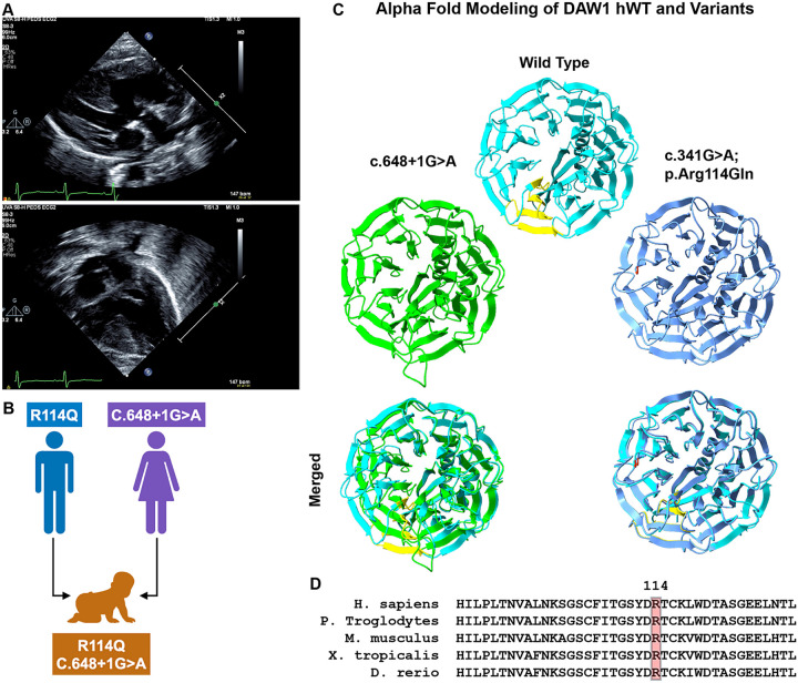 Figure 1
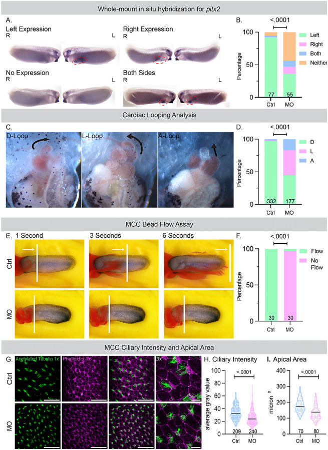 Figure 2
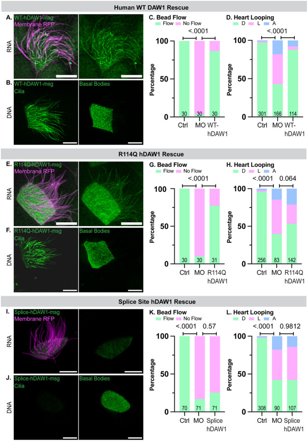 Figure 3
Peer Reviews
No public reviews on file for this paper yet. If you reviewed it on a platform where reviews are public (OpenReview, ICLR, NeurIPS, ICML), you can paste yours below so the community can read it here.
Videos
No videos yet. Explain this paper in a talk, walkthrough, or lecture? Add one.
Taxonomy
TopicsGenetic and Kidney Cyst Diseases · Cystic Fibrosis Research Advances · Genomics and Rare Diseases
INTRODUCTION
Cilia are highly conserved, microtubule-based projections that extend from the plasma membrane into the extracellular space and are critical in vertebrate development and physiology. They can be broadly divided into immotile (primary) cilia, which function in sensory and signaling processes, and motile cilia, which generate directional fluid flow. During embryogenesis, motile monocilia located at the left-right organizer (LRO), also known as the node in mammals and the gastrocoel roof plate (GRP) in frogs, generate a leftward extracellular flow that is essential for establishing left-right (LR) body asymmetry(1–5). Motile cilia are also present on specialized multiciliated cells (MCCs) that generate unidirectional fluid flow in the respiratory tract, cerebral ventricles, and fallopian tubes in mammals(4, 6, 7). Defects in ciliary assembly or motility can lead to motile ciliopathies, including primary ciliary dyskinesia (PCD), heterotaxy (HTX) syndrome, and congenital heart disease (CHD)(8–10).
PCD is a rare disorder characterized by impaired mucociliary clearance, leading to persistent respiratory complications. PCD’s pulmonary effects are variable but significant(11–17). More than 75% of neonates with PCD experience respiratory distress at birth, necessitating oxygen supplementation for extended periods(15, 18, 19). Despite these early signs, PCD diagnoses in neonates are rare(15, 20). As children grow, they commonly develop symptoms like chronic cough, sputum production, and wheezing, often progressing to obstructive lung disease or bronchiectasis(14, 15, 20). Comorbidities associated with PCD are recurrent otosinopulmonary infections and male infertility (Mirra et al., 2017). About half of PCD patients also exhibit situs inversus (mirror-image reversal of internal organs) without significant physiological effects(21). However, around 12% experience HTX, leading to complex congenital heart disease that can be life-threatening(22, 23).
HTX arises from disrupted left-right (LR) body patterning, resulting in discordant organ positioning, frequently accompanied by a wide range of complex CHD phenotypes(10). These include atrioventricular septal defects, atrial isomerism, transposition of the great arteries, double-outlet right ventricle, anomalous pulmonary venous return, single ventricle, and left ventricular outflow tract obstruction(24–26). These anomalies often require early surgical intervention, yet outcomes remain poor, with high neonatal mortality and lifelong cardiovascular morbidity in survivors. Extra-cardiac manifestations such as splenic abnormalities (asplenia or polysplenia), intestinal malrotation, and pulmonary isomerism may further complicate clinical management(26). Notably, a substantial portion of patients with HTX also present with respiratory complications resembling PCD(27–31), indicating a shared genetic basis. Indeed, several genes involved in ciliary function, including dynein heavy and intermediate chains (e.g., DNAH5, DNAH11, DNAI1, DNAI2), structural regulators (e.g., CCDC39, CCDC40), and transcription factors such as FOXJ1, have dual roles in both mucociliary clearance and LR patterning, illustrating how a single variant can manifest as both PCD and HTX(12, 32–38).
Dynein arms are essential for ciliary motility: outer dynein arms (ODAs) generate propulsive force and control beat frequency, while inner dynein arms (IDAs) adjust waveforms and bending(39–41). Proper ODA assembly involves cytoplasmic preassembly and transport into the ciliary axoneme, facilitated by intraflagellar transport (IFT) proteins and specific adaptor molecules(40, 42). Defects in ODA assembly account for the majority of cases in which PCD co-occurs with HTX(16, 21, 32, 35). Genetic studies in animal models revealed that the protein DAW1 is crucial for the structure and function of motile cilia(36, 42–45). In Chlamydomonas, DAW1 (ODA16) interacts with IFT46 to mediate ODA transport, although it remains unclear whether mammalian DAW1 functions similarly remains unclear(42, 45). In animal models, the loss of DAW1 disrupts ODA assembly, leading to laterality defects(45). In humans, predicted pathogenic DAW1 variants have been associated with HTX, CHD, and chronic respiratory symptoms(36).
Here, we report on a proband with HTX and complex CHD with no evidence of PCD, who carries compound heterozygous DAW1 variants: a paternally inherited missense mutation (c.341G > A; p.Arg114Gln) and a maternally inherited splice-site mutation (c.648 + 1G > A). Both variants are classified as variants of uncertain significance (VUS) and are rare in population databases such as gnomAD. To investigate their pathogenicity, we used Xenopus tropicalis as a model system to examine their effects on L-R patterning, cardiac looping, and mucociliary flow. This study provides experimental evidence that patient-derived DAW1 mutations disrupt laterality development and highlight tissue-specific requirements of DAW1 in human disease.
RESULTS
Clinical presentation of the patient
The proband is a male infant born at 37 + 6 weeks of gestation to a 35-year-old mother with type 2 diabetes, treated with insulin during pregnancy, and a 51-year-old father. Birth weight was 3130 g (78th percentile), length 49 cm (90th percentile), and head circumference 32 cm (21st percentile). The pregnancy was notable for prenatal concerns of congenital heart disease, and the infant was transferred to the NICU after delivery. Echocardiography demonstrated a double-outlet right ventricle with D-malposed great arteries, a large subpulmonic VSD with inlet extension, a posterior muscular VSD, moderate PDA, and a small secundum ASD/PFO with combined left-to-right shunting. RV systolic function was qualitatively normal, and LV size and function were preserved. Cardiac MRI confirmed these findings and additionally demonstrated a thin membranous structure attached to the interventricular septum and directed toward the tricuspid valve. The infant underwent balloon atrioseptostomy on the 9th day, followed by pulmonary artery band placement. Head ultrasound revealed a left grade 1 germinal matrix hemorrhage without ventriculomegaly, and the abdominal ultrasound was normal. On physical examination, the patient was nondysmorphic with no extracardiac malformations.
Family history was negative for consanguinity. A maternal half-brother died suddenly at 8 months of age following acute respiratory distress. The father had childhood-onset hearing loss, and a paternal half-sister had asthma. There were no additional congenital anomalies, intellectual disability, recurrent pregnancy losses, or known genetic conditions in the family. Given the proband’s presentation with complex congenital heart disease and family history, whole genome sequencing (WGS) with parental samples was pursued through GeneDx after informed consent.
Whole Genome Sequence analysis and findings
Analysis of variants identified from whole-genome sequencing (WGS) data and prioritized using Genomiser(46) identified DAW1 as the top candidate gene, with a phenotype similarity score of 0.683, driven by concordance between the proband’s clinical features and Primary ciliary dyskinesia 52. Two rare compound heterozygous variants in DAW1 were detected:
DAW1 (ENST00000309931.3): c.648 + 1G > A, p.? (rs927376980). This splice donor variant has not been previously reported in ClinVar and was classified as a variant of uncertain significance (VUS) by both Genomiser and Exomiser according to ACMG/AMP guidelines(47) (Exomiser ACMG: UNCERTAIN_SIGNIFICANCE [PM2_Supporting, PP4]). The variant is extremely rare in population databases, with a maximum allele frequency of 1.33 × 10^−5^ observed in individuals of African/African American ancestry. LOFTEE(48) predicts this variant to be a high-confidence loss-of-function allele. Splice prediction analyses indicate that the most likely consequence is exon 7 skipping, resulting in an in-frame deletion (p.Val181_Arg216del). A less likely alternative outcome is the activation of a cryptic splice donor site approximately 459 bp downstream, potentially leading to premature truncation.
DAW1 (ENST00000309931.3): c.341G > A, p.(Arg114Gln) (rs759511456). This missense variant has not been reported in ClinVar and was also classified as a VUS by Genomiser and Exomiser (Exomiser ACMG: UNCERTAIN_SIGNIFICANCE [PP4]). The variant is rare in population databases, with a maximum allele frequency of 5.13 × 10^−4^ observed in East Asian ancestry. The substitution has a Phred-scaled CADD(49) score of 28, placing it among the top 0.16% of predicted deleterious variants in the human genome.
Both variants segregated in the proband in a compound heterozygous configuration, consistent with autosomal recessive inheritance, and were therefore prioritized for downstream structural modeling and functional interpretation. Additional candidate genes with non-zero phenotype similarity scores were identified but were considered less likely contributors to the phenotype, as these genes were predicted to act in an autosomal-dominant manner, and neither parent exhibited overlapping clinical features. The secondary candidates included RYR1 (phenotype score 0.522; similarity to King–Denborough syndrome), KAT8 (phenotype score 0.522; similarity to Li–Ghorgani–Weisz–Hubshman syndrome), and CCDC22 (phenotype score 0.538; similarity to Ritscher–Schinzel syndrome type 2).
In silico predictions and AlphaFold modeling
We assessed the predicted pathogenicity of the compound heterozygous DAW1 variants using multiple in silico algorithms and AlphaFold modeling. For the missense variant c.341G > A (p.Arg114Gln; R114Q) inherited from the father, Arginine is highly conserved across taxa (Fig. 1). Computational predictors yielded inconsistent results, producing an overall classification of uncertain significance (Table 1). Revel (0.39), SIFT (0.004), FATHMM (0.1), MetaLR (0.27), and PrimateAI (0.54) indicated an uncertain or benign effect (Table 1). The MetaLR logistic regression–based ensemble score, which integrates ten independent predictors (SIFT, PolyPhen-2 HDIV, PolyPhen-2 HVAR, GERP++, MutationTaster, MutationAssessor, FATHMM, LRT, SiPhy, PhyloP) with population allele frequencies, also supported a benign effect (0.27). In contrast, AlphaMissense (0.78), MutationAssessor (2.75), MutationTaster (1), and DANN (1) predicted a deleterious impact. AlphaFold structural modeling showed no discernible difference relative to the wild-type protein (Fig. 1). Overall, although some algorithms showed partial support for a deleterious effect, these findings suggest that R114Q remains of uncertain pathogenicity.
In contrast, the canonical splice-site variant c.648 + 1G > A, inherited from the mother, was consistently predicted to be deleterious. DANN (0.97), SpliceAI (0.95), dbscSNV Ada (1), and dbscSNV RF (0.95) all strongly suggest splice disruption (Table 1). Exon 7 skipping was predicted, although AlphaFold modeling of the resulting transcript did not show major perturbation of the β-propeller structure of DAW1 (Fig. 1). This may be because the splicing of exon 7 does not change the reading frame or introduce a premature stop codon, despite being expected to impair splicing.
Together, these findings indicate that the R114Q missense variant remains a variant of uncertain significance, while the splice-site variant c.648 + 1G > A is strongly predicted to disrupt normal DAW1 splicing. Detailed scores and categorical outputs from all in silico prediction tools are provided in Table 1.
Daw1 knockdown in vivo affects left-right patterning and cilia motility in X. tropicalis embryos.
HTX was the predominant phenotype in the patient. Therefore, we investigated the effects of a daw1 knockdown on LR patterning in X. tropicalis embryos using a morpholino oligonucleotide (MO). We performed the whole-mount in situ hybridization for pitx2, a marker of LR asymmetry at embryonic Stage 28(50). pitx2 is normally expressed on the left side of the lateral plate mesoderm downstream of ciliamediated leftward flow in the LRO(51). While control embryos showed normal left-sided expression, Daw1 morphants showed significantly more abnormal pitx2 expression (right-sided, bilateral, or absent) (Fig. 2A, B). To further evaluate LR patterning and cardiac development, we assessed cardiac looping at embryonic stage 48 (72–96 hours post fertilization, hpf)(52, 53). A D-loop of the outflow tract (left to right, situs solitus) was classified as normal, whereas an L-loop (right to left, situs inversus) and an A-configuration (straight back, HTX) were classified as abnormal. Daw1 depletion resulted in a significant increase in heart-looping defects compared with controls (Fig. 2C, D). Together, these findings suggest that the absence of Daw1 disrupts LRO flow, as indicated by abnormal pitx2 expression, and leads to defects in laterality and organ morphogenesis, as demonstrated by aberrant heart looping.
Beyond its role in LR development, motile cilia are also vital for airway mucociliary clearance. Indeed, patients with CHD, and especially HTX, often suffer from chronic respiratory issues due to ciliary dysfunction(29–31). A previous study with patients with DAW1 variants described chronic respiratory dysfunction, suggesting that DAW1 may play a significant role in mucociliary clearance(36). We therefore examined the function of Daw1 in the multiciliated cells (MCCs) of the X. tropicalis embryonic epidermis, a well-established in vivo system for analyzing mucociliary flow.
We visualized mucociliary flow by adding fluorescent latex microspheres (beads) to the culture medium for time-lapse imaging of bead movemen(54). In control embryos, fluorescent beads placed on the anterior of the embryo were rapidly transported toward the posterior, reflecting coordinated ciliary beating. Daw1 depletion with MO led to a significant loss of cilia-generated fluid flow, indicating either a significant loss of cilia motility or assembly (Fig. 2E). To test these possibilities, we performed immunofluorescence using acetylated tubulin to label the ciliary axoneme and phalloidin to label F-actin and the apical size of the cells. Compared with controls, Daw1-depleted embryos exhibited a small but significant reduction in the apical surface area of MCCs and lower normalized ciliary fluorescence intensity, consistent with impaired ciliogenesis (Fig. 2G–I). These results demonstrate that Daw1 is essential for both L-R patterning and mucociliary clearance function in vivo.
Wild-type human DAW1 rescue of left-right patterning and mucociliary flow
To test the specificity of the MO and establish the function of wild-type (WT) - human DAW1 in Xenopus, we performed a rescue of the LR patterning and loss of mucociliary flow phenotypes. First, we coinjected an RNA construct encoding WT-hDAW1 fused to an MStayGold (msg) fluorescent tag at the C-terminus with Membrane RFP RNA to label the ciliary axonemes of MCCs in control embryos and assessed its localization. WT-hDAW1-msg localized to the basal bodies and ciliary axonemes of MCCs (Fig. 3A). We also expressed the DNA of WT-hDAW1-msg and observed the same localization as DNA (Fig. 3B). Next, we co-injected the WT-hDAW1-msg with daw1 MO to assess the rescue. WT-hDAW1-msg significantly rescued both mucociliary flow and heart-looping (LR patterning) phenotypes relative to MO embryos lacking WT-hDAW1-msg (Fig. 3C, D). These results laid the foundation for testing the function of DAW1 variants in LR patterning and mucociliary flow.
Context-specific rescue with the missense mutation
Individual mutant constructs tagged with the C-terminus-msg were then generated for each of the patient’s DAW1 mutations. The paternally inherited missense variant c.341 G > A, p.(Arg114Gln) (R114Q) was first examined. Injections of both RNA and DNA showed that localization of the R114Q protein at the basal bodies and ciliary axoneme of MCCs was similar to WT-hDAW1 (Fig. 3E, F). Given that localization was unaffected, we performed functional assays to assess pathogenicity. At the developmental Stage 28 (24 hpf), R114Q-hDAW1-msg-injected embryos showed improved mucociliary flow relative to MO embryos lacking R114Q-hDAW1-msg (Fig. 3G). To assess LR patterning, we raised the same embryos to developmental stage 48 (72 hpf) to analyze heart looping. Interestingly, LR patterning was not rescued relative to MO embryos lacking WT-hDAW1 (Fig. 3H), suggesting that the R114Q mutation affects cilia function in a tissue-specific context.
Loss-of-function splice-site variant
A second variant, the maternally inherited splice-site mutation c.648 + 1 G > A, was predicted to be more disruptive than R114Q due to skipping of entire exon 7 (E7) as a most likely output (Fig. 1). Injection of both RNA and DNA of Splice-hDAW1-msg showed a consistent loss of localization at the basal bodies and ciliary axonemes of MCCs suggesting complete loss of function (LOF) (Fig. 3I, J). To functionally confirm our localization results, we examined both LR patterning and mucociliary flow using the rescue experiments described above for the missense mutation. As expected, the Splice-hDAW1-msg did not rescue either phenotype, confirming the complete LOF in both LR patterning and mucociliary clearance contexts.
DISCUSSION
Cilia are essential organelles that regulate fluid movement during development and homeostasis. Motile cilia in the LRO generate leftward flow needed for proper LR patterning during embryogenesis, while motile cilia of MCCs mediate mucociliary clearance in the respiratory tract and fluid circulation in other organ systems. Defects in these processes can cause a wide range of motile ciliopathies, including PCD, HTX, and CHD(8). Here, we identify compound heterozygous DAW1 variants in a patient presenting with HTX and complex CHD, but notably did not exhibit features of PCD. This phenotype, CHD/HTX in the absence of respiratory disease, was precisely recapitulated in Xenopus tropicalis, providing strong experimental validation of the genetic findings.
In silico predictors failed to reach consensus on the R114Q missense variant, with results ranging from benign to deleterious. AlphaFold modeling similarly showed no structural disruption, highlighting the limitations of current computational tools in determining the pathogenicity of subtle variants. Therefore, functional assays were crucial. Notably, R114Q displayed a context-specific effect: it completely restored mucociliary flow in MCCs but did not rescue LR patterning (heart looping) defects, closely reflecting the proband’s phenotype of HTX without chronic respiratory disease. These findings illustrate that DAW1 function can diverge across ciliary subtypes and that single amino acid substitutions can selectively affect LRO cilia without impairing MCC function. Such specificity cannot currently be captured by in silico prediction models, emphasizing the importance of functional validation in relevant developmental contexts.
In contrast, the splice-site variant c.648 + 1G > A functions as a complete loss-of-function allele. Both in silico splicing tools and functional assays indicated exon 7 skipping, loss of proper localization, and failure to rescue either L–R patterning or MCC flow. These findings confirm c.648 + 1G > A as a deleterious variant and demonstrate that compound heterozygosity for R114Q and c.648 + 1G > A can fully explain the proband’s phenotype. Notably, the Xenopus model mirrored the clinical presentation, underscoring its utility as a rapid and reliable model for analyzing genotype–phenotype relationships in ciliopathies. Together, these data support reclassification of c.648 + 1G > A as pathogenic and identify p.Arg114Gln as a context-dependent hypomorphic allele whose effects are not detected by current in silico or ACMG/AMP guidelines (Table 2).
Our results also build upon and expand previous reports of DAW1-related disease. In the largest published series, Leslie et al. described several families with different DAW1 genotypes and varying effects on laterality and respiratory features(36). For example, homozygous p.(Asn143Asp) variants were associated with situs inversus without respiratory symptoms in two patients, whereas another patient exhibited respiratory symptoms without laterality defects. A homozygous p.(Trp119*) nonsense variant resulted in complex CHD with situs ambiguous, a transverse liver, and a right-sided spleen, but no respiratory symptoms. Compound heterozygous variants p.(Leu66*)/p.(Trp372Cys) and homozygous p. (Ser364Thr) were both reported in patients with complex CHD resembling HTX, although respiratory involvement was not specified. Functional studies in zebrafish further supported allele-specific effects, with p.(Asn143Asp) and p.(Ser364Thr) showing complete loss of function, while p.(Trp372Cys) was a hypomorph based on the rescue of cardiac looping and cilia motility in Kupffer’s vesicle(36).
In this context, our study offers the first direct evidence that patient-derived DAW1 variants can differentially affect LRO and multiciliated cilia in X. tropicalis, explaining why the proband exhibited HTX and CHD without respiratory disease. The R114Q allele acted as a context-specific hypomorph (affecting LR patterning but not mucociliary flow), whereas the splice-site variant resulted in complete loss of function, together creating the compound heterozygous state observed clinically. This accurate replication of the human phenotype highlights both the tissue-specific functions of DAW1 and the value of Xenopus as a translational model.
While our findings demonstrate functional effects for DAW1 variants, several aspects warrant further exploration. The Xenopus tropicalis model provides a rapid and robust system for studying DAW1 function in vivo; however, additional validation using human respiratory or cardiac cell models would enhance its translational significance. Similarly, direct RNA analysis of patient-derived tissue is necessary to confirm the predicted exon 7 skipping due to the splice-site variant. Lastly, given the family history of respiratory and auditory features, the influence of genetic background and modifier alleles warrants further investigation. Future research involving patient iPSCs, human airway cultures, and larger clinical cohorts will help clarify the phenotypic spectrum and genotype–phenotype correlations of DAW1 variants.
In summary, this study provides functional validation for compound heterozygous DAW1 variants in a patient with HTX and complex CHD. The ability of Xenopus tropicalis to reproduce this precise phenotype underscores its unique capacity to link patient genotypes to mechanistic outcomes. We demonstrate that relying solely on in silico tools is insufficient to predict the pathogenicity of DAW1 variants, and that developmental models are crucial for revealing context-specific requirements of ciliary assembly factors.
METHODS
IRB protocol
The family was recruited under the IRB protocol HSR210285.
Sequence analysis
Sequence reads were processed using a Nextflow workflow (https://github.com/aakrosh/PedigreeVarFlow). As part of the workflow, genome reads were aligned to the GRCh38 reference genome with BWA-MEM(55) (v. 0.7.19-r1273), and SAMBLASTER (v. 0.1.26)(56) was used to flag putative PCR duplicates and add MC/MQ tags to paired-end alignments. Resulting SAM files were converted to BAM format and coordinate-sorted using samtools (v. 1.21.42)(57). Alignment statistics and quality metrics were generated with alignstats (v. 0.11, https://github.com/jfarek/alignstats).
Variants were called using FreeBayes(58) (v. 1.3.9 with default parameters) and filtered with bcftools (v. 1.21) to remove low-confidence calls. We applied the following filters to retain variants with strong read support:
QUAL > 1 && QUAL/INFO/AO > 10 && INFO/SAF > 0 && INFO/SAR > 0 && INFO/RPR > 1 && INFO/RPL > 1. Variants overlapping known problematic genomic regions were flagged during annotation. Variant-level quality metrics were summarized using Variant QC(59).
Variants were left-aligned and normalized using bcftools prior to downstream analysis. Candidate variants were prioritized with Genomiser using recommended best practices (REVEL, MVP, AlphaMissense, and SpliceAI variant pathogenicity prediction sources and human-only hiPHIVE gene:phenotype associations, ClinVar whitelist, inheritance filters) and the following human phenotype ontology terms: Double outlet right ventricle, Dextrotransposition of the great arteries, and Subarterial ventricular septal defect(60). In parallel, variants were annotated using AutoGVP(61), which integrates germline pathogenicity data from ClinVar and applies ACMG guideline-based classifications using a modified version of InterVar.
Sequence Alignment and Alphafold modelling
Multiple sequence alignment was performed using the MultAlin web server. Sequences corresponding to residues 91–130 were obtained from human (UniProt: Q8N136–1), Xenopus tropicalis (UniProt: Q6P2Y2), Pan troglodytes (UniProt: H2QJJ9), mouse (UniProt: D3Z7A5), and Zebrafish (Danio rerio, UniProt: Q1LV15). Predicted structural models of human DAW1-WT (UniProt: Q8N136–1), R114Q, and the splice-site mutant were generated using AlphaFold3. Structural visualization and annotation were performed in UCSF ChimeraX.
Animal Husbandry and microinjections
Xenopus tropicalis were bred, housed, and cared for in our aquatics facility according to established protocols (ACUC# 4295) that were approved by the University of Virginia Institutional Animal Care and Use Committee (IACUC). Embryos needed for experiments were produced by in vitro fertilization according to previously established protocols(50, 62). Briefly, testes are removed from the male and crushed in 1xMBS + 0.2%BSA and added to eggs obtained from hCG-injected female frogs. The eggs and sperm are incubated for 3 minutes before being flooded with 0.1x MBS (pH 7.8–8) for 10 minutes. Fertilized eggs were then dejellied using 3% Cysteine in 1/9MR (pH 7.8–8) for 6 minutes. Embryos were then washed using 0.1xMBS and used for microinjections in 1/9MR+Gentamicin. Staging of Xenopus tadpoles was as previously described(63).
Cloning and mRNA synthesis
The full-length human DAW1 (NM 178821.3) and mStayGold was subcloned into the pCS2 + vector using PCR amplification using Gibson assembly to generate DAW1-mStayGold. The primers used for PCR are provided in the resource table. The missense and splice-site variants were generated by site-directed mutagenesis using the WT plasmid as a template. For mRNA synthesis, the plasmids were linearized with NotI and used as templates. Capped mRNAs were synthesized in vitro using the mMessage and mMachine SP6 transcription kit following the manufacturer’s instructions.
Morpholino and mRNA microinjections
Morpholino oligonucleotides (MO) or mRNA were injected into one-cell or four-cell embryos as described previously(64). For most experiments, the translation-blocking MO for Daw1 (AAGGAATCGCTTTAGCCGCATCGTG) was injected at 20 ng at the one-cell stage, along with Oregon green 488-labelled Dextran (10 kDa, non-fixable), a tracer for all flow and heart looping trials (described below). The DAW1 mRNAs were injected at 200pg in the one-cell stage. For some experiments, the plasmid DNA (WT-hDAW1-msg and the variants) was injected at a 100 pg concentration with the membrane RFP mRNA (100 pg) in one of the 4 blastomere. Post-injection, the embryos were allowed to develop to the appropriate stage for further experiments. For rescue experiments, the embryos were injected with 20ng of DAW1 MOmixed with 200pg of either WT, or variant mRNA.
Immunofluorescence, image analysis and statistics
Confocal imaging was done on embryos once they reached stage 28 either live or fixed.
For fixation, 4% paraformaldehyde (PFA) was used then the embryos were washed three times with PBST (1× PBS with 0.2% Triton X-100) for 10 min each and then incubated in a blocking solution (3% BSA in PBST) for 1 hour. The primary antibody Mouse Monoclonal Anti-Acetylated α-tubulin was added to the embryos, incubated for 1 hour at room temperature, and washed three times for 10 min each with PBST. Dilutions of the secondary antibody Chicken anti-mouse conjugated to Alexa fluor 488 and the Actin stain Phalloidin in PBST were used to stain embryos for 1 hour. All live imaging was done with Stage 28 embryos in 1/9MR+Gentamicin and a drop of Benzocaine (0.05% in 1/9x MR). Confocal imaging was performed using the Leica DMi8 SP8 microscope with a 40x or 63x oil immersion objective (1.3 NA). Images were captured at 1x, 3x, or 5x zoom and adjusted (brightness and contrast), analyzed, cropped in Fiji, and assembled in Adobe Illustrator software.
All the experiments were repeated three times. All measurements and analyses were performed on at least three embryos per trial, for a total of 3 trials. Sample size, indicated by “n” values, and number of trials, indicated by “N” values, is included in the figure legends. The Fiji freehand selection tool was used to measure ciliary intensity, in which embryos were first thresholded, and the mean gray value within a standard 100×100 pixel box over individual ciliary bundles was then measured and compiled. For analysis of the apical area, the rectangle tool was used to outline the perimeter of five MCCs per embryo to measure the area in microns^2^ and compile the data in Microsoft Excel. Statistical analysis was performed using Prism version 10, where a Welch’s t-test was performed with a significance level of 0.05.
Flow Analysis in Xenopus tropicalis and DAW1 Rescue
To measure mucociliary flow on (uninjected controls and injected) embryos were raised to Stage 28 and anesthetized with benzocaine, 1μL of latex beads was placed at the anterior end of the embryo and visualized under a dissecting scope. If the beads were moved (classified as ‘Flow’) or not (classified as ‘No Flow’) was recorded.
Cardiac Looping in Xenopus tropicalis
The injected X. tropicalis embryos that were examined for the presence of mucociliary flow were then allowed to develop to Stage 48 for examination of cardiac formation. The embryos were treated with benzocaine, examined ventrally, and scored for cardiac looping using a light dissection microscope as previously described(52, 65). Loop direction is defined by the position of the outflow tract relative to the inflow of the heart: outflow to the right: D loop; outflow to the left: L loop; outflow midline, fails to loop: A loop.
RNA in situ hybridization
X. tropicalis embryos (control and MO injected) were collected at Stage 28 for in situ hybridization. A digoxigenin-labeled antisense probe for pitx2 was in vitro transcribed with T7 High Yield RNA Synthesis Kit. Embryos were collected and fixed in MEMFA for 2 hours at room temperature and dehydrated for 4–6 hours in 100% EtOH. Briefly summarized, whole mount in situ hybridization of digoxigenin-labeled antisense probes was performed overnight, the labeled embryos were then washed, incubated with anti-digoxigenin-AP Fab fragments, and signal was detected using BM-purple, as previously described(5).
Resource list
Supplementary Material
This is a list of supplementary files associated with this preprint. Click to download.
The reference list from the paper itself. Each links out to its DOI / PubMed record.
- 1Schweickert A, Weber T, Beyer T, Vick P, Bogusch S, Feistel K, Cilia-driven leftward flow determines laterality in Xenopus. Current biology: CB. 2007;17(1):60–6.17208188 10.1016/j.cub.2006.10.067 · doi ↗ · pubmed ↗
- 2Hirokawa N, Tanaka Y, Okada Y, Takeda S. Nodal flow and the generation of left-right asymmetry. Cell. 2006;125(1):33–45.16615888 10.1016/j.cell.2006.03.002 · doi ↗ · pubmed ↗
- 3Mc Grath J, Somlo S, Makova S, Tian X, Brueckner M. Two populations of node monocilia initiate left-right asymmetry in the mouse. Cell. 2003;114(1):61–73.12859898 10.1016/s 0092-8674(03)00511-7 · doi ↗ · pubmed ↗
- 4Rao VG, Kulkarni SS. Xenopus to the rescue: A model to validate and characterize candidate ciliopathy genes. Genesis. 2021;59(1–2):e 23414.33576572 10.1002/dvg.23414 · doi ↗ · pubmed ↗
- 5Kulkarni SS, Khokha MK. WDR 5 regulates left-right patterning via chromatin dependent and independent functions. Development. 2018.
- 6Stannard W, O’Callaghan C. Ciliary function and the role of cilia in clearance. J Aerosol Med. 2006;19(1):110–5.16551222 10.1089/jam.2006.19.110 · doi ↗ · pubmed ↗
- 7Spassky N, Meunier A. The development and functions of multiciliated epithelia. Nature reviews Molecular cell biology. 2017;18(7):423–36.28400610 10.1038/nrm.2017.21 · doi ↗ · pubmed ↗
- 8Wallmeier J, Nielsen KG, Kuehni CE, Lucas JS, Leigh MW, Zariwala MA, Motile ciliopathies. Nat Rev Dis Primers. 2020;6(1):77.32943623 10.1038/s 41572-020-0209-6 · doi ↗ · pubmed ↗