Preserving integrity: innovative in vitro methods for extracellular matrix decellularization and collagen purification
Uliana Bashtanova, Rui Li, Ieva Goldberga, Sneha Bansode, Kathryn Gerl, Kristen Paige Burgess, Annika Janine Wegner-Repke, Melinda Jane Duer

TL;DR
This paper introduces new methods to purify collagen and remove cells from extracellular matrix while preserving their structure, which could improve tissue engineering and drug development.
Contribution
The novel methods focus on preserving the structural and biochemical integrity of collagen and extracellular matrix during purification and decellularization.
Findings
Cytoskeleton-targeting drugs effectively dislodge intact cells from ECM-producing cultures.
Collagen purified with chymotrypsin retains its native triple-helical structure and post-translational modifications.
The methods are broadly applicable to various cell types for producing species- and tissue-specific ECM and collagen.
Abstract
In tissue engineering and cell therapy development, synthetic biomaterials are frequently supplemented with collagen or other extracellular matrix (ECM) components to enhance biocompatibility. To support these applications, novel methods for collagen purification and ECM decellularization were developed, with a focus on preserving the structural and biochemical integrity of the final products. The effectiveness of these methods was validated using solid-state NMR and fluorescence spectroscopy, bright-field and confocal microscopy, amino acid analysis, proteomics and transmission electron microscopy. Intact cells were dislodged from ECM-producing cultures through the application of cytoskeleton-targeting drugs, while the native protein composition of the ECM was maintained. In parallel, collagen purified using chymotrypsin was shown to retain its native triple-helical structure and…
Genes, proteins, chemicals, diseases, species, mutations and cell lines named across the full text — each resolved to its canonical identifier and authoritative record.
Click any figure to enlarge with its caption.
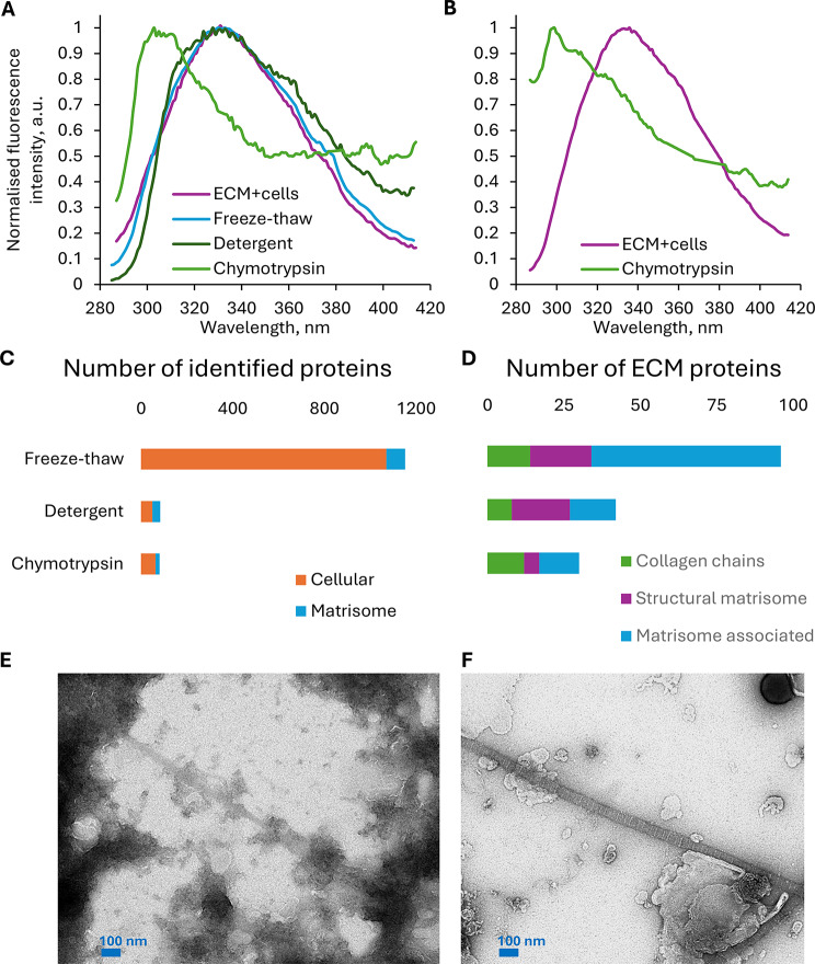 Figure 1
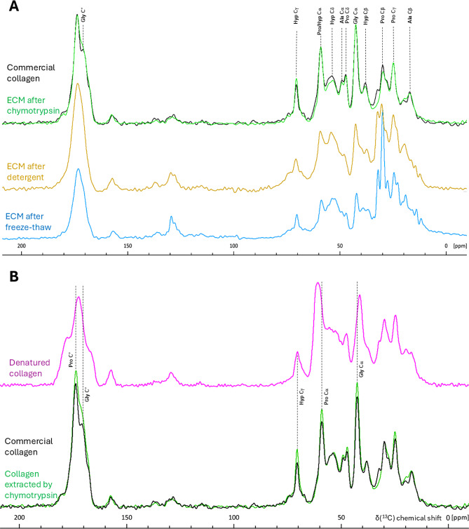 Figure 2
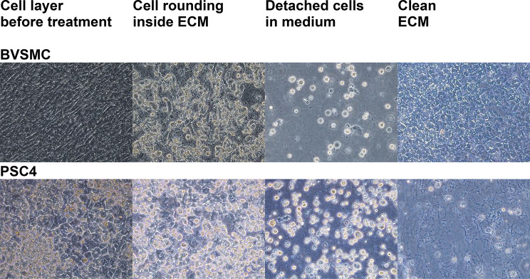 Figure 3
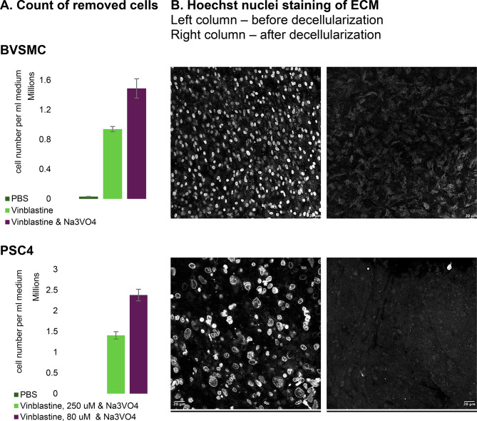 Figure 4
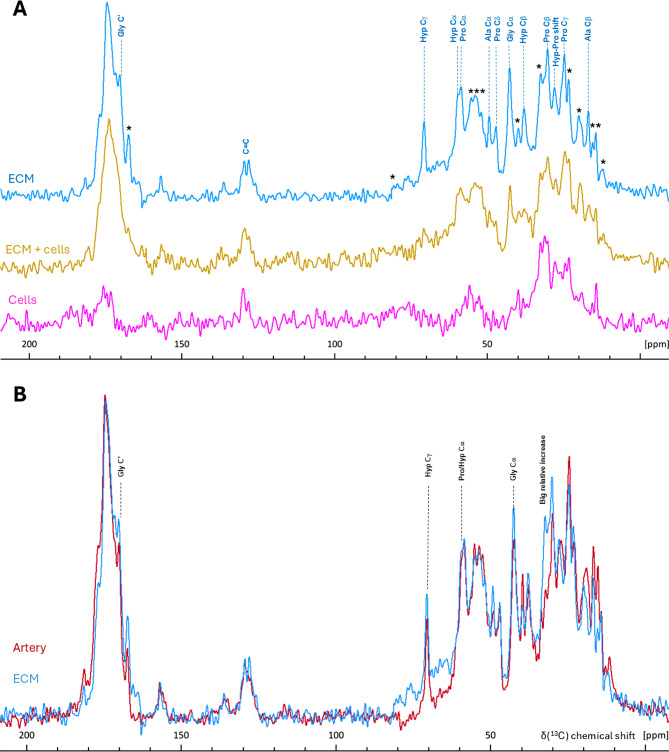 Figure 5
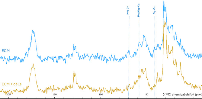 Figure 6
- —China Scholarship Council–Cambridge Trust
- —UK EPSRC studentship
- —Royal Society Newton Trust Fellowship
- —the Gates Foundation
- —the EPFL Women in Science and Humanities Foundation
- —the Leducq Foundation
- —the European Union
Peer Reviews
No public reviews on file for this paper yet. If you reviewed it on a platform where reviews are public (OpenReview, ICLR, NeurIPS, ICML), you can paste yours below so the community can read it here.
Videos
No videos yet. Explain this paper in a talk, walkthrough, or lecture? Add one.
Taxonomy
TopicsTissue Engineering and Regenerative Medicine · Collagen: Extraction and Characterization · Proteoglycans and glycosaminoglycans research
Introduction
The extracellular matrix (ECM) is the non-cellular component present in all tissues and organs, providing essential physical scaffolding for cells. The main structural components of the ECM are fibrillar collagen proteins, triple helical molecules that organize into ordered arrays forming fibrils 10 –100 s of nanometres in diameter, providing stiffness and regulating cell adhesion or migration [1, 2]. The fibrous ECM network is further supported by other fibril and filament-forming proteins, including elastin, fibrillin, fibronectin, and laminin. These protein networks are coupled together by linker ECM proteins including tenascin, non-fibrous collagens and certain types of proteoglycans. The associations among these proteins not only maintain the 3D architecture of the ECM, as well as the orientation of collagen and elastin fibres, but also preserve the ECM’s elasticity, tensile strength, and porosity required by each tissue. Equally important, they regulate ligand presentation for cell surface integrins, growth factors, and signalling molecules. Proteoglycans, together with hyaluronic acid, form a hydrated gel that provides pH buffering, tissue hydration, reservoirs of cations and chemokines, and lubrication [3, 4]. The types of ECM proteins expressed, along with their folding, crosslinking, and post-translational modifications, are species-, organ-, tissue-, disease- and age-specific [5]. The complexity of these protein combinations and their characteristic 3D architecture within the ECM is difficult to replicate using fabrication methods. As a result, seeding cells onto protein beds derived from native ECM has become increasingly desirable over the past decade [6].
The first step in the ECM preparation process is a removal of cellular material. Numerous decellularization methods have been developed, which can be divided into physical, chemical, and enzymatic approaches [6]. Physical methods include freeze-thaw cycling and/or high hydrostatic pressure. They preserve structure of ECM, but on their own, are ineffective for complete decellularization and typically combined with chemical and enzymatic approaches [7]. Chemical reagents include acids and bases, hypo- or hypertonic solutions, and detergents. They facilitate the solubilization of cellular membranes and dissociation of DNA, which otherwise bind to the ECM after cell lysis, and the disruption of lipid-lipid/protein interactions [8]. Proteolytic enzymes are also used to ensure the removal of specific cellular protein or genetic material and sample enrichment of the ECM protein of interest [6].
Recovering the complete ECM—i.e., retaining its key proteins, glycans, and lipids that influence cell physiology—is enormously challenging and is difficult to achieve with current physical and chemical approaches. This is partly because these methods rely on cell lysis via physical disruption and/or chemical solubilization of cellular membranes, which leads to contamination of the ECM with cellular lipids and DNA, as well as the probable loss of soluble ECM proteins such as proteoglycans [9] and tenascins. We have previously demonstrated that conventional approaches, including freeze-thaw cell lysis, detergent treatment, and DNase digestion, are insufficient for the complete removal of DNA and nuclear membranes from the ECM [10]. The extraction of collagen alone—as a major scaffolding and anchoring protein—is also neither a straightforward nor chemically mild process. It is typically achieved either by solubilization in acid (for tissues where immature aldimine crosslinks predominate) or by pepsin proteolysis of non-triple-helical domains (for tissues with predominantly mature crosslinks) [1, 2] and then reconstituted. Clearly, reconstituted collagen, which was initially denatured by acid or digested by pepsin, cannot be considered native because pepsin proteolysis destroys the initial fibril structure, and acid hydrolysis breaks glycosidic bonds introduced during post-translational modifications [1, 2].
In this paper we report a new method of collagen purification from in vitro-grown ECM. Our collagen clean-up method uses chymotrypsin to remove non-collagenous proteins while preserving both the collagen fibril structure and the glycosidic bonds [12], which are crucial for the immunological applications of biological scaffolds [11]. This is achieved by performing the process at a non-acidic pH [1, 2, 12]. Such collagen can be further processed into gels, sponges, films, fibres, and scaffolds or combined with synthetic biomaterials to tailor their mechanical and biological properties.
Beyond this, we propose a completely novel approach to ECM decellularization. Our method employs cytoskeleton-targeting drugs to induce loss of adhesion in living cells, followed by their dislocation from the ECM—leveraging the cells’ physiological capacity for detachment. This approach delivers the most structurally and biochemically ‘unspoiled’ native ECM reported to date. We anticipate that the preserved ECM will be highly valuable for studies requiring a native matrix [7]. Additionally, it may serve as a reference material for the development of synthetic ECM-like biomaterials, or be combined with synthetic polymers in applications where 3D ECM scaffolds are essential [6, 7, 13, 14].
Materials and methods
Cell cultures
The bovine aorta vascular smooth muscle cells (BVSMC) were obtained from Prof. Catherine Shanahan, King’s College London. Passages #4–8 were used for the experiments. Cell culture flasks were incubated at 37 °C in a humidified atmosphere with 95% air and 5% CO2, and the culture medium was refreshed every 2–3 days. Dulbecco’s Modified Eagle Medium (DMEM; Gibco) with 4.5 g/L glucose was supplemented with 10% foetal calf serum (Pan-Biotech) and 1% L-Penicillin-Streptomycin-Glutamine solution (Gibco). A stock solution of L-ascorbic acid (Sigma-Aldrich) was prepared in water, aliquoted, and stored frozen at -20 °C for no longer than 2 months. It was added to the flask at a final concentration of 50 µg/mL with each media change, after the cells reached 100% confluency. ECM was harvested when the cells produced a dense matrix that began to peel off the surface of the tissue culture flask, typically around 25 days after cell seeding.
The parent cells of the foetal sheep osteoblasts (FSOB) were obtained from Dr Rakesh Rajan, Department of Chemistry, University of Cambridge and grown the same way as BVSMC.
The mouse pancreatic stellate cells (PSC4) were obtained from Dr Giulia Biffi, CRUK [15]. DMEM with 4.5 g/L glucose was supplemented with 5% foetal calf serum (Pan-Biotech) and 1% L-Penicillin-Streptomycin-Glutamine solution (Gibco). As described above, the media was supplemented with 50 µg/mL ascorbic acid throughout the 3-week treatment post-confluence.
Freeze-thaw cell lysis
The flask with cells was placed in a freezer at -80 °C overnight, and the cells were lysed by thawing the flask at room temperature for 30 min. The cell debris was then removed by repeated washes with PBS. The collected ECM in PBS was transferred to a Falcon tube. Samples were stored at -20 °C prior to any further procedures.
Detergent cell lysis
Medium was aspirated and cell washed twice with PBS. 10 mL of detergent solution [16] (0.5% Triton X-100, 20 mM NH₄OH in PBS) was added to a flask with cell cultures and incubated at 37 °C for 10 min. ECM was dislodged by gently swirling the flask in the presence of detergent, transferred to a 50 mL Falcon tube, and supplemented with 10 mL of PBS. The tube was then left at 4 °C overnight. To remove detergent and cell debris, the ECM was washed with PBS three times and then incubated with DNase (Thermo Fisher Scientific, 10 µg/mL, 1:1000 dilution in its reaction buffer) at 37 °C for 30 min. DNase was washed out with PBS before transferring the ECM to a fresh Falcon tube. Samples were stored at -20 °C prior to any further procedures.
Collagen purification by chymotrypsin
To remove non-collagenous proteins and extract collagen, a flask of ECM, partially decellularized either by freeze-thaw lysis or detergent lysis, was incubated with chymotrypsin (0.1 mg, Sigma) in 2 mL of buffer (100 mM Tris-HCl, 10 mM CaCl₂, pH 7.8 in water) at 37 °C for different time intervals (ranging from 5 min to 24 h). The ECM was then thoroughly washed with PBS. To remove digested proteins, the sample was incubated with a Tween-20 solution (0.25% in 2 mL PBS) at 37 °C overnight and thoroughly washed again. Tween-20 was an essential step in BVSMC collagen cleanup, but not in FSOB. The sample was stored at -20 °C in a Falcon tube until further processing.
ECM decellularization by cytoskeleton-targeting drugs
It was important to keep the cells alive during the whole decellularization procedure. Therefore, during the procedure, the growth medium supplemented with glucose and FBS was used, and the flasks were kept in the cell incubator. To remove as many rounded cells as possible, on the second day of treatment the flask was firmly tapped for 5 min against the palm of the hand, but care was taken not to tear the ECM apart.
Vinblastine sulphate (Fisher Scientific; Bio-Techne) was dissolved in Milli-Q water at room temperature and then filter sterilized. The 12 mM stock solution was diluted to final concentrations by directly adding it to the flasks with cells. Stock solution was kept in the fridge and discarded after 3 days.
Na₃VO₄ (Fisher Scientific) was solubilized in cell growth medium preheated to 37 °C. The maximal solubility could be different in different media. For example, it was found to be 35 mM in a-MEM and 50 mM in DMEM growth medium and full solubilization could take up to 60 min at 37 °C. The 35 mM Na₃VO₄-containing medium was added to flasks in place of the usual medium, then spiked with vinblastine to achieve a final concentration of 360 µM for BVSMC and 80 for PSC. The treatment usually started before midday and lasted overnight. The following morning, rounded cells were shaken off and counted, and the drug-containing medium was replaced with normal medium. After 3–4 h, cells were shaken off again, counted, and then treated for a second overnight time period. Typically, 3 overnight treatments were sufficient to dislodge all cells from the ECM.
Fluorescence spectroscopy
Measurements were performed using a Cary Eclipse Fluorescence Spectrophotometer (Agilent, Santa Clara, CA, USA) with excitation at 275 nm and at 295 K. ECM was gently placed in a micro quartz cuvette with a path length of 10 mm and a chamber volume of 700 µL (SUPRASIL^®^, Hellma Analytics, Mülheim, Germany). The water solvent background was recorded and subtracted from the ECM spectra.
Sample staining for confocal microscopy
The control, non-decellularised sample, which was attached to the bottom of the flask, was carefully detached using a cell scraper as described in the previous section. The decellularised sample was already suspended in the culture medium and did not require scraping. Both samples, now floating in cell culture medium, were transferred to separate 100mm^2^ petri dishes, and rinsed once with 1X PBS (pH 7.4). The matrix samples were then suspended in fresh PBS (pH 7.4), and tweezers were used to gently spread them out, removing any folds or creases. Once the matrices were evenly spread with minimal wrinkles, a glass microscope slide was inserted into each petri dish. The dishes were carefully tilted to allow the matrix to evenly spread across the surface of the slide. Next, a working solution of 10 µM Hoechst 33,342 (Thermo Fisher Scientific) was prepared in 1X PBS (pH 7.4). The microscope slides, with matrices on top, were briefly removed the dishes, and the remaining PBS was discarded. The microscope slides were returned to the edges of the petri dishes, and the Hoechst solution was carefully added until the matrices were fully submerged. To ensure complete submersion, a small support was placed under one corner of each dish to slightly tilt it. The samples were incubated in Hoechst for 10 min in the dark. Following incubation, the slides were removed, and the matrices were gently adjusted to remove any remaining wrinkles. Finally, a large rectangular coverslip was placed over each sample, taking care to avoid introducing air bubbles. The samples were imaged using 405 nm excitation with a STELLARIS 5 inverted fluorescent microscope (Leica microsystems).
Transmission electron microscopy (TEM) imaging
ECM samples are gentled sonicated to better disperse sample in solution. 5 µl of solution was adsorbed onto glow-discharged 400 mesh copper/carbon-film grids (EM Resolutions) for about 2 min. Grids were rinsed on two drops of DIW and negative staining was performed using a 2% aqueous uranyl acetate solution. Grids were viewed in a FEI Tecnai G^2^ electron microscope run at 200 keV using a 10 μm objective aperture. Images were acquired using AMT Camera software.
Amino acid analysis
Amino acid analysis was provided by the Department of Biochemistry, University of Cambridge. Dried samples were provided to the service for analysis. The procedure is described in [17] with slight modifications for handling the extracellular matrix samples. Briefly, to carefully weighted, lipolyzed sample a solution containing L-norleucine (100 µL, 250 nM in 0.1 HCl, Sigma) was added as an internal standard. The mixture was incubated for 1 h at 4 °C and centrifuged at 3000 g for 15 min. The sample was transferred to a pyrolysed tube, where the residual liquid is removed via centrifugal evaporator (SpeedVacc). Then the sample was placed in a hydrolysis vial containing a mixture of concentrated HCl and phenol (0.5 mL) together with dodecanthiol (0.68 µL, Sigma) at 4 °C. The hydrolysis vial was evacuated and flushed with argon four times. Finally, the vial was placed in an oven at 115 °C for 22 h for amino acid hydrolysis. The sample was placed in a desiccator over solid NaOH for 40 min until the acid was removed. The sample was then dissolved in sodium citrate loading buffer (pH 2.2, Sigma), centrifuged at 3000 g for 15 min and then filtered through a 0.2-µm filter. The filtrate was injected into a loading capsule placed in a Pharmacia Alpha Plus series amino acid analyser (Biochrom Ltd, Cambridge, UK). Chromatography was performed on a sodium system ion exchange resin eluting with buffers over the pH range 3.2 to 5.45. Peak detection was achieved by mixing eluate with ninhydrin at 135 °C and measuring the absorbance at 570 and 440 nm. The area of the signals was taken of each amino acid and compared with respect to the internal standard and scaled against it to find mol%.
Proteomics
ECM was washed with excess PBS to fully remove the medium and then drained. 200 µl of SDS buffer (5% SDS, 10% glycerol, 60 mM Tris–HCl, pH 6.8) was added to assist protein solubilisation. The SDS lysates were then boiled at 95 °C for 5 min, mixed gently, and spun down at 16,000 g for 10 min. The supernatant was collected, designated the ‘SDS-soluble fraction’, and placed on ice. The pellet was solubilised in ten volumes of urea buffer (8 M urea, 4% SDS, 60 mM Tris–HCl, 12.5 mM EDTA) for 30 min at room temperature, pipetted repeatedly to break it apart, and spun down at 16,000 g for 5 min. The supernatant was collected, combined with the SDS-soluble fraction, and referred to as the ‘SDS/urea-soluble fraction’. Protein content in this fraction was estimated using a bicinchoninic acid protein assay kit and concentrated by precipitation with chloroform and methanol. Briefly, 400 µl methanol was added to 100 µl of starting protein solution and vortexed well. Then, 100 µl chloroform was added and vortexed again. Finally, 300 µl Milli-Q water was added and mixed thoroughly. The mixture was centrifuged at 14,000 g for 2 min. The top aqueous layer was removed. Protein precipitated between the layers and appeared as a thin visible wafer.
Then, 400 µl methanol was added and mixed thoroughly, after which the mixture was centrifuged at 14,000 g for 3 min and the methanol was removed. The protein pellet was air-dried, and processed for LC–MS/MS as described in [18].
Scaffold (version Scaffold_5.1.1, Proteome Software Inc., Portland, OR) was used to validate MS/MS based peptide and protein identifications. Peptide identifications were accepted at greater than 95.0% probability by the Scaffold Local FDR algorithm. Protein identifications were accepted if they could be established at greater than 99.0% probability and contained at least 2 identified peptides. Protein annotation according to Gene Ontology terms was also performed in Scaffold using NCBI web site: http://www.ncbi.nlm.nih.gov/ and UniProt GOA: http://www.ebi.ac.uk/GOA and additional check-up of matrisome proteins was performed in MatrisomeDB 2.0 [19].
Sample preparation for ssNMR
Collagen samples were freeze-dried for 72 h, yielding a white powder that could be easily packed into 4 mm zirconia MAS rotors (Bruker). As a reference pure collagen type I from bovine Achilles tendon (Sigma) was used.
However, as freeze-dry conditions could compromise the structural integrity of decellularized ECM, a gentler sample preparation method was used for our decellularized ECM samples, which involved freezing them in a hydrated state, as detailed below.
The non-decellularized ECM was attached to the bottom of the culture flask, so it was carefully stripped, starting from the edges and progressing toward the centre. In most cases, the ECM could be lifted in one piece. Minimal disruption was critical to avoid biochemical alterations caused by cellular proteases. Following decellularization, the ECM became free-floating in the medium, so there was no need for careful stripping.
Both non-decellularized and decellularized ECM samples were washed twice in serum- and glucose-free medium, followed by deionized (DI) water to remove residual salts. Excess liquid was removed using lint-free paper. Samples were then packed into a 3.2 mm zirconia Bruker MAS rotors with Vespel caps, frozen on dry ice, and stored at − 80 °C. All steps were performed with care to prevent thawing and potential protein unfolding.
For NMR analysis of dislodged cells, samples were collected after the first day of treatment, centrifuged at 1400 rpm for 5 min, washed with medium lacking serum and glucose, centrifuged again, and packed into rotors. They were then frozen on dry ice and stored at − 80 °C. As with ECM samples, every precaution was taken to prevent defrosting.
ssNMR
All NMR data was acquired on a Bruker 9.4 T wide bore NMR magnet system with a ^1^H Larmor frequency of 400 MHz, which was equipped with an Avance I or Neo console. For the NMR spectra of commercial collagen and isolated collagen a 4 mm HX double resonance MASDVT probe (Bruker) was utilised. Whereas, NMR experiments on the ECM and detached cells were conducted with a 3.2 mm HCP triple resonance MASDVT Efree probe whose coil design is known to minimise sample heating of salt-rich samples.
The ^13^C chemical shifts were indirectly referenced to the trimethylsilane scale by using α-glycine as external standard [δ(^13^Cα) = 43.1 ppm]. Probe sensor temperatures were corrected for MAS-induced frictional heating by measuring the temperature-dependent T_1_(^79^Br) relaxation time to determine the actual sample temperature [20]. All temperatures stated in this study refer to the calibrated sample temperatures at the respective MAS rates.
^13^ C CP/MAS experiments were employed to enhance the sensitivity of the natural abundance samples by transferring polarisation from high-abundance ^1^H spins to the low-abundance, low gyromagnetic ratio ^13^C nuclei via dipolar coupling. A ^1^H 90° pulse of 2.1–2.5 µs was used, followed by a ramped contact pulse of 2.0 or 2.5 ms at radiofrequency field strengths of ~ 90 kHz for ^1^H and 50–70 kHz for ^13^C, optimised to fulfil the Hartmann-Hahn matching condition. During acquisition SPINAL-64 ^1^H decoupling was applied with 90 kHz nutation frequency [21]. Recycle delays of 2–5 s were utilised depending on the sample.
NMR spectra were processed in TopSpin 4.5.0.
Results
Collagen purification
Extracting collagen from a cell culture offers the advantage of full control over the animal species, cell type, growth conditions, and medium composition, so the resulting collagen exhibits the specific structural and biochemical characteristics, including post-translational modifications, relevant to the tissue which are impossible to achieve with the very limited selection of commercially-available collagen preparations. However, in vitro ECM contains many proteins other than collagen, and their removal is challenging, because commonly used proteolytic enzymes such as trypsin, papain, and pepsin can digest collagen along with the target proteins [22].
Thus, our first step was to identify an enzyme capable of removing non-collagenous proteins, including cellular proteins, from in vitro cell cultures with minimal disruption to collagen fibrils. Chymotrypsin was ultimately chosen because it is active at non-acidic pH, which preserves collagen secondary modifications – glycosidic bonds [1, 2, 12] - and because it preferentially cleaves on the carbonyl side of amide bonds in large hydrophobic or aromatic amino acids such as phenylalanine, tyrosine, tryptophan, and leucine, unless this site is protected by a neighbouring proline [23]. Collagen contains far fewer aromatic amino acids compared to other ECM proteins, and of those few, many have a neighbouring proline or hydroxyproline residue, resulting in fewer chymotrypsin cleavage sites than in other proteins. For example, in the amino acid sequence of bovine collagen type I, only 2.5% of the amide bonds are theoretically cleavable, while in fibronectin isoform 1, this number increases to 12% (Table 1). An additional advantage is that most enzymes do not readily cleave intact triple helical collagen, as the steric hindrance provided by the triple helix protects the proteolytic cleavage sites [1] and for effective proteolysis, collagen must first be denatured—through heat treatment or acid hydrolysis—to separate the collagen a chains in the triple helix [1]. Chymotrypsin activity is maximal at pH 7.8–8 and remains effective at the physiological pH of 7.4 [24]. The use of this gentle pH condition minimized the risk of collagen denaturation and reduced the exposure of cleavage sites to chymotrypsin.
Table 1. Chymotrypsin-cleavable sites in selected fibrillar bovine ECM proteinsChymotrypsinproteolytic sitesNumber of amino acidsDigestiblesitesper sequenceDigestible sitesper 1 kDaTotalProtected by ProDigestible Fibronectin Isoform 1 MW 269,111 Da2,446Phe51645Tyr101893Trp40040Leu13525110Sum 288 11.8%
1.07 sites
Elastin Isoform 1 MW 61,356 Da721Phe21912Tyr752Trp000Leu431231Sum 45 6.2%
0.73 sites
Collagen I a1 chain MW 94,673 Da1,056Phe1358Tyr505Trp000Leu221012Sum 25 2.3%
0.26 sites
Collagen I a 2 chain MW 93,415 Da1,038Phe16412Tyr312Trp000Leu341618Sum 32 3.1%
0.34 sites
Collagen type I triple helix Sum 25 × 2 + 32 = 82 2.5%
0.28 sites
For demonstration purposes, the calculation assumes that chymotrypsin cleaves only at four specific amino acids. Residue counts were determined from sequences published in the UniProt protein sequence database [25]. Identifiers for Bovine Collagen Type I were P02453 (a1) and P02465 (a2); for Bovine Fibronectin Isoform 1 – P07589-1; and for Bovine Elastin Isoform 1 – P04985-1.
For the specific purpose of collagen purification, perfect decellularization was not essential, as the nucleus and membrane proteins were obviously sensitive to chymotrypsin digestion, along with non-collagenous ECM proteins. Therefore, for collagen purification, we chose to use a conventional physical/chemical decellularization method – freeze-thaw cell lysis followed by detergent washing (see Methods), as it is quick and requires minimal resources.
Collagen autofluorescence was used to determine whether chymotrypsin could selectively remove non-collagenous proteins from ECM-producing cell cultures and to identify the optimal clean-up conditions. There are three fluorescent amino acids in proteins: tyrosine (Tyr), tryptophan (Trp) and phenylalanine (Phe). Tryptophan is absent in collagen, and phenylalanine has a low quantum yield, therefore its fluorescence is not detectable in the presence of tyrosine or tryptophan [26–28]. Thus, collagen autofluorescence in the UV-A region originates from excitation and emission from its tyrosine residues which are predominantly located within the collagen telopeptides [29]. Tyrosine and tryptophan differ substantially in their emission wavelengths: tyrosine has an excitation/emission maximum at 275/303 nm at neutral pH, while tryptophan has an excitation/emission maximum at 280/330–355 nm (depending on the polarity of the environment [26–28], resulting in a difference in the emission wavelength of at least 25 nm. Thus, monitoring tryptophan autofluorescence enabled us to track the chymotrypsin digestion of non-collagenous proteins, while monitoring tyrosine autofluorescence allowed us to stop the reaction if collagen began to be digested; the non-triple helical collagen telopeptides where most of the collagen tyrosine residues reside are the most susceptible part of the collagen molecule to chymotrypsin cleavage, so we expected tyrosine fluorescence to be a sensitive monitor of any unintended collagen digestion.
As expected, neither freeze-thaw cell lysis nor subsequent detergent washes of the ECM resulted in the removal of tryptophan autofluorescence, which had a very broad peak with maximum around 330 nm (Fig. 1A). Different chymotrypsin incubation times were tested, and for BVSMC and FSOB ECM overnight incubation was found to be optimal for the selective digestion of non-collagenous proteins. After chymotrypsin digestion, tryptophan autofluorescence was no longer observed in the ECM, while tyrosine autofluorescence remained (Fig. 1A, B), indicating the presence of collagen fibrils with preserved telopeptides. In the case of BVSMC ECM, the emission maximum of tyrosine autofluorescence was sharp at 295 nm (Fig. 1B), whereas in the case of FSOB ECM, the fluorescence peak was broader with the maximum ranging from 300 to 310 nm (Fig. 1A).
This likely reflected differences in tyrosine environment in collagen in the two cell cultures.
Fig. 1. Collagen isolation from in vitro-grown ECM. The effectiveness of collagen extraction was assessed by autofluorescence emission spectra (excitation at 275 nm), qualitative proteomics or transmission electron microscopy (TEM). The extraction steps included either freeze-thaw cell lysis (“freeze-thaw”), or Triton X-100 cell lysis (“detergent”), followed by chymotrypsin digestion (“chymotrypsin”). The control spectrum was represented by the cellular sheet before decellularization (“ECM + cells”). A-B. Tyrosine fluorescence (emission maximum at 300 nm), specific for collagen, was only detected after the chymotrypsin step in FSOB (A) and BVSMC (B). C-D. Proteomic analysis showed that freeze–thaw lysis yielded 1,070 identified cellular proteins and 82 matrisome proteins. In contrast, detergent treatment and chymotrypsin digestion markedly reduced the number of identified cellular proteins to 50 and 63, respectively, and matrisome proteins to 34 and 18 (C). Within the matrisome, matrisome-associated proteins were the most affected by these treatments. Chymotrypsin effectively removed structural proteoglycans and glycoproteins from core matrisome (only 5 proteins were identified) without reducing the number of identified collagen chains (12), whereas detergent treatment caused a reduction across all core matrisome components (D). E-F. TEM images of BVSMC ECM demonstrate that after the freeze-thaw or detergent steps, fibrils, possibly collagen, were visible, but their banding pattern was obscured by numerous non-fibrillar proteins (E). After chymotrypsin digestion, which removed a substantial amount of non-fibrillar proteins, the collagen banding pattern became visible (F)
We then used amino acid analysis to confirm that, after chymotrypsin digestion, the ECM was substantially enriched in collagen. The amino acid composition of collagen differs significantly from that of other proteins because every third amino acid in the collagen triple helix is glycine (Gly) [25, 30]. Proline (Pro) and alanine (Ala) are also unusually abundant in collagen [30]. Hydroxylated amino acids—hydroxyproline (Hyp) and hydroxylysine (Hyl)—are characteristic of collagen and exceptionally rare in non-collagenous proteins [30]. Therefore, after chymotrypsin treatment, an increase in the relative content of glycine, proline, alanine, hydroxyproline, and hydroxylysine were expected in the ECM, alongside a decrease in leucine (Leu), isoleucine (Ile), phenylalanine, and tyrosine, as these amino acids are less abundant in collagen compared to non-collagenous ECM proteins [30]. Indeed, amino acid analysis showed that with each cleaning step, the ECM became progressively enriched in collagen with the relative content of collagen-specific amino acids gradually approaching their expected values (Table 2). The slight discrepancies in amino acid composition of the chymotrypsin-treated samples compared to purified collagen type I could be attributed to the presence of different types of collagen in the ECM and the different species each ECM was derived from. Thus, amino acid analysis correlated well with fluorescence measurements, demonstrating that chymotrypsin successfully digested all proteins except collagen.
Table 2. The molar percentage (mol%) of amino acids in FSOB and BVSMC ECMAminoAcidresiduesCollagen type Ifrom Achilles tendonBVSMCFSOBChymotrypsinDetergentFreeze-thawChymotrypsinDetergentFreeze-thaw High relative abundance or specific for collagen Ala 8.27
7.94 5.635.59 7.79 6.565.41Gly 22.00
20.25 9.909.89 19.41 13.397.81Pro 12.22
12.24 7.527.62 11.42 8.946.43Hyp 11.16
11.94 4.154.21 11.09 7.172.54Hyl 1.00
1.61 0.440.39 1.73 0.770.37 Low relative abundance in collagen His 0.77
0.83 2.192.18 0.88 1.582.26Ile 1.58
1.68 3.673.82 1.79 2.984.20Met 0.09
0.94 1.971.89 0.97 1.611.91Phe 2.35
2.27 3.694.15 2.53 3.304.36Thr 1.91
2.00 4.394.50 2.34 3.454.63Tyr 0.79
0.70 3.313.46 1.06 2.363.84Val 2.61
2.60 4.885.05 2.86 3.985.61 Other Arg 8.74
8.96 8.838.34 8.87 9.178.84Asp 5.88
5.90 8.658.82 6.33 8.049.45Glu 10.66
10.76 13.4512.81 10.83 12.1112.49Leu 3.42
3.38 7.016.99 3.74 5.717.88Lys 3.43
2.93 5.945.79 3.33 5.127.30Ser 3.11
3.07 4.394.52 3.03 3.764.68Mol% was measured by amino acid analysis as described in Methods. ECM was progressively cleaned by freeze-thaw cell lysis, detergent disruption of lipid-lipid/protein interactions, and chymotrypsin digestion of non-collagenous proteins (see in Methods). Due to hydrolysis, amino acids aspartate and asparagine (Asn) were combined as Asn, and glutamate and glutamine (Gln) as Glu. Data were normalized against L-norleucine, which was used as an internal standard. Collagen type I, purified from bovine Achilles tendon, was obtained from Sigma
In addition to amino acid quantification, proteomics was performed in qualitative (discovery) mode to assess whether detergent and chymotrypsin effectively removed cellular proteins as well as non-collagenous core matrisome components—such as proteoglycans, glycoprotein linkers, chaperones, and adhesion mediators—and matrisome-associated proteins, including enzymes, inhibitors, and growth factors. The analysis showed that most of these proteins, although present in the untreated control, were no longer detectable in the treated samples (Table 3; Fig. 1C, D), supporting the findings from the amino acid analysis. Chymotrypsin and detergent treatments both reduced the number of identified cellular proteins by approximately 20-fold (Fig. 1C) and matrisome proteins by approximately 4-fold (Fig. 1D). Chymotrypsin treatment was more effective than detergent at removing non-collagenous core matrisome proteins while better preserving the set of collagen chains (Fig. 1D; Table 3).
Table 3. Core matrisome protein profile of BVSMC-derived ECM following different treatments aimed at collagen purificationProteinSample treatmentProtein, GENESample treatmentFreeze-thawDetergentChymotrypsinFreeze-thawDetergentChymotrypsin Collagen chains
Proteoglycans I a1 +
+
+ Perlecan, HSPG2 +
+
- I a2 +
+
+ Biglycan, BGN +
+
+ II a1 -
-
+ Chondroitin sulfate proteoglycan-4, CSPG4 +
-
- III a1 +
+
- Fibromodulin, FMOD +
-
- IV a1 -
-
+ Decorin, DCN +
+
- IV a2 -
-
+ Glypican-1, GPC1 +
-
- V a1 +
-
+ Nidogen-1, NID1 +
+
- V a2 +
+
+ Nidogen-2, NID2 +
+
- VI a1 +
+
+ Dystroglycan, DAG1 +
-
- VI a2 +
+
+ Lumican, LUM +
-
- VI a3 +
+
+ Versican, VCAN -
+
- VIII a1 +
-
+ Structural adaptors: linkers,* chaperons*,* and adhesion mediators*XI a1 +
-
+ Fibronectin, FN1 +
+
+ XII a1 +
+
- Fibronectin type III domain containing 3B, FNDC3B +
-
- XIV a1 +
-
- Vitronectin, VTN +
+
+ XV a1 +
-
- Laminin a5, LAMA5 +
+
- Laminin b1, LAMB1 +
+
- Laminin g1, LAMC1 +
+
- Fibulin-1, FBLN1 -
+
- Fibulin-2, FBLN2 +
+
+ Tenascin C, TNC +
+
- Prolargin, PRELP +
-
+ Matrillin-4, MATN4 -
+
- Proteins were identified in ECM samples that had been decellularized using freeze–thaw lysis, detergent, or chymotrypsin treatments, and followed by SDS/urea extraction, trypsin digestion, and LC–MS/MS peptide detection (see Methods). Only proteins belonging to the core matrisome are shown in the table. “+” indicates that a protein was detected in the sample, while “–” indicates that it was not
Although fibrous collagen chains were detected in our samples, proteins from elastin fibres were not, and it cannot be expected that all fibrous collagens would be successfully detected when extracted using the same method as for non-collagenous proteins. Reliable detection of insoluble ECM fibres by proteomics can be achieved through digestion with Lys-C, trypsin, and elastase during protein extraction [31]; however, the most informative method for characterising heavily crosslinked insoluble proteins and confirming their native folding is not proteomics, but solid-state NMR spectroscopy (ssNMR). It provides structural information about the solid bulk of the sample, as different amino acid residues produce ^13^C signals at distinct resonance frequencies, and are sensitive to secondary protein structures [32]. Triple-helical collagen fibres exhibit distinctive signal patterns due to their specifically high glycine, proline, and hydroxyproline content and their unique helical packing [33–35]. Thus, we used ^13^C ssNMR to demonstrate that the collagen triple helix structure was indeed preserved after chymotrypsin treatment. Firstly, after each step of the clean-up procedure, the ^13^Cα and ^13^C’ NMR signals from Gly and Pro, along with the ^13^Cg and ^13^Cβ signals of Hyp, became progressively more prominent (Fig. 2A and S1), with every ^13^C chemical shift for Pro, Hyp, Ala, and Gly becoming increasingly typical of the collagen triple helix [33–35]. Secondly, and most importantly, we compared ssNMR ^13^C spectra of ECM-extracted collagen with commercial collagen type I, which had been partially denatured by collagenase digestion (Fig. 2B). In the denatured collagen spectrum, significant signal shifts of Gly ^13^Cα and Pro ^13^Cα, as well as overall peaks’ broadening, were observed, revealing the spectral signature of the denatured collagen (Fig. 2B). However, no obvious changes in ^13^C chemical shifts or relative signal intensities were observed in the ^13^C ssNMR spectrum of ECM-derived collagen, purified by chymotrypsin (Fig. 2B), indicating that in bulk the triple helical structure was indeed preserved and no significant denaturation occurred during the chymotrypsin treatment.
Fig. 2¹³C CP/MAS ssNMR spectra of unlabelled/natural abundance BVSMC ECM following collagen isolation steps. (A) Collagen isolation steps included either freeze-thaw cell lysis or Triton X-100 cell lysis (“detergent”), followed by chymotrypsin digestion. The control spectrum was represented by commercially available bovine collagen type I. After the chymotrypsin step, collagen from the ECM was clean enough to correlate well with that of commercial collagen. (B) After partial digestion of commercial collagen with collagenase, the resulting denatured collagen shows shifted proline and glycine signals compared to both commercial collagen and ECM collagen extracted via chymotrypsin. All samples were freeze-dried and spectra were recorded on a 400 MHz spectrometer at room temperature using a 10 kHz MAS rate, and the following number of scans: 20k (freeze-thaw), 10k (denatured collagen), 8k (both detergent and chymotrypsin), and 256 (commercial collagen)
Transmission electron microscopy (TEM) can visually demonstrate the presence of intact collagen fibrils. In TEM images, collagen fibrils appear as long (typically longer than the field of view), relatively straight (i.e., not kinked) fibril structures and exhibit a distinctive banding pattern [33, 36]. These unique features of collagen fibrils enable their accurate recognition in TEM images. Images of the in vitro ECM after freeze-thaw cell lysis and detergent washes showed that a substantial amount of non-fibrillar protein remained in the sample (Fig. 1E), as expected from fluorescent, amino-acid, proteomics and ssNMR-studies: collagen fibrils were observed, but the banding pattern was not visible, as it was obscured by non-fibrillar proteins attached to the collagen fibrils (Fig. 1E). After the inmajority of non-collagenous proteins were removed by chymotrypsin treatment (Fig. 1F), collagen fibrils could be easily identified not only by their straightness and length, but most importantly, by their unique banding pattern.
To summarize, collagen with a preserved triple helix structure in well-defined fibrils was the bulk protein remaining in the FSOB and BVSMC ECMs after purification, with the crucial step being the digestion of non-collagenous proteins by chymotrypsin. Protein autofluorescence enabled us to determine the optimal digestion duration, while amino acid analysis confirmed that after digestion the ECM had amino acid ratios typical of collagen. NMR spectroscopy confirmed the bulk preservation of the collagen triple helix structure, while TEM visualized the depletion of non-collagenous proteins, with collagen fibrils retaining their characteristic banding.
Decellularization to obtain native ECM
We then developed a decellularization procedure tailored for studies in which a native ECM is essential [6, 7, 13, 14]. Our goal was to preserve the ECM’s composition and protein intertwining as closely as possible to the pre-decellularization state. To achieve this, we designed a strategy that dislodges unruptured living cells from the ECM using cytoskeleton-targeting drugs, thereby avoiding contamination with intracellular material. This approach eliminates the need for the relatively harsh treatments typically used to remove cell debris following lysis. Such treatments that result in significant loss of ECM proteins (Fig. 1C, D; Table 3) and thus compromise the retention of native protein network in ECM [7].
Initially, we tested vinblastine, which is known to depolymerize microtubules [37]. Microtubules interact with both actin and keratin filaments [38, 39], which in turn support adhesion structures such as focal adhesions and hemidesmosomes. Therefore, microtubule collapse could potentially lead to the disassembly of all adhesion complexes. Indeed, addition of vinblastine induced filopodia retraction and cell rounding in cell cultures (Fig. 3).
Fig. 3. Brightfield microscopic observations of the ECM decellularization process using cytoskeleton-targeting drugs. After reaching confluency, cell cultures were maintained for 3–4 weeks in the presence of ascorbic acid to promote collagen production. Cells were then treated with 35 mM sodium orthovanadate and 80 µM vinblastine sulphate (PSC4) or 360 µM vinblastine sulphate (BVSMC). Loss of cell–ECM adhesion and subsequent cell rounding were observed as birefringence at the edges of cell bodies. Detached cells appeared rounded, while the cleaned ECM appeared as an acellular sheet. Representative images were captured such that each field of view measured 475 μm × 624 μm (height × width)
However, treatment with vinblastine alone was insufficient for complete removal of cells (Fig. 4), as many rounded cells remained attached to the ECM. We hypothesized that while the actin cytoskeleton in the filopodia was sufficiently depolymerized—evidenced by filopodia retraction—the depolymerization of keratin filaments in hemidesmosomes was not achieved, causing rounded cells to either stay attached to the ECM or be dislodged together with ECM fragments. To assist keratin depolymerization, we decided to add sodium orthovanadate, a disruptor of the keratin filament network [40]. Co-application with vinblastine resulted in effective ECM clean-up in both PSC4 and BVSMC cultures. Compared to vinblastine alone the number of detached cells increased by 60% (Fig. 4, BVSMC). Decellularized ECMs contained many structures that could be mistaken for residual cells. However, Hoechst staining revealed no nuclei in either ECM following the most effective cell-removal treatments (Fig. 4, BVSMC and PSC4), making the presence of cells unlikely, as nuclei—whether healthy, apoptotic, or damaged—typically produce a strong Hoechst signal. Increasing the laser excitation intensity threefold revealed numerous oval or angular structures, consistent with weak autofluorescence from amino acids such as tyrosine and tryptophan and their derivatives. While these could theoretically represent apoptotic bodies retained within the ECM, the complete absence of Hoechst-positive nuclear debris makes this interpretation unlikely.
It is important to note that vinblastine concentration played a critical role in BVSM and PSC4 decellularization. When testing for the optimal dose, we found that in the case of PSC4, a concentration of 80 µM resulted in greater cell detachment than 250 µM (Fig. 4, PSC4), most likely because the higher dose was cytotoxic, leading to cell death rather than physiological microtubule disruption. In contrast, for BVSMCs, lower vinblastine concentrations were ineffective at inducing cell rounding (data not shown), and a higher concentration (360 µM) was required to achieve microtubule depolymerization. This highlights that each cell culture has a unique tubulin sensitivity to co-crystallisation with vinblastine (see more in Discussion), necessitating careful optimization in each case. We also briefly tested various concentrations of sodium orthovanadate (data not shown) and found 35 mM to be optimal for all cultures. This is possibly because the intracellular orthophosphate concentration is controlled by its level in the medium, thereby also determining the required concentration of its competing ion, orthovanadate.
Fig. 4. Validation of ECM decellularization. (A) Cell cultures required different combinations and concentrations of cytoskeletal-targeting drugs for maximal removal of cells from the ECM. Bar charts represent the average and standard error of two to four independent experiments. A two-tailed Student’s t-test revealed a statistically significant difference between number of BVSMC cells removed with and without sodium orthovanadate (t = 4.9048, df = 3, p = 0.015 < 0.05), and PSC4 cells removed with low and high vinblastine concentration (t = 4.6823, df = 5, p = 0.005461 < 0.05). (B) Effective decelllarization was confirmed by Hoechst 33,342 staining of nuclei in the ECM before (left column) and after (right column) treatment with cytoskeleton-targeted drugs. The most effective treatments were as follows: 35 mM sodium orthovanadate with 80 µM vinblastine sulphate (PSC4) or 360 µM vinblastine sulphate (BVSMC). Representative images were captured using a Leica STELLARIS 5 confocal microscope
Importantly, following decellularization, the BVSMC ECMs remained structurally intact and could be handled as single pieces of tissue. In contrast, the PSC4 ECM, which lacks fibrillar collagen and was not expected to remain fully cohesive, fragmented into several pieces. However, these fragments could still be handled with tweezers and did not disintegrate. This suggests that, within the matrix, ECM proteins remained held together by weak intermolecular interactions, indicative of a preserved protein network.
To confirm that our new decellularization procedure preserved non-collagenous proteins within the ECM, we employed ssNMR—this time applying it to frozen ECM rather than freeze-dried collagen samples, in order to better preserve networks composed of different proteins and stabilized by weak hydrophobic and hydrophilic interactions. Performing ssNMR on fully hydrated tissue is challenging, as the high salt content limits the radiofrequency power that can be applied and leads to sample heating [41]. Additionally, the magic-angle spinning (MAS) required for ssNMR can distort the structure of softer ECM components. To overcome these limitations, we froze the ECM samples, which allowed us to record cross-polarization CP/MAS ¹³C ssNMR spectra with good signal-to-noise ratios.
The ^13^C CP/MAS ssNMR spectrum of properly decellurised BVSMC ECM contained all the characteristic features of fibrillar collagen (Fig. 5A). First, all hydroxyproline signals (Hyp ^13^Cα, ^13^Cβ, ^13^Cγ, and ^13^Cδ) were well-defined in the decellularized ECM spectra, confirming the presence of collagen. Notably, the Hyp ^13^Cβ and ^13^Cδ peaks were better resolved in the frozen ECM than in the completely purified dry collagen control (compare Figs. 2A and 5A), evidencing greater molecular organization of collagen in the decellularized ECM sample. This is consistent with the retention of essential weak hydrophilic and hydrophobic interactions between collagen and other ECM proteins, for example, proteoglycans, as previously demonstrated [42]. In the ^13^C ssNMR spectrum of non-decellularized ECM, the relative intensity of the Hyp signals was diminished, and these signals were completely absent in the spectrum of cells, confirming the effective removal of cells without dislodging the ECM (Fig. 5A). Second, the Gly ^13^Cα was the most prominent signal in the decellularized ECM (Fig. 5A), but was absent in the cell spectrum, consistent with the low glycine abundance in intracellular proteins. Third, ^13^C signals from proline and alanine, were well-defined in the decellularized ECM spectrum (Fig. 5A), consistent with the high relative abundance of these residues in ECM proteins, and collagens in particular. Fourth, the ^13^C chemical shifts of Gly, Pro, and Hyp in the decellularized BVSMC ECM spectrum (Fig. 5B) were typical of those for the collagen triple helix [34, 35].
Fig. 5¹³C CP/MAS ssNMR spectra of unlabelled detached cells, non-decellularized ECM, and decellularized ECM from BVSMCs, compared with bovine tail artery. A. Cells showed no collagen-specific spectral features, whereas these features were prominent in the decellularized ECM. Asterisk (*) marks the presence of non-collagen signal in the spectrum of the decellularized ECM. B. The spectrum of decellularized ECM closely matched that of the bovine tail artery. Spectra were acquired on a 400 MHz spectrometer using MAS rate of 11 kHz at − 8 °C (corrected for friction heating) and the following number of scans: 46k (cells) and 16k (ECM or ECM + cells), and 24k (artery)
Importantly, the ^13^C ssNMR spectrum of the decellularized BVSMC ECM contained features that were absent in pure collagen type I, such as signals at 12.2, 14.4, 15.4, 20, 23.3, 32.4, 39.7, 51.8, 53.9, 55.2, 79.3 and 167.5 ppm (Fig. 5A). By using our decellularization procedure, which dislodges whole cells or, towards the end, apoptotic bodies [43], we can be certain that the observed signals originate from ECM proteins, not cellular debris. Our proteomics data demonstrated the potential presence of up to 82 matrisome proteins in ECM samples after gentle decellularisation by freeze–thaw lysis (Fig. 1C), a method that, in terms of matrix exposure to chemicals, resembles cell removal by cytoskeleton-targeting drugs. These 82 matrisome proteins included 20 non-collagenous structural proteins (Table 3). Additionally, elastic fibres were detected immunochemically in BSMC-derived ECM (data not shown), even though elastin and fibrillin were not present in the proteomic dataset for reasons described above. Binding among core matrisome proteins within native ECM is expected to restrict their molecular motion, producing ¹³C ssNMR signals with chemical shifts distinct from those of the collagen triple helix. Assigning every unknown feature in the spectrum could take several years of research. However, a simpler approach to confirm that the chemical shifts listed above indeed represent non-collagenous ECM proteins was to compare them with those from ex vivo tissue, where the ¹³C ssNMR signal from ECM overwhelmingly dominates over cellular components. A good example of such tissue is the artery wall, where the ECM produced by the same VSMC is the major constituent. Serial block-face scanning electron microscopy 3D volume images have shown that the medial lamellar unit (MLU)—the fundamental structural and functional unit of the aorta— was comprised, by volume, of 47% collagen, 29% elastic fibre, and only 24% VSMCs [44]. Comparison of the ^13^C ssNMR spectral features of the ox tail artery with the decellularized BVSMC ECM showed that the spectra were nearly identical with two small discrepancies: a broad signal in the region at 60–80 ppm, and a very high intensity of an aliphatic signal at 32.4 ppm (Fig. 5B). However, neither of these features were observed in the dislodged cells (Fig. 5A), suggesting that they represented differences between in vitro-grown ECM and the ex vivo matrix of the ox tail artery. Thus, the unassigned, non-collagen spectral features in the ^13^C ssNMR spectrum of decellularized ECM (Fig. 5A) indeed corresponded to non-collagenous ECM proteins. Furthermore, the decellularized ECM appears to be a good in vitro model of the artery MLU in terms of the ECM protein composition and structure.
The ¹³C ssNMR spectrum of the decellularized, non-activated PSC4 ECM was dominated by features arising from non-collagenous proteins with some collagen features—particularly Pro/Hyp ^13^Cα and Hyp ^13^Cγ—present but of relatively weak intensity (Fig. 6). Notably, the characteristic Gly ^13^Cα signal at 42.5 ppm, typically associated with fibrillar collagen, was almost absent, suggesting that Pro/Hyp signals come from flexible, non-fibrillar collagens such as, for example, types IV and VI. We expected to observe such a “collagen-poor” spectrum because PSC4 was cultured without activation factors and, under these conditions, did not show any staining for procollagen III or collagen I [45]. A variety of core matrisome (structural) proteins in PSC ECM would include tenascin, fibronectin, laminins, fibrillins, and ECM1, as well as non-fibrillar collagens (types IV, VI, XVI, and XXIII) and fibrillar collagens (types I, II, III, and V), as shown by proteomics [46]. All of these proteins, except fibrillar collagens, can potentially generate ¹³C ssNMR signals in the decellularized ECM, produced by non-activated PSC4.
Thus, we can conclude that our novel decellularization procedure, which dislodges whole living cells using cytoskeleton-targeting drugs, yields a clean ECM while preserving its native complexity—whether it is collagen-rich or collagen-poor.
Fig. 6¹³C CP/MAS ssNMR spectra of unlabelled ECM from PSC4 culture. The non-decellularized ECM showed no collagen-specific spectral features, whereas collagen specific signals (Hyp Cα, Hyp Cγ, Pro Cα, and Gly Cα) became detectable after decellularization. However, non-collagen features continued to dominate the spectrum. Spectra were acquired using a 400 MHz spectrometer with MAS rate of 12 kHz, at − 10 °C (corrected for frictional heating), and 10k scans
Discussion
Many extracellular matrix-related diseases, such as atherosclerosis, arteriosclerosis, fibrosis, cancer spread, and metastasis, require molecular cell biology studies with cells in their native or near-native ECM environment. Novel cell therapies like stem cell transplantation and tumour-infiltrating lymphocyte (TIL) and CAR T-cell therapies, require testing of their effectiveness within native extracellular matrix settings [47–50]. For instance, studies have shown that increased rigidity of the ECM can impair the function of T cells [50], highlighting the necessity of the ECM in the design of therapies.
Though ECM is never composed solely of collagen (Fig. 1), it is often a major protein that determines cell anchoring, mobility and fate, as collagen contributed to the evolution of multicellular animals [51]. It is no wonder that cells have numerous receptors which can bind directly to collagen or its sugar chains. Collagen sensors – tyrosine receptor kinases discoidin domain receptors 1 and 2 (DDR1 and DDR2) - are present in many tissues [52]. The largest family of collagen adhesion receptors consists of αI domain-containing integrins [51], which are present in focal adhesion sites in nearly every vertebrate cell. Collagen binding to annexin V regulates mineralisation in chondrocytes [53]. Collagen binding to platelet receptors glycoprotein Vi (GPVI) and CD36 is an important stage in thrombosis [54] The high avidity collagen receptor - inhibitory leukocyte-associated immunoglobulin-like receptor-1 (LAIR-1) - sets threshold for activation of natural killer cells, effector T cells, B cells and dendritic cells, and also may down-regulate responses directed against a tumour by various effector cells [55]. Cell surface heparan sulphate proteoglycans, such as syndecan-1 and − 2, and glypican-1, bind to sugar residues on collagens [56] providing sugar-specific type of binding.
Therefore, seeding cells onto collagen matrices is used in numerous research studies: a basic search in Science Citation Index Expanded yields over 63,000 papers containing phrase “cells in collagen matrix” [57] highlighting the importance of the subject. However, commercially-available reconstituted collagens (mainly bovine and rat) cannot be considered native because harsh proteolysis damages the fibril structure, and acid hydrolysis breaks glycosidic bonds in post-translational modifications. Moreover, in most cases, such collagen is neither species- nor tissue-specific. In this paper, we report a new method for collagen purification from in vitro-grown ECM, which could potentially be applied to any type of cell capable of producing collagen in vitro. This method expands the range of species- and tissue-specific collagens available for research, making it especially important for human-related studies and drug discovery. We show that this method preserves the collagen fibril structure (Figs. 1F and 2). When produced from patient-derived cells, such collagen—being a syngeneic biological material—can offer the highest possible biocompatibility [58].
For applications where native ECM is essential [47–50], we developed a novel decellularization procedure, exploiting a physiological approach to cell detachment from the ECM, yielding a clean native ECM. Methods that use cell lysis result in DNA and other nuclear material adventitiously adhered to the ECM. We have previously demonstrated that it is impossible to completely remove DNA and nuclei from the ECM using conventional methods [10]. Furthermore, methods which involve cell lysis exposes the ECM to aggressive intracellular enzymes (e.g., proteases, phosphatases, etc.), which leads to ECM modification and also involve aggressive chemicals that can break ester and glycosidic covalent bonds in the ECM, leading to ECM degradation [1, 2].
To drive the detachment of live cells from the ECM, we chose a strategy based on disrupting the cytoskeletal architecture, leading to the loss of cell adhesion. In general, cell adhesion is mediated by focal adhesions (FAs) and podosomes supported by actin filaments [59], as well as hemidesmosomes anchored by keratin intermediate filaments [60]. Both types of filaments engage in cross-talk with microtubules through molecular components that provide a physical crosslink [38, 39]. Hence, our initial approach was to trigger microtubule depolymerization and, consequently, collapse the actin- and keratin-based adhesive structures.
Although the majority of studies on tubulin focus on the inhibition of microtubule elongation, an interesting study on vinblastine-treated cancer cell lines demonstrated that, at concentrations well above cancer-related IC50, it was possible to induce fragmentation of already assembled microtubules [61]. Furthermore, in 1968, it was shown that vinblastine, at isodesmic concentrations, induced the formation of paracrystalline tubulin aggregates in oocytes, fibroblasts, and leukocytes [37]. A similar effect was observed with extracted tubulin: vinblastine at stoichiometric concentrations induced paracrystals similar to those formed in oocytes [62]. In our study, the range of concentrations of vinblastine (80–360 µM, Fig. 1) required for cell rounding correlated with the mode of action of vinblastine reported back in 1968, where it was shown to induce the formation of paracrystalline tubulin aggregates.
However, vinblastine alone did not dislodge all cells from the matrix, and forcing them out by vigorous shaking resulted in numerous ECM ruptures. These observations led us to hypothesize that cell-ECM junctions—specifically keratin component of hemidesmosomes [63] —were not fully disassembled during vinblastine treatment. The balance between the different organizational states of keratins (filamentous, granular, soluble) depends on the state of cellular phosphorylation. Therefore, tyrosine kinase inhibitor sodium orthovanadate and the serine/threonine phosphatase inhibitor okadaic acid were considered, as both are known to disrupt the keratin filaments and lead to reversible formation of granular aggregates [39–65]. For ECM decellularization, we decided to use sodium orthovanadate, because upon this treatment, plectin was shown to colocalize with keratin granules, suggesting a better chance of hemidesmosome disassembly [64]. Indeed, the combination of sodium orthovanadate with vinblastine resulted in decellularized ECM in the case of BVSMC and PSC4 cultures (Figs. 3 and 4).
The most widely used in vitro model of native ECM is Matrigel^®^, an extract from Engelbreth–Holm–Swarm mouse sarcoma. It has been utilized in over 12,000 publications [66] and has provided scientists with the opportunity to study embryonic, normal, stem, and malignant cells in a more natural 3D environment compared to growing them on a plastic surface in liquid growth media. However, the composition of Matrigel^®^ (60% laminins LAMB1, LAMA1, and LAMC1; 20% nidogen NID1; and 20% fibrinogens FGG, FGB, and FGA [67], does not reflect the structural complexity of ECMs, nor can it reflect ECM changes in matrix-related diseases and therapies. For instance, it has been shown that gastrointestinal organoids grow and transplant better when cultured in hydrogels derived from decellularized gastrointestinal tissues compared to Matrigel^®^ [67]. This is understandable because decellularized gastrointestinal tissues mainly contained collagens and proteoglycans, which Matrigel^®^ lacked.
Our method enables the production of native ECM—either collagen-rich or collagen-poor—from in vitro-grown cells. This significantly expands the range of native matrices available for 3D studies. The resulting ECM can be dried, powdered, and reconstituted into a gel, or used as a sheet for repopulation with cells of interest. It also offers potential for tissue engineering applications, where it can be combined with synthetic polymers to address the limitations of polymer-only biocompatibility and the insufficient mechanical strength of ECM-only constructs.
Conclusions
In this study, two novel methods for isolating native extracellular proteins were developed. Collagen with a preserved triple-helical structure and well-defined fibrillar organization was successfully purified from FSOB and BVSMC cell cultures, with the digestion of non-collagenous proteins by chymotrypsin identified as a critical step. In parallel, intact ECM with preserved protein complexity was isolated from BVSMC and PSC4 cell cultures using a newly developed decellularization strategy that removes entire living cells via the application of cytoskeleton-targeting agents.
Supplementary Information
Below is the link to the electronic supplementary material.
Supplementary Material 1
The reference list from the paper itself. Each links out to its DOI / PubMed record.
- 1Uni Prot. https://www.uniprot.org (accessed August 2025).
- 2Web of Science. https://www.webofscience.com/wos/woscc/basic-search (accessed March 2025).