Impaired systemic antibody response against gut microbiota pathobionts in critical illness and susceptibility to nosocomial infections
Nicole A. Cho, Jared Schlechte, Ian-ling Yu, Ish Bains, Tanner Fahlman, Colin Mackenzie, Braedon McDonald

TL;DR
Critically ill patients have weakened antibody defenses against gut bacteria, increasing their risk of hospital infections.
Contribution
This study reveals a link between impaired antibody responses to gut pathobionts and increased susceptibility to ICU-acquired infections.
Findings
ICU patients showed reduced IgM and IgG reactivity against gut pathobionts like E. coli and K. pneumoniae.
Low antibody responses were associated with B cell lymphopenia and microbiota dysbiosis.
Reduced antibody reactivity to Gram-negative gut bacteria increased risk of nosocomial infection or death.
Abstract
Critically ill patients in intensive care units (ICUs) experience high rates of hospital-acquired (nosocomial) infections, commonly caused by translocation and dissemination of pathogenic microorganisms that colonize the intestinal tract (pathobionts). Multiple immune barriers protect the host against commensal and pathogenic colonizers, including a repertoire of circulating anti-commensal antibodies. The integrity of this systemic antibody-mediated defense system, its relationship with gut microbiota dysbiosis, and its impact on nosocomial infections in the ICU have not been explored. We performed a longitudinal cohort study of 46 critically ill patients at day 1 and day 3 of their ICU admission compared to 28 healthy volunteer controls. Circulating IgM, IgG, and IgA responses against 10 common gut and extra-intestinal pathobionts were quantified by flow cytometry, together with…
Genes, proteins, chemicals, diseases, species, mutations and cell lines named across the full text — each resolved to its canonical identifier and authoritative record.
Click any figure to enlarge with its caption.
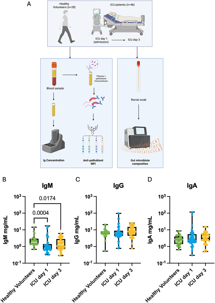 Figure 1
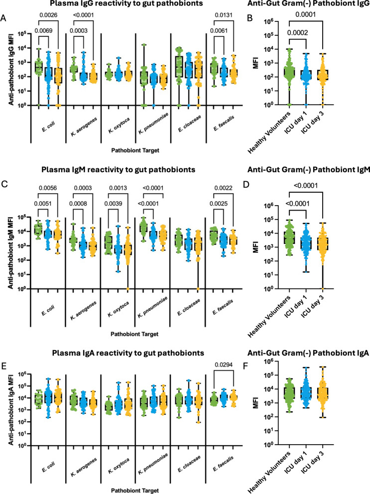 Figure 2
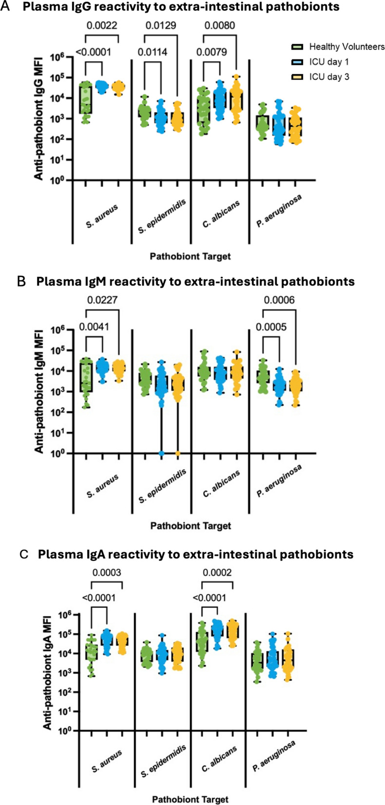 Figure 3
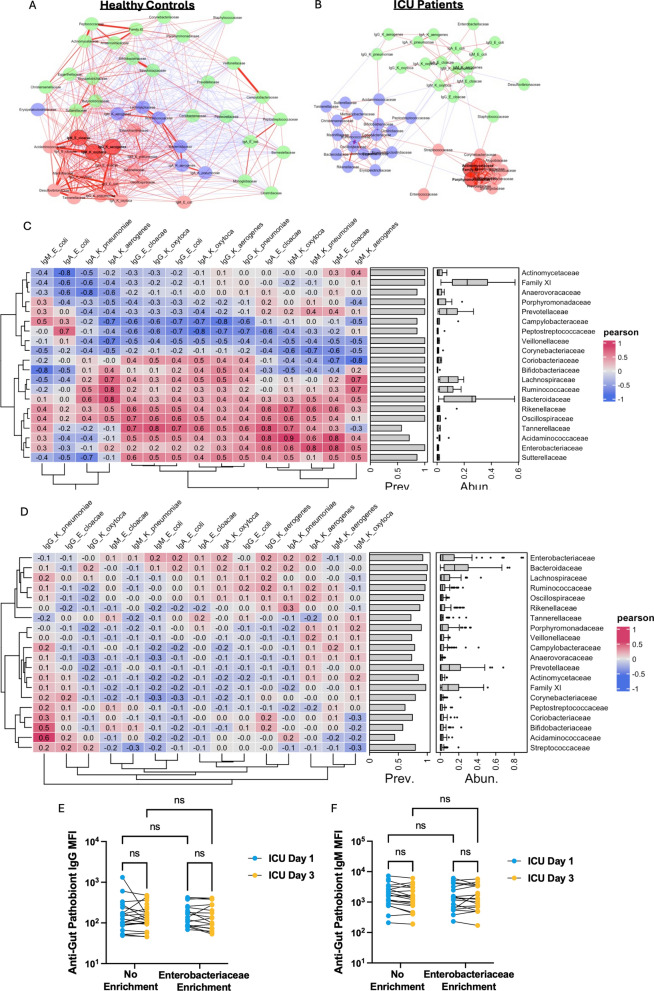 Figure 4
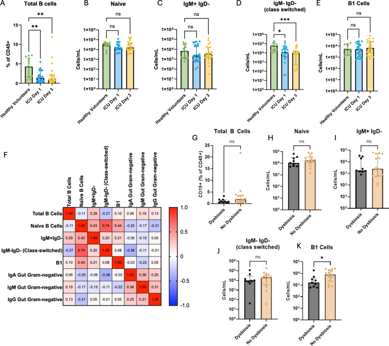 Figure 5
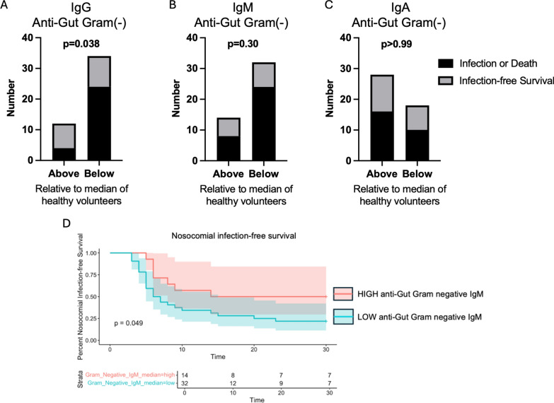 Figure 6
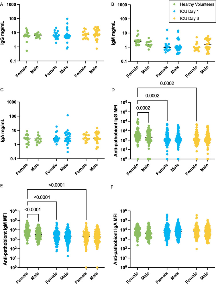 Figure 7
- —http://dx.doi.org/10.13039/501100000024Canadian Institutes of Health Research
- —Canadian Foundation for Innovation
Peer Reviews
No public reviews on file for this paper yet. If you reviewed it on a platform where reviews are public (OpenReview, ICLR, NeurIPS, ICML), you can paste yours below so the community can read it here.
Videos
No videos yet. Explain this paper in a talk, walkthrough, or lecture? Add one.
Taxonomy
TopicsClostridium difficile and Clostridium perfringens research · Gut microbiota and health · Nosocomial Infections in ICU
Background
Critically ill patients treated with life-support interventions in intensive care units (ICU) are at very high risk of nosocomial (hospital-acquired) infections, including ventilator-associated pneumonia (VAP), bloodstream infections (BSI), urinary tract infections (UTI), and Clostridium difficile infections (CDI), among others [1, 2]. Between 20 and 50% of mechanically ventilated ICU patients develop nosocomial infections [1, 3, 4], which is associated with a 2.5-fold increased risk of death compared to patients who do not develop nosocomial infections [5]. This marked susceptibility to deadly infections in critical illness is often attributed to invasive interventions that breach normal barriers and create portals of entry for pathogens, such as intravascular devices, indwelling catheters, and endotracheal intubation. It is now well established that susceptibility to infections in critical illness is also the result of immune dysfunction causing impaired host defense against invading microbes, as well as pathological alterations to the host’s microbiome resulting in a reservoir of potential pathogens in the gut and other mucosal sites [6–10]. As nosocomial infections remain a common and deadly threat to critically ill patients, there has been much attention to therapeutic and prophylactic strategies to reduce the burden of infectious complications in the ICU. Many of these interventions have been aimed at putative biological drivers including pathological microbiome alterations (digestive decontamination strategies, antimicrobial prophylaxis, probiotics), immune dysfunction, and care bundles [11–16]. However, our incomplete understanding of the interplay between the gut microbiome and immune defenses in critical illness have likely contributed to the limited effectiveness of current strategies, yielding substantial opportunities to advance clinical care through improved understanding of microbiome–immune interactions.
The importance of microbiome dysbiosis in the pathogenesis of nosocomial infections is highlighted by the microbiology of infecting pathogens in the ICU. First, the majority of nosocomial infections are caused by organisms that are resident members of the microbiome, including Gram-negative pathobionts that reside in the gut (e.g., Escherichia coli, Klebsiella spp., Enterobacter spp., Acinetobacter spp.) and airway microbiota (e.g., P. aeruginosa), Gram-positive pathobionts of the gut (Enterococcus spp.) and skin and airways (Staphylococcus spp.), and fungal pathobionts (notably Candida albicans) [17, 18]. Secondly, these pathobionts are often residents of the human microbiota during homeostasis, and rarely cause serious infections in healthy people due to the presence of effective immune defense barriers that prevent translocation and dissemination of microbiota organisms [19, 20]. This suggests that microbiota pathobionts act as “opportunistic” pathogens in the context of critical illness, possibly due to a breakdown of normal mechanisms that prevent translocation and dissemination of pathobionts during homeostasis [21]. One such mechanism is the production of antibodies in the blood directed against pathogenic microorganisms that colonize the gut and other mucosal surfaces (anti-pathobiont antibodies), which have been shown in animal models to serve as a crucial systemic immune barrier to pathobiont translocation and dissemination [22–25]. However, the importance of anti-pathobiont antibody responses in humans is less understood.
Therefore, we conducted a study to test the hypothesis that healthy humans harbor anti-pathobiont antibodies in the blood, and that this mechanism of immune defense is impaired during critical illness in the setting of gut microbiota dysbiosis, thus contributing to the marked risk of nosocomial infections caused by gut pathobionts in critically ill patients.
Methods
Study design and participants
This study was approved by the health research ethics boards of the University of Calgary and Alberta Health Services (REB18-1294). Male and female participants were included in this study, and sex as a biological variable was considered. The current study represents a secondary analysis of participants in the MICRO-ICU study, from which additional gut microbiome and immunological analyses are available in a prior publication [6]. Enrollment occurred between July 23, 2019 and July 20, 2021, with delays and disruptions in enrollment due to the COVID-19 pandemic between March 2020 and April 2021. Written informed consent was obtained from all study participants or appropriate surrogate decision-makers for patients who were unable to provide consent due to incapacitating illness. Patients admitted to the medical, surgical, neurological and trauma ICUs at the Foothills Medical Center in Calgary were screened for the following inclusion criteria (adapted from elsewhere[12]): adult (> 18 years of age) with an index admission to ICU, requiring mechanical ventilation, expected to require continuous mechanical ventilation for > 72 h as judged by the treating ICU specialist. Patients were excluded if they had a pre-existing immunocompromised state (systemic immunomodulatory therapy, chemotherapy, HIV infection or other congenital or acquired immunodeficiency), had been hospitalized > 48 h before ICU admission in the previous 3 months, had received systemic antimicrobial therapy in the previous 3 months, had inflammatory bowel disease or active GI malignancy, previous surgery leaving a discontinuous GI tract, pregnancy, goals of care that excluded life-support interventions or moribund patients not expected to survive > 72 h. At the onset of the COVID-19 pandemic, the study team added SARS-CoV-2 infection as an exclusion criterion and therefore no patients with COVID-19 were included in this study.
Blood and rectal swab samples were serially collected from ICU patients (N = 46) on day 1 of ICU admission and again from survivors who remained in the ICU on day 3 (N = 42). For reference comparison, plasma samples were obtained from healthy volunteers (N = 28). Population information is detailed in Supplementary Table 1. Data from both male and female patients and healthy volunteers were collected and analyzed. Details of infection diagnostic criteria and nosocomial endpoints in this cohort are previously reported [6].
Plasma antibody isotyping
The MSD Multi-Array Assay System (Human/NHP Isotyping Kit) Multiplex kit was used to determine concentrations of IgA, IgG, and IgM in plasma samples according to the manufacturer’s instructions.
Bacterial and fungal FACS
To determine whether circulating antibodies against microbiome pathobionts are reduced in critical illness, we quantified IgG, IgM, and IgA binding in patient plasma samples to a library of 10 pathobionts representing the most common causative pathogens in ICU nosocomial infections using flow cytometry. Bacterial and fungal strains used in this study are listed in Supplementary Table 2. Subcultures were made from overnight monocultures of each listed pathobiont and diluted to 2 × 10^7^ CFU/mL in FACs wash buffer. Serum was incubated at 56°C for 30 min, and centrifuged at 4000g for 10 min to inactivate complement. 100ul of subcultures were plated in a 96 well V-bottom plate, and 5ul of serum was added to each well to incubate for 1 h at room temperature, shaking at 300rpm. The wells were washed twice with FACs wash buffer, then cells were resuspended in 50 ul FACs wash buffer containing 1:50 IgA-PE (Miltenyi Biotec), 1:50 IgG-AF647 (Biolegend), and 1:50 IgM-PE/Cy7 (Biolegend) and incubated overnight at 4°C. Fluorescence minus 1 (FMO) as well as negative controls were also included. The next day, wells were washed with PBS and resuspended in 100ul of 4% PFA/PBS for 15 min at room temperature to fix cells, then washed again with PBS. Cells were resuspended in 100ul of 1:200 DAPI/PBS for bacteria or 1:2000 CFW/PBS for C. albicans cultures and acquired on CytoFlex LX (Beckman Coulter). Data were analyzed in Flowjo Software to determine median fluorescence intensity (MFI) for each sample and isotype (if negative values occurred, they were assigned a value of 1 to be included in analysis).
16S rRNA gene amplification and sequencing, sequence data processing and analysis
This study contains a secondary analysis from a previously published dataset, where additional methods details are available [6]. Briefly, rectal swabs were collected and stored in sterile tubes at − 80 °C, and DNA was isolated using the DNeasy PowerSoil kit (QIAGEN) according to the manufacturer’s protocol. PCR amplification of the 16S V4 region was performed and sequenced using an Illumina MiSeq platform. De-multiplexed Illumina MiSeq paired-end reads (FASTQ) were processed in R v4.1.2 following the DADA2 pipeline v.1.14. Taxonomy of unique amplicon sequence variants (ASVs) was assigned in DADA2 using the SILVA v.138.1 database. ASVs and sample data were combined using the Phyloseq package v.1.38.0 for further downstream analysis.
Time-of-flight mass cytometry
Single cell time-of-flight mass cytometry (CyTOF) was acquired as previously described [6]. Whole blood was cryopreserved in PROT1 proteomic stabilizer (SmartTube Inc.) at – 80 ºC following sample collection. For acquisition, samples were thawed and red blood cells were lysed using Thaw-Lyse buffer (SmartTube Inc.), and the single cell suspension was then passed through a 70-uM cell strainer. Cells were then fixed and permeabilized and subsequently barcoded with a unique 3 metal barcode using the Cell-ID 20-Plex Pd Barcoding Kit following manufacturers protocol (Standard Biotools). Following barcoding, samples were pooled and then stained with a cocktail of metal-labeled antibodies, then fixed overnight with DNA intercalator—Cell-ID™ Intercalator-Ir—and the following day the pooled samples were acquired on a Helios II. The acquired data were de-barcoded and analyzed using Cytobank (Beckman Coulter).
Statistical analysis
Antibody data are presented as median and IQR. Normality was assessed using the Shapiro–Wilk test in GraphPad Prism. Differences between groups were assessed using Kruskal–Wallis test with post hoc Dunn’s or Tukey’s test for multiple comparisons, or mixed-effects model with post hoc Tukey’s test for multiple comparisons. Multiple linear regressions and correlations were performed in GraphPad Prism. Results were considered significant at p < 0.05. E. coli IgM MFI cutoff and survival analysis was performed in R using the survminer R package. Microbiome correlation networks with antibody concentrations and MFI were generated using the NetCoMi R package (60). Microbiome data were filtered using a 25% prevalence filter and the remaining taxa were aggregated to the family level and log transformed. Antibody concentration and MFI data were log transformed, and a Pearson correlation was calculated between microbiome data and antibody data.
Results
Participant population
Demographic, clinical, and outcome data are displayed in Supplementary Table 1. The median age of ICU patient participants was 61 years, and 39.1% of patients were female. Inclusion and exclusion criteria (see details in Methods) yielded a population of participants experiencing multi-system critical illness (median SOFA score of 8), all of whom were receiving mechanical ventilation support and enteral nutrition, and were free of overt confounders to immune and microbiome analyses. Enrolled participants had a range of admission diagnoses including sepsis (45.7%), trauma (26.1%), neurological emergencies (19.6%), and other (8.7%). Median duration of mechanical ventilation was 6 days, median ICU length of stay (LOS) was 7 days, and median duration of hospitalization was 17 days. Adverse outcomes were analyzed from ICU admission to day 30, including nosocomial infections that were diagnosed and treated in 56.5% of participants, and an overall 30-day mortality rate of 32.6% (Supplementary Table 1).
Critically ill patients demonstrate a sustained decrease in circulating IgM compared to healthy individuals
Sample collection schedule and experimental design are outlined in Fig. 1A. Consistent with the known circulating isotype distributions in humans, IgG concentrations were higher than IgM and IgA in the circulation in both healthy and critically ill patients (Fig. 1B–D) [26]. However, the median concentration of total IgM in ICU patients on day 1 of admission was significantly lower than healthy volunteer controls (Fig. 1B), while no differences were observed in IgG nor IgA concentrations (Fig. 1C, D). We next analyzed the temporal progression of total antibody concentrations in the circulation of ICU patients between admission and day 3, and observed stability of IgM, IgG, and IgA concentrations, with IgM concentrations on day 3 remaining significantly below levels seen in healthy volunteers (Fig. 1B–D). Importantly, when stratified by admission diagnosis (sepsis, trauma, neurological, other), there were no differences in IgM, IgG, or IgA concentrations between these sub-groups of patients (Supplementary Fig. 1).Fig. 1. Systemic antibody responses in critically ill patients. A Flowchart of study design. B–D Quantitative analysis of B IgG, C IgM, and D IgA concentrations in the plasma of healthy volunteers (N = 28) and critically ill patients (N = 46) on day 1 and day 3 of ICU admission. Dots represent individual patients, central line indicates median, box shows interquartile range (IQR) and whiskers show range; analyzed by Kruskal–Wallis test with a post hoc Dunn’s test for multiple comparisons, significant p values as shown
ICU patients have impaired antibody responses against gut microbiome pathobionts
In healthy volunteers, we observed the presence of anti-pathobiont reactivity for all isotypes, indicating that healthy adults possess circulating antibodies that are reactive to microbiome pathobionts (Supplementary Fig. 2). Interestingly, MFI (median fluorescence intensity) of IgM and IgA reactivity was consistently higher than MFI of IgG reactivity for all pathobionts tested (Supplementary Fig. 2), despite lower total IgA and IgM concentrations compared to IgG (Fig. 1).
Gut pathobionts including Gram-negative bacteria (E. coli, Klebsiella spp., Enterobacter spp.) as well as Gram-positive bacteria (Enterococcus spp.), have previously been reported to dominate the gut microbiome of many ICU and other acutely ill patients, which has been associated with immune dysfunction, as well as increased risk of nosocomial infections [6, 27, 28]. These gut pathobionts are frequently causative pathogens in ICU nosocomial infections [1]. Next, we analyzed circulating antibody reactivity against common gut pathobionts in ICU patients compared to healthy volunteers (Fig. 2). IgG reactivity against E. coli, K. aerogenes, and E. faecalis were all reduced in ICU patients at admission, with sustained reduction at day 3, compared to healthy volunteers (Fig. 2A), whereas no differences were observed in IgG binding towards K. oxytoca, K. pneumoniae, and E. cloacae (Fig. 2A). Summation of plasma IgG binding towards all Gram-negative gut pathobionts tested revealed an overall reduction in the cumulative anti-Gram negative pathobiont IgG reactivity at day 1 that was sustained to day 3 (Fig. 2B). Compared to IgG, IgM binding to gut pathobionts was even more prominently impaired, with reduced plasma IgM binding to all gut pathobionts tested except E. cloacae, which demonstrated a non-significant trend towards reduction at admission and day 3 (Fig. 2C). Overall anti-Gram negative pathobiont IgM reactivity was markedly lower at both day 1 and day 3 of ICU admission compared to levels observed in healthy volunteers (Fig. 2D). Conversely, there were no reductions in plasma IgA binding to gut pathobionts between ICU patients (either day 1 or 3) and healthy controls (Fig. 2E, F).Fig. 2. Systemic antibody responses against gut pathobionts in critically ill patients. A Flow cytometry was used to quantify plasma IgG binding to 6 individual gut pathobionts (expressed as median fluorescence intensity, MFI) in healthy (N = 28) and critically ill patients (N = 46) on days 1 and 3 of ICU admission, and B shows pooled MFI values for IgG binding to all gut Gram-negative pathobionts together (E. coli, K. pneumonia, K oxytoca, K. aerogenes, E. cloacae). The same analyses are shown for plasma IgM (C, D), and IgA (E, F) binding to gut pathobionts. Dots represent individual patients, central line indicates median, box shows interquartile range (IQR) and whiskers show range; analyzed by Kruskal–Wallis test with a post hoc Dunn’s test, significant p values as shown
Having identified important alterations in anti-gut pathobiont antibody responses in our diverse cohort of ICU patients, we sought to determine whether these antibody responses were associated with any important patient variables. Multiple linear regression analyses found that IgG, IgM, and IgA binding to gut pathobionts, as well as their total plasma concentrations were not significantly associated with age, admission diagnosis, illness severity (SOFA score), burden of co-morbidities (Charlson index), nor duration of antibiotic exposure prior to sample collection (Supplementary Table 3).
ICU patients demonstrate variable systemic antibody responses towards extra-intestinal and multi-site colonizing pathobionts
Nosocomial infections in ICU patients are also commonly caused by pathobionts that reside outside the gut, in niches such as the skin (S. aureus, and coagulase-negative staphylococci like S. epidermidis), as well as airway colonizers (Pseudomonas aeruginosa), and the fungal pathobiont Candida albicans, which frequently resides at multiple body sites [1, 29, 30]. To determine whether systemic anti-pathobiont antibody responses differed against extra-intestinal and multi-site colonizing pathobionts, we quantified plasma IgG, IgM, and IgA binding towards S. aureus, S. epidermidis, C. albicans, and P. aeruginosa. Of note, we utilized a protein A (SpA)- and Sbi-deficient strain of S. aureus (ΔspAΔsbi SH1000) to avoid non-specific Fc binding of IgG in our assays. Contrary to what we observed towards gut pathobionts, there was a significant and sustained increase of plasma IgG, IgM, and IgA binding towards S. aureus in ICU patients on both days 1 and 3 compared to healthy controls (Fig. 3A–C). Alternatively, IgG binding to the less pathogenic and less invasive skin pathobiont S. epidermidis was reduced in ICU patients compared to healthy volunteers, whereas other isotopes were no different (Fig. 3A–C). Like anti-S. aureus antibody responses, plasma reactivity towards the fungal pathobiont C. albicans was significantly increased in ICU patients for both IgG (Fig. 3A) and IgA (Fig. 3C), but not IgM (Fig. 3B). For the airway pathobiont P. aeruginosa, decreased IgM reactivity (Fig. 3B) was observed, without any change in IgG (Fig. 3C) or IgA (Fig. 3A) in ICU patients. Taken together, these data reveal an intriguing dichotomy between anti-pathobiont antibody responses in critically ill patients, with impaired responses towards gut pathobionts (Fig. 2) yet augmented responses towards the skin pathobiont S. aureus and fungal pathobiont C. albicans (Fig. 3).Fig. 3. Systemic antibody responses against extra-intestinal pathobionts in critically ill patients. Flow cytometry was used to quantify plasma A IgG, B IgM, and C IgA binding to 4 extra-intestinal pathobionts (expressed as median fluorescence intensity, MFI) in healthy (N = 28) and critically ill patients (N = 46) on days 1 and 3 of ICU admission. Dots represent individual patients, central line indicates median, box shows interquartile range (IQR) and whiskers show range; analyzed by Kruskal–Wallis test with a post hoc Dunn’s test, significant p values as shown
Circulating anti-pathobiont antibody responses and gut microbiota dysbiosis in critical illness
Hallmarks of gut microbiota dysbiosis in critical illness include reduced microbial biodiversity, and taxonomic alterations characterized by accumulation of pathobionts (most commonly Enterobacteriaceae such as E. coli, Klebsiella spp., Enterobacter spp., as well as Enterococcaceae Enterococcus spp.), as shown in previously published studies including prior gut microbiota profiling from the patients in this study [6]. Given the prominent impairment of circulating antibody responses against gut pathobionts observed in ICU patients, we next investigated if these humoral immune alterations were associated with composition of the fecal microbiota in each participant using matched samples. Correlation networks between anti-pathobiont antibody MFI and the relative abundance of the top 20 taxa in the gut microbiota is shown for healthy volunteers in Fig. 4A, C, and ICU patients in Fig. 4B, D. Healthy volunteers were found to have strong and stereotyped correlations between plasma antibody reactivity against gut pathobionts and composition of the gut microbiota. In healthy volunteers there were strong positive correlations between plasma anti-pathobiont antibody quantities and intestinal abundance of Enterobacteriaceae and many anaerobic fermenter taxa, and negative correlations with Campylobacteriaceae, Peptostreptococcaceaea, and Veillonelaceae. In stark contrast, very few significant correlations were observed in ICU patients (Fig. 4B, D). These data suggest that the relationships between gut microbiota composition and systemic anti-pathobiont antibody responses during health are disrupted in the context of gut microbiota dysbiosis in critical illness.Fig. 4. Relationships between plasma antibody binding to gut pathobionts and the composition of the gut microbiota in ICU patients and healthy volunteers. Pearson correlation networks of bacterial families (relative abundance) and plasma antibody binding to intestinal pathobionts (MFI) in A healthy volunteers and B ICU patients with matched antibody and gut microbiota datasets (N = 7 and 42, respectively). Lines represent Pearson correlations between factors (red = positive correlation, blue = negative correlation) and line thickness representing strength of Pearson correlation. Nodes are colored by cluster membership. C, D Heatmaps of Pearson correlation coefficients between the 20 most abundant bacterial families (relative abundance) and plasma antibody binding to gut pathobionts (MFI) in C healthy volunteers (N = 7) and D ICU patients (N = 42). Bacterial family prevalence and relative abundance are represented as bar graphs and box-and-whisker boxplots, respectively. E Median MFI of plasma IgG and F IgM antibody binding to gut pathobionts in ICU patients with and without gut microbiota Enterobacteriaceae enrichment. Dots represent individual patients, lines link day 1 and day 3 values for individual patients, analyzed by mixed-effect model with a post hoc Tukey’s test for multiple comparisons, no significant p values
Previous work has demonstrated that patients with intestinal dysbiosis characterized by progressive Enterobacteriaceae enrichment have impaired innate immune function and increased risk of nosocomial infection or death [6]. To investigate whether gut Enterobacteriaceae enrichment was also associated with alteration of humoral immunity against gut pathobionts, we compared anti-pathobiont reactivity between days 1 and 3 of ICU admission in patients with versus without gut microbiota Enterobacteriaceae enrichment (as defined in Schlechte et al. [6]). However, we observed no difference in anti-gut pathobiont IgG, IgM (nor IgA, not shown) reactivity in plasma of ICU patients with gut Enterobacteriaceae enrichment versus those without (Fig. 4E, F).
Impaired anti-pathobiont antibody responses in critical illness are associated with dysregulated B cell responses
We observed profound B lymphopenia in ICU patients at admission that was sustained to day 3 compared to healthy volunteers (Fig. 5A). Analysis of B cell subsets (naïve, IgM+, class-switched IgM−IgD−, and putative B1 (CD19+CD1d+CD43+, designated “putative” due to the lack of consensus surface marker definition for this rare population in human blood), revealed no difference in the number of naïve (Fig. 5B), IgM + (Fig. 5C), nor putative B1 cells between ICU and healthy participants (Fig. 5E). However, class switched (IgM−IgD−) antigen experienced B cells were significantly decreased in ICU patients on day 1, and this reduction was sustained at day 3 (Fig. 5D).Fig. 5. Systemic B cell responses and antibody reactivity against gut pathobionts in critically ill patients. Mass cytometry was used to quantify A total B cells (CD19 + B220 + cells), B naïve B cells, C IgM + IgD- B cells, D class-switched (IgM-IgD-) B cells, and E putative B1 cells in available blood samples from healthy volunteers (N = 13) and ICU patients (N = 24) on day 1 and 3 of ICU admission. F Spearman correlation matrix showing the relationships between quantities of B cell populations and anti-gut pathobiont IgG, IgA, and IgM reactivity (MFI values) in the blood of ICU patients (numbers indicate Spearman’s correlation coefficient). G–K ICU patients were dichotomized into patients with gut microbiota dysbiosis (Enterobacteriaceae enrichment as defined in ref [6], N = 9) versus those without dysbiosis (N = 15), and the quantities of the indicated B cell populations were compared. Dots represent individual patients, bars indicate median, error bars show interquartile range (IQR); analyzed by Kruskal–Wallis test with a post hoc Dunn’s test (A–E) or Mann–Whitney U test (G–K). ***p < 0.001, **p < 0.01, *p < 0.05, ns (non-significant, p > 0.05)
Next, we interrogated the relationship between plasma IgM, IgG, and IgA binding towards gut Gram-negative pathobionts and circulating B cell populations, to understand the potential relationship between altered circulating B cells and impaired humoral response towards gut pathobionts. We observed negative correlations between IgM anti-gut pathobiont reactivity (MFI) and the quantity of circulating B1, class-switched (IgM-IgD-), IgM + , and naïve B cells, while IgA and IgG against gut pathobionts were negatively correlated with class-switched and Naïve B cells (Fig. 5F). Together, these data suggest that reduced anti-pathobiont antibody responses in ICU patients, particularly IgM responses, are associated with alterations of circulating B cell populations reflective of B cell dysregulation.
Prior literature, primarily from mouse models, have demonstrated that systemic anti-commensal antibody responses arise from microbiota-dependent regulation of multiple B cell compartments, including germinal center reactions in secondary lymphoid organs as well as natural antibody production by B1 B cells. Therefore, we investigated whether patients with gut microbiota dysbiosis (Enterobacteriaceae enrichment, as defined in Schlechte et al. [6]) mounted differential B cell responses comparted to those without this signature of intestinal dysbiosis (no enrichment). Interestingly, while conventional B cell populations were equivalent between ICU patients with and without gut Enterobacteriaceae enrichment (Fig. 5G–J), we observed significantly reduced quantities of circulating putative B1 cells in patient with Enterobacteriaceae dysbiosis (Fig. 5K). These data raise the interesting possibility that gut microbiota composition may impact systemic B cell responses in critical illness specifically within the natural antibody producing B1 B cell compartment, which may contribute to the prominent dysregulation of circulating IgM responses in ICU patients.
Impaired anti-gut pathobiont antibody responses in critically ill patients are associated with adverse outcomes including nosocomial infections and death
Having observed reduced plasma levels of IgM and IgG binding towards multiple gut pathobionts that commonly cause nosocomial infections in the ICU [6], we hypothesized that impairment of this humoral immune defense system may be associated with a higher risk of infections and related adverse outcomes including mortality. To test this hypothesis, we first stratified patients into 2 groups based on their antibody reactivity towards gut Gram-negative pathobionts—those with anti-gut pathobiont antibody reactivity (MFI) above “normal” (median value of healthy volunteers) versus those with levels below normal. We found that patients with low anti-gut Gram-negative IgG reactivity (MFI below median of healthy controls) had significantly higher rates of nosocomial infections or death compared to patients with normal or elevated levels of anti-gut Gram-negative IgG reactivity (Fig. 6A). We saw no significant differences in rates of nosocomial infections or death between patients with high versus low IgM nor IgA reactivity (Fig. 6B, C). Time-dependent analysis of nosocomial infection-free survival using maximal log rank statistic determination showed that ICU patients with low levels of anti-Gut Gram-negative IgM plasma reactivity (MFI < 2466) had significantly lower infection-free survival compared to ICU patients with higher plasma levels of anti-gut Gram-negative IgM reactivity (MFI > / = 2466) (p = 0.049, Fig. 6D). Collectively, these data suggest that reduced anti-gut pathobiont IgG responses are associated with an increased risk of nosocomial infections or death during critical illness.Fig. 6. Association between systemic antibody responses against gut pathobionts and nosocomial infection-free survival. A–C Graphs show the number of ICU patients (N = 46) who survived without nosocomial infections (infection-free survival, grey bars) versus those who developed nosocomial infections or died within 30 days of ICU admission (black bars), comparing between patients with plasma antibody reactivity against gut Gram-negative pathobionts (MFI) above or below the “normal” level (median level of healthy volunteers); A IgG, B IgM, or C IgA. Data were analyzed using Fisher’s exact test, p values as shown. D Kaplan–Meier curve displaying the proportion of ICU patients (N = 46) with nosocomial infection-free survival to 30 days post-ICU admission comparing individuals with high plasma IgM reactivity against gut Gram-negative pathobionts (MFI > / = 2466) versus those with low plasma anti-gut Gram-negative IgM (MFI < 2466). Analyzed by log rank statistic test, p = 0.049
Impairment of anti-pathobiont antibody responses may disproportionately affect critically ill females
It is well established that many immunological diseases demonstrate sex-biased epidemiology, and in particular that B cell and antibody responses differ between males and females. In critical illness, the prevalence, severity, and outcomes of infections and sepsis differ by sex, but is overall understudied, underreported, and poorly understood. Therefore, we performed a sex-stratified analysis of our data as an exploratory analysis, acknowledging the limited samples size in this cohort. We observed no differences in total IgG, IgM, or IgA concentration between males and females in healthy volunteers nor critically ill participants (Fig. 7A–C). However, sex-stratified analysis of anti-pathobiont antibody reactivity in plasma revealed that healthy females possessed higher anti-pathobiont IgG and IgM reactivity compared to healthy males, and that in critically ill patients, reduced anti-gut Gram-negative pathobiont IgG and IgM reactivity was seen only in females but not males (Fig. 7D, E). This reduction in anti-pathobiont IgG and IgM was sustained from admission to day 3 in female patients (Fig. 7D, E). Anti-gut Gram-negative IgA did not differ between female and male healthy volunteers or critically ill patients (Fig. 7F). Additional sex-stratified analyses of individual pathobiont antibody binding are shown in Supplementary Fig. 3. Collectively, these data suggest that impaired anti-pathobiont antibody responses during critical illness may disproportionately impact females as compared to males.Fig. 7. Sex disaggregated analysis of systemic antibody responses against intestinal pathobionts in critically ill patients. Flow cytometry was used to quantify plasma. A IgG, B IgM, and C IgA binding to 6 intestinal pathobionts (expressed as median fluorescence intensity, MFI) in healthy (N = 16 females, N = 12 males) and critically ill patients (N = 18 females, N = 28 males) on days 1 and 3 of ICU admission. Dots represent individual patients, line indicates median; analyzed by mixed-effects model with a post hoc Tukey’s test for multiple comparisons, significant p values as shown
Discussion
In this study, we report that humans mount systemic antibody responses against gut microbiome pathobionts that are impaired during acute critical illness. Prominent reductions of circulating IgM and IgG antibodies against intestinal pathobionts were observed in critically ill patients, and reduced IgG and IgM responses towards gut Gram-negative pathobionts were associated with increased susceptibility to nosocomial infections or death. Matched analysis of fecal microbiota composition as well as systemic B cell responses revealed that gut microbiota dysbiosis, characterized by enrichment of Enterobacteriaceae pathobionts, was coupled with reduced circulating (putative) B1 cells, a unique population of B cells well known to produce natural IgG and IgM antibodies, including against microbes that colonize mucosal surfaces.
Prior studies, primarily in mouse models, have established that systemic anti-microbiota antibodies in the bloodstream form a crucial barrier that protects against dissemination of microbes that translocate from mucosal surfaces into the blood [23, 24]. Indeed, acute and critically ill patients have been shown to display impaired gut barrier integrity, and translocation of gut microbes followed by dissemination through the bloodstream represents an important contributor to infections and end-organ dysfunction in these patient populations [10, 31–33]. Microbiological epidemiology of nosocomial infections in the intensive care setting consistently reports that more than half of ICU-acquired infections are caused by gut pathobionts, including Enterobacteriaceae and Enterococcaceae organisms [1]. Therefore, our observation of significantly reduced circulating IgM and IgG reactivity towards Enterobacteriaceae and Enterococcaceae pathobionts may help to explain why ICU patients experience such a high risk of infections caused by these gut pathobionts, due to breakdown of this antibody-mediated barrier against the spread of microbiota organisms in the blood.
Dysbiosis of the gut microbiota has been linked with adverse outcomes including nosocomial infections in critically ill patients. In particular, expansion of Enterococcaceae and Enterobacteriaceae in the gut of critically ill patients has been associated with infections and in-hospital mortality [6, 34, 35]. Mechanistically, pathobiont expansion in the gut may potentiate infections during critical illness due to the establishment of a pathogen reservoir in the gut that can translocate in the setting of impaired mucosal barrier integrity, as well as adverse microbiota–immune interactions and impaired host defense [36]. Indeed, our previous work linked Enterobacteriaceae expansion with innate immune dysfunction in critical illness, most notably neutrophil dysfunction [6]. In the present study in which we performed a secondary analysis of this same patient cohort, matched analysis of individual patient circulating antibody, B cell, and gut microbiota data revealed that intestinal dysbiosis in critically ill patients was associated reduced anti-pathobiont antibody levels as well as a reduction in putative B1 cells. B1 cells are innate-like lymphocytes that are important producers of natural antibodies, the repertoire of which is known to include IgM and IgG anti-commensal antibodies [37]. Germ-free mice harbor reduced titres of microbiota-reactive serum IgM, suggesting that the gut microbiota directs B1 cells to expand and secrete antibodies against mucosal colonizers [38]. Interestingly, a recent study in critically ill patients as well as mice reported that depletion of B-1a lymphocytes and their natural antibodies resulted in profound susceptibility to infections by the environmental mucosal colonizer Aspergillosis fumigatus [39]. This aligns with our observation of reduced B1-like cells in ICU patients with gut dysbiosis, and its association with increased risk of nosocomial infections. Similar to prior reports, we also observed total B lymphopenia with a prominent loss of class-switched (IgM−IgD−) B cells in critically ill patients compared to healthy controls. Therefore, the loss of anti-pathobiont IgM and IgG from the circulation of critically ill patients may be the result of both dysbiosis-related loss of natural antibody producing B1 cells, as well as the widespread reduction in classical B cell populations in critically ill patients.
Interestingly, we observed differences between circulating anti-pathobiont antibody responses directed against gut versus extra-intestinal pathobionts. While gut pathobiont-directed IgG responses were reduced in critically ill patients towards E. coli, K. aerogenes, and E. faecalis, we observed the opposite for skin-colonizing pathobionts S. aureus, multi-site colonizer C. albicans, and no difference in IgG reactivity towards lung pathobiont P. aeruginosa. Categorization of these organisms as gut, skin and lung pathobionts is a generalization, as S. aureus, C. albicans, and P. aeruginosa are often multi-site colonizers, but the unique antibody responses against these microbes may relate to unique microbiota changes that occur between the gut and extra-intestinal sites in critically ill patients. In addition, we also observed a striking difference between antibody isotype responses between IgG/M and IgA. IgA produced at mucosal sites is well known for its reactivity towards commensal organisms and central role in shaping the gut microbiota composition and protecting against pathobiont invasion [40]. Interestingly, we observed no differences in systemic IgA reactivity towards any gut pathobionts between critically ill and healthy controls, while observing an increase in systemic IgA reactivity towards skin colonizer S. aureus and the fungal pathobiont C. albicans. It will be of interest for future studies to investigate mucosal Ig responses in critically ill patients, to understand the relationship between microbiota dysbiosis and anti-pathobiont antibody responses at mucosal barrier sites versus the systemic circulation.
Meta-analysis of prior clinical trials of pooled donor immunoglobulin supplementation (IVIg) in sepsis has shown reduced mortality and length of hospitalization [41]. IVIg contains a polyreactive repertoire of antibodies including against microbiota organisms, as well as many other proposed mechanisms of immunomodulatory activity, therefore the potential role of anti-commensal Igs within IVIg is unknown. Interestingly, our data showed prominently impaired IgM anti-pathobiont responses in ICU patients, which is consistent with prior findings that IgM-enriched formulations of IVIg showed the greatest efficacy in sepsis [41]. Furthermore, monoclonal antibody therapies targeting selected pathobionts have also been investigated as prophylaxis against nosocomial infections by S. aureus and P. aeruginosa in the ICU [42–44]. Clinical trials of supplemental monoclonal antibodies against these pathogens did not reduce nosocomial infections in the ICU [42–44], which is consistent with our data showing no deficiency of endogenous IgG responses against S. aureus or P. aeruginosa in ICU patients. To date, the impact of therapeutic antibodies targeting of gut pathobionts has not been addressed in humans, and may represent an interesting avenue for therapeutic development.
Finally, we observed sex-specific anti-pathobiont responses in healthy volunteers as well as critically ill patients. Healthy females were found to express higher levels of anti-gut Gram-negative IgG and IgM reactivity compared to healthy males. Among critically ill patients, impairment of these antibody responses was seen in female participants but not males. Prior studies have reported that healthy females have higher levels of circulating B cells [45], as well as more robust antibody responses to vaccines compared to males [46–48]. Studies in both animal models as well as humans with severe infections and sepsis have reported sex-biased outcomes, with many studies finding higher incidence, severity, and mortality in males [49]. Future studies of larger cohorts are needed to determine whether the observed sexual dimorphism of pathobiont-specific antibody responses are associated with the sex-biased clinical outcomes in critical illness.
This study has a number of limitations. First, the sample size was insufficient to explore the granular relationship between anti-pathobiont antibody responses and the specific pathogens causing nosocomial infections in ICU patients. It is unknown whether our findings reflect dysregulation of antibody responses directed at specific pathobionts versus polyreactive antibodies that broadly protect against diverse pathobionts, and whether the antibody repertoire continues to evolve over the course of ICU admission. These questions will be important to explore in future studies that are larger and incorporate additional sampling beyond day 3 of admission. Next, our investigation was limited to a panel of 10 common pathobionts, and therefore it will be of interest for future studies to investigate antibody responses against additional pathobionts as well as commensal microbes. Dysregulation of systemic antibody responses against non-pathogenic commensals have been associated with inflammatory bowel disease pathogenesis [50]. Furthermore, investigating additional anti-commensal antibody responses and their relationship with microbiota dysbiosis in critical illness may uncover further insight into the contribution of the intestinal dysbiosis towards nosocomial infections and outcomes in the ICU. In particular, future studies using deep metagenomic sequencing analysis of gut microbes may help dissect species and strain-level regulation of microbiota-antibody relationships. Similarly, our study investigated only the bacterial microbiota and its relationship with anti-pathobiont antibody and B cell responses. Given that the fungal mycobiome in the human gut shapes systemic antifungal IgG binding, and individuals with systemic candidiasis mount a robust IgG response to Candida albicans [51], future studies should also look at how the mycobiome, and even virome, are linked to antibody production and patient outcomes.
Conclusions
In summary, this study reports that the systemic antibody responses against common gut pathobionts are compromised in critically ill patients, and is associated with an increased risk of nosocomial infections. These findings contribute to our understanding of the high rates of nosocomial infections caused by mucosal pathobionts in critical illness.
Supplementary Information
Additional file1 (PDF 897 kb)
The reference list from the paper itself. Each links out to its DOI / PubMed record.
- 1Zarb P, Coignard B, Griskeviciene J, Muller A, Vankerckhoven V, Weist K et al (2025) The European Centre for Disease Prevention and Control (ECDC) pilot point prevalence survey of healthcare-associated infections and antimicrobial use. Eurosurveillance 17(46). Available from: 10.2807/ese.17.46.20316-en 10.2807/ese.17.46.20316-en 23171822 · doi ↗ · pubmed ↗
- 2Schlechte J, Zucoloto AZ, Yu I ling, Doig CJ, Dunbar MJ, Mc Coy KD et al (2023) Dysbiosis of a microbiota–immune metasystem in critical illness is associated with nosocomial infections. Nat Med: 1–1110.1038/s 41591-023-02243-5PMC 1011564236894652 · doi ↗ · pubmed ↗
- 3Yang C, Chen-Liaw A, Spindler MP, Tortorella D, Moran TM, Cerutti A et al (2022) Immunoglobulin A antibody composition is sculpted to bind the self gut microbiome. Sci Immunol 7(73):eabg 320810.1126/sciimmunol.abg 3208 PMC 942156335857580 · doi ↗ · pubmed ↗
- 4Burkhard R, Koegler M, Brown K, Wilson K, Mager LF, Zucoloto AZ et al (2023) Intestinal colonization regulates systemic anti-commensal immune sensitivity and hyperreactivity. Front Immunol 14. Available from: 10.3389/fimmu.2023.103039510.3389/fimmu.2023.1030395 PMC 1023994637283756 · doi ↗ · pubmed ↗
- 5Schwartz DJ, Shalon N, Wardenburg K, De Veaux A, Wallace MA, Hall-Moore C et al (2023) Gut pathogen colonization precedes bloodstream infection in the neonatal intensive care unit. Sci Transl Med 15(694):eadg 556210.1126/scitranslmed.adg 5562 PMC 1025920237134153 · doi ↗ · pubmed ↗
- 6Kreuk LS, Koch MA, Slayden LC, Lind NA, Chu S, Savage HP et al (2019) B cell receptor and Toll-like receptor signaling coordinate to control distinct B-1 responses to both self and the microbiota. In: Mac Pherson AJ, Garrett WS, editors. e Life. 2019;8:e 47015.10.7554/e Life.47015 PMC 670385531433298 · doi ↗ · pubmed ↗
- 7Sarden N, Sinha S, Potts KG, Pernet E, Hiroki CH, Hassanabad MF et al (2022) A B 1a–natural Ig G–neutrophil axis is impaired in viral- and steroid-associated aspergillosis. Sci Transl Med 14(674):eabq 668210.1126/scitranslmed.abq 668236475902 · doi ↗ · pubmed ↗
- 8Chastre J, François B, Bourgeois M, Komnos A, Ferrer R, Rahav G et al (2022) Safety, efficacy, and pharmacokinetics of gremubamab (MEDI 3902), an anti-Pseudomonas aeruginosa bispecific human monoclonal antibody, in P. aeruginosa-colonised, mechanically ventilated intensive care unit patients: a randomised controlled trial. Crit Care Lond Engl 26(1):35510.1186/s 13054-022-04204-9PMC 966693836380312 · doi ↗ · pubmed ↗