A CLIP on the Ear: Spitz Melanocytoma Harbouring a CLIP2-BRAF Gene Fusion
Karwan A. Moutasim, Mohammed Atweh, Jeffery M. Theaker

TL;DR
This paper reports a rare case of a spitzoid melanocytoma with a CLIP2-BRAF gene fusion, challenging the assumption that BRAF mutations are incompatible with Spitz tumors.
Contribution
The novel contribution is the identification of a rare CLIP2-BRAF gene fusion in a spitzoid melanocytoma.
Findings
A spitzoid melanocytoma was found to harbor a CLIP2-BRAF gene fusion.
This finding challenges the conventional view that BRAF mutations are incompatible with Spitz tumors.
Abstract
Spitzoid lesions represent one of the most challenging areas in melanocytic pathology. Many such lesions are characterised by key gene alterations including ALK, ROS and NTRK fusions. BRAF mutations are generally considered incompatible with the diagnosis of Spitz tumours. Here, we present the case of a spitzoid melanocytoma harbouring a rare BRAF gene fusion. A brief overview of the literature is also touched upon.
Genes, proteins, chemicals, diseases, species, mutations and cell lines named across the full text — each resolved to its canonical identifier and authoritative record.
Click any figure to enlarge with its caption.
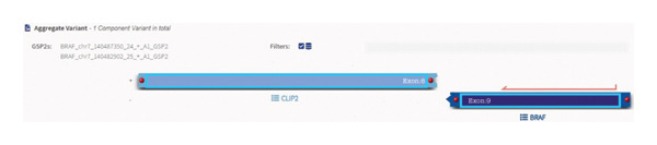 Figure FIGURE 2
Peer Reviews
No public reviews on file for this paper yet. If you reviewed it on a platform where reviews are public (OpenReview, ICLR, NeurIPS, ICML), you can paste yours below so the community can read it here.
Videos
No videos yet. Explain this paper in a talk, walkthrough, or lecture? Add one.
Taxonomy
TopicsCutaneous Melanoma Detection and Management · Melanoma and MAPK Pathways · Ocular Oncology and Treatments
1. Case
Spitzoid lesions represent one of the most challenging areas in melanocytic pathology. The original case series published by Sophie Spitz highlighted a series of paediatric Spitz lesions [1], the majority of which were likely Spitz naevi. It has now been over 75 years since her landmark contribution to the literature, and our understanding of these histologically heterogeneous lesions has been substantially improved by the advent of molecular and genomic techniques that the histopathologist in categorising and ultimately providing a clinically meaningful diagnosis.
It is now accepted that Spitzoid melanocytic tumours represent a spectrum ranging from benign Spitz naevi at one end and Spitzoid melanomas at the other with the intermediate category of Spitz melanocytoma (atypical Spitz tumour) also increasingly recognised. The majority of Spitz lesions are driven by fusions in receptor tyrosine kinase genes (e.g., ALK, ROS1, NTRK1-3 and RET) [2, 3]. Less commonly, serine threonine gene fusions may also be seen (e.g., BRAF) [4].
A 37‐year‐old male presented with a polypoidal lesion in the right postauricular area clinically thought to be neurofibroma. Macroscopic description showed a skin ellipse 13 × 12 × 3 mm with a centrally attached polypoidal nodule, 18 × 15 × 9 mm. Histological examination showed a predominantly intradermal melanocytic proliferation with a symmetrical, wedge‐shaped silhouette (Figure 1(a)). Epithelioid melanocytes with voluminous cytoplasm and prominent nucleoli were seen (Figure 1(b)). A prominent sclerotic stroma was noted (Figure 1(c)). Occasional multinucleated cells were present. Mitotic activity was rare and appeared confined to the superficial aspect of the lesion.
Figure FIGURE 1(a) A polypoidal melanocytic lesion with a fairly symmetrical outline. (b) Epithelioid melanocytes with voluminous cytoplasm, vesicular nuclei and identifiable nucleoli. (c) Fibrous stroma harbouring scattered epithelioid melanocytes, with occasional multinucleated cells.(a)(b)(c)
ALK and ROS1 immunohistochemistry were negative, as was PRAME. The MIB1 proliferation index was low. A retained, checkerboard pattern p16 pattern was seen. An initial provisional diagnosis of atypical Spitz tumour (Spitz melanocytoma) was issued, pending comprehensive targeted next generation sequencing (NGS) utilising the ArcherFusion panel.
NGS results showed a CLIP2-BRAF gene fusion (Figure 2). BRAF fusions in general are rare in Spitz tumours and are found in less than 5% of these lesions [4]. A recent case report had also interestingly highlighted similar morphological features, including a sclerotic stroma [5].
Schematic of CLIP2-BRAF gene fusion.
A limited re‐excision together with discussion at multidisciplinary meeting (MDT) and appropriate surveillance was performed. Sentinel lymph node biopsy was not performed. To date, the patient remains well with no evidence of recurrent or metastatic disease.
Botton and colleagues recently performed a systematic review of BRAF fusions in melanocytic tumours, identifying 100 such cases [4]. These had varying morphological patterns ranging from banal naevi to frank melanomas. Spitzoid features were seen in more than half of the cases reviewed. Here, we present a case of Spitz melanocytoma with a rare CLIP2-BRAF gene fusion. The key learning point is to interpret this correctly as a BRAF gene fusion as the majority of these lesions behave in an indolent manner, with a rare propensity to progression to melanoma. BRAF V600E gene mutations are rare in Spitz melanocytic lesions and should be interpreted with caution, as the majority of these may represent frankly malignant melanomas [6].
Further studies are required to elucidate the exact role of BRAF gene fusions and mutations in the spectrum of Spitzoid melanocytic lesions.
Author Contributions
K.A.M. wrote the manuscript. M.A. and J.M.T. also contributed to writing the manuscript.
Funding
No funding was received for this manuscript.
Consent
No written consent has been obtained from the patient as there are no patient identifiable data included in this case report.
Conflicts of Interest
The authors declare no conflicts of interest.
The reference list from the paper itself. Each links out to its DOI / PubMed record.
- 1Spitz S. , Melanomas of Childhood, The American Journal of Surgical Pathology. (1948) 24, no. 3, 591–609.PMC 194279818859360 · pubmed ↗
- 2Cheng T. W. , Ahern M. C. , and Giubellino A. , The Spectrum of Spitz Melanocytic Lesions: From Morphological Diagnosis to Molecular Classification, Frontiers Oncology. (2022) 12, 10.3389/fonc.2022.889223.PMC 920974535747831 · doi ↗ · pubmed ↗
- 3WHO Classification of Skin Tumours, 2020, 5th edition, World Health Organisation.
- 4Botton T. , Talevich E. , Mishra V. K. et al., Genetic Heterogeneity of BRAF Fusion Kinases in Melanoma Affecting Drug Responses, Cell Reports. (2019) 29, no. 3, 573–588.e 7, 10.1016/j.celrep.2019.09.009, 2-s 2.0-85073031706.31618628 PMC 6939448 · doi ↗ · pubmed ↗
- 5Donati M. , Kastnerova L. , Ptakova N. , Michal M. , and Kazakov D. V. , Polypoid Atypical Spitz Tumor With a Fibrosclerotic Stroma, CLIP 2-BRAF Fusion and Homozygous Loss of 9p 21, The American Journal of Dermatopathology. (2020) 42, no. 3, 204–207, 10.1097/dad.0000000000001502.31449064 · doi ↗ · pubmed ↗
- 6Chatzopoulos K. , Syrnioti A. , and Linos K. , Spitz Melanocytic Tumors: A Fascinating 75-Year Journey, Genes. (2024) 15, no. 2, 10.3390/genes 15020195.PMC 1088781338397186 · doi ↗ · pubmed ↗