From Technical Pitfall to Clinical Consequences: Leadless Pacing as a Rescue Solution
Fulvio Cacciapuoti, Ciro Mauro, Flavia Casolaro, Antonio Torsi, Salvatore Crispo, Mario Volpicelli

TL;DR
This case study shows how leadless pacing can be a safe solution when traditional pacemakers fail due to complications like stroke and venous occlusion.
Contribution
The paper presents a novel clinical application of leadless pacing as a rescue strategy in complex, high-risk scenarios.
Findings
Leadless pacing was successfully used in a patient with early pacemaker failure and venous occlusion.
Device interrogation helped identify lead insulation failure and battery depletion as causes of malfunction.
The patient showed favorable neurological recovery after the leadless pacemaker was implanted.
Abstract
Background and Clinical Significance: Early lead failure after dual-chamber pacemaker implantation is rare but clinically significant, particularly when associated with thromboembolic complications. Technical pitfalls at the time of implantation, such as suture fixation without protective sleeves, may be predisposed to premature lead damage and abrupt device malfunction. This case highlights the role of device interrogation in diagnosing arrhythmia-related stroke, the challenges of reimplantation in the setting of venous occlusion and anticoagulation, and the value of leadless pacing as a safe rescue strategy. Case Presentation: A 78-year-old man with a history of complete atrioventricular block underwent dual-chamber pacemaker implantation one year earlier. He presented to the emergency department with acute aphasia, right-sided hemiparesis, and facial asymmetry. Stroke was diagnosed,…
Genes, proteins, chemicals, diseases, species, mutations and cell lines named across the full text — each resolved to its canonical identifier and authoritative record.
Click any figure to enlarge with its caption.
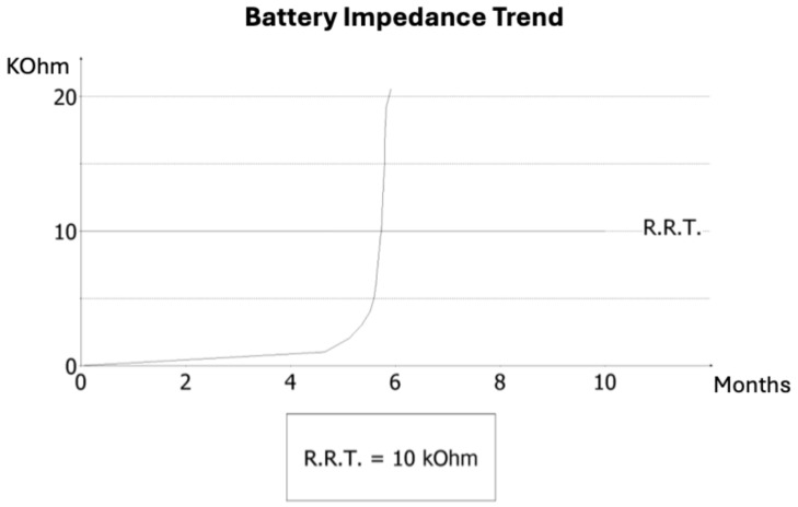 Figure 1
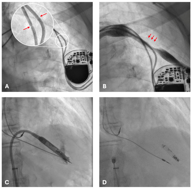 Figure 2
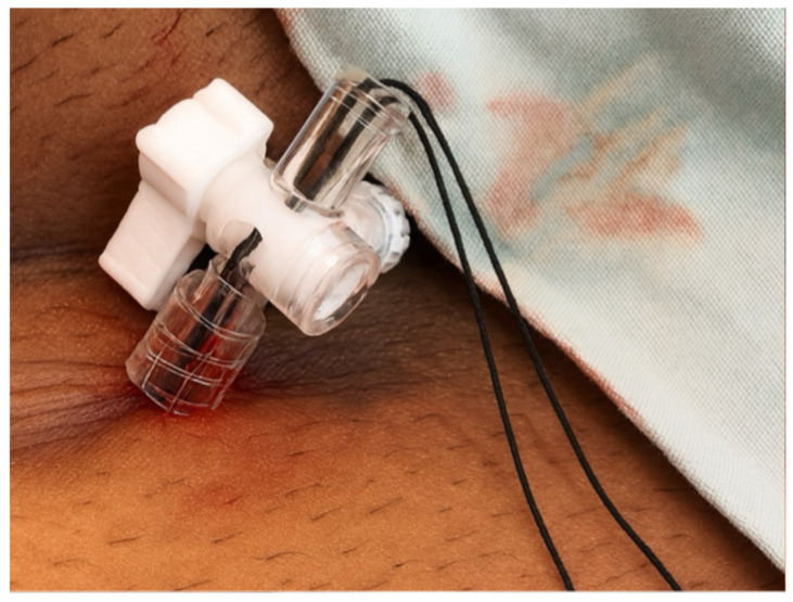 Figure 3
Peer Reviews
No public reviews on file for this paper yet. If you reviewed it on a platform where reviews are public (OpenReview, ICLR, NeurIPS, ICML), you can paste yours below so the community can read it here.
Videos
No videos yet. Explain this paper in a talk, walkthrough, or lecture? Add one.
Taxonomy
TopicsCardiac pacing and defibrillation studies · Cardiac Arrhythmias and Treatments · Atrial Fibrillation Management and Outcomes
1. Introduction and Clinical Significance
Dual-chamber pacemakers are an established therapy for the management of bradyarrhythmias, particularly in patients with atrioventricular conduction disturbances [1]. By preserving atrioventricular synchrony, these devices improve cardiac output, exercise tolerance, and quality of life [2]. Modern systems are highly reliable, yet complications can still occur, often as a result of technical issues at the time of implantation [3]. Small details, such as the proper placement of sutures or the use of protective sleeves, may appear minor during the procedure but can have long-term consequences on lead integrity. Mechanical stress, insulation damage, and conductor fracture are well-documented outcomes of such technical pitfalls, and while they may remain silent for months, they can ultimately manifest as sudden lead failure or premature device depletion [4].
Another dimension in the management of patients with pacemakers is the detection of arrhythmias, including atrial fibrillation (AF). Device interrogation has become a powerful diagnostic tool, capable not only of identifying technical dysfunctions but also of uncovering arrhythmic events that may otherwise remain asymptomatic [5]. The recognition of AF is particularly relevant because of its established role as a major risk factor for ischemic stroke. Early detection of AF burden provides clinicians with an opportunity to initiate anticoagulation in accordance with international guidelines, thereby significantly reducing the risk of thromboembolic events [6].
In situations where lead revision becomes necessary, venous access problems or anatomical limitations may preclude conventional transvenous reimplantation [7]. Leadless pacemakers have emerged as a valuable alternative in this setting. Implanted directly into the right ventricle through femoral venous access, these devices bypass the need for leads and subcutaneous pockets, reducing the risks of infection, lead fracture, and hematoma [8]. Furthermore, registry data support the safety of leadless implantation in patients on uninterrupted oral anticoagulation, a feature of particular relevance in those with recent thromboembolic stroke [9].
Clinical Significance: The following case illustrates not only how technical pitfalls can result in unusually early device failure but also how a rare convergence of complications—stroke related to new atrial fibrillation, complete venous occlusion, and the need for uninterrupted anticoagulation—created an exceptionally complex therapeutic scenario. In this setting, leadless pacing provided the only safe and effective solution.
2. Case Presentation
A 78-year-old man with a history of hypertension, type 2 diabetes mellitus, and coronary artery disease underwent dual-chamber pacemaker implantation one year earlier for complete atrioventricular block. He presented to the emergency department with sudden onset of aphasia, right hemiparesis, and mouth angle deviation. Neurological examination confirmed a disabling focal deficit, with a National Institutes of Health Stroke Scale (NIHSS) score of 12. Urgent brain computed tomography (CT) excluded intracranial hemorrhage, and CT angiography revealed an occlusion of a left middle cerebral artery branch. The patient presented outside the therapeutic window for intravenous thrombolysis or mechanical thrombectomy. He was managed conservatively with acute stroke care and subsequently initiated on secondary prevention, including oral anticoagulation (Apixaban 5 mg twice daily) and optimization of cardiovascular risk factors.
On admission, AF was documented for the first time. Pacemaker interrogation was performed to clarify arrhythmic burden and device status. Analysis revealed that AF had been persistent for several weeks prior to admission, temporally consistent with the occurrence of the cerebrovascular event. Additionally, diagnostic review identified striking electrical abnormalities: three months earlier, the ventricular lead impedance had dropped abruptly to 145 Ω, suggesting insulation breach or conductor damage, followed by a sharp rise to values above 30 kΩ, consistent with an open circuit (Figure 1). This electrical instability accelerated battery consumption, and the device entered elective replacement indicator (ERI) mode within months of implantation—a highly unusual finding, as large registries report lead failure rates of <1% at one year. Notably, at previous follow-up visits, no signs of lead malfunction or abnormal impedance trends had been detected and, unlike implantable loop recorders, conventional dual-chamber pacemakers do not systematically provide continuous remote monitoring, and in this case no remote alerts were available. However, despite the abrupt impedance drop and premature ERI, no loss of capture, pauses, or syncope were documented before replacement.
Fluoroscopic examination demonstrated the pacemaker generator in situ with both atrial and ventricular leads displaying focal narrowings in the infraclavicular region (Figure 2A). These findings were consistent with chronic mechanical damage, most likely related to suture fixation without protective sleeves at the time of implantation.
Conventional transvenous reimplantation was not feasible because of complete left subclavian vein occlusion (Figure 2B) and arterial overlap. Extraction of the damaged leads was deemed high risk. Leadless pacemaker implantation represented the only feasible and safe alternative, given the presence of complete venous occlusion, the high hemorrhagic risk of contralateral reimplantation, and the prohibitive risk of extraction. A leadless pacemaker (Aveir, Abbott, Abbott Park, IL, USA) was therefore selected as the optimal solution.
The procedure was performed via ultrasound-guided right femoral venous access without interruption of anticoagulation therapy. The presence of pre-existing intracardiac leads posed technical challenges, obstructing advancement of the delivery system (Figure 2C) and requiring multiple repositioning attempts. After careful manipulation and sheath angulation adjustments, stable fixation of the leadless device was achieved on the apico-septal region of the right ventricle (Figure 2D).
Final pacing parameters were optimal (threshold 0.5 V at 0.4 ms, R-wave 10 mV, impedance 1070 w Ω). No procedural complications occurred, and no bleeding events were observed despite uninterrupted anticoagulation.
The patient underwent neurological rehabilitation with partial functional recovery. At discharge, his modified Rankin Scale (mRS) score was 2, indicating slight disability but preserved independence.
At two-months follow-up, the leadless device demonstrated stable function with excellent electrical performance, a ventricular pacing percentage of 41%, and the patient showed ongoing neurological recovery.
3. Discussion
Contemporary registries consistently show that leadless systems perform well even in higher-risk cohorts. In the 12-month Micra AV Post-Approval Registry (n = 801), implantation success was 99.4% and major complications were significantly lower than with transvenous dual-chamber systems (3.7% vs. 8.8%) [10]. Five-year data from the Micra post-approval registry also demonstrate durable safety with low revision and infection rates [11]. For dual-chamber leadless pacing, pivotal investigations of the AVEIR DR system reported very high implant success (98–99%) with robust atrioventricular synchrony and acceptable 1-year complication profiles [12,13].
What makes this case distinctive is the convergence of challenges: an abrupt lead-impedance drop–rise with premature ERI, device-detected weeks of AF coinciding with stroke, complete ipsilateral subclavian occlusion with abandoned leads obstructing delivery, and the strict requirement for uninterrupted anticoagulation after ischemic stroke. Such a combination has rarely been reported in prior series. Our report therefore expands current evidence by illustrating a real-world rescue strategy when multiple constraints intersect, a scenario not captured in registries or larger studies. It was this clustering of adverse factors—rather than any single element—that defined the therapeutic complexity and guided management. Notably, the most striking feature was the abrupt failure of a dual-chamber system within only one year of implantation, an exceptionally early event compared with expected device longevity. In large national registries, lead failure rates during the first year remain well below 1%, with most malfunctions occurring after several years [14,15]. This strongly suggests a preventable technical cause rather than intrinsic device defect.
The fluoroscopic finding of focal narrowings along the lead trajectory supports the hypothesis of suture-related compression injury. Such injury is avoidable with meticulous implant technique and routine use of protective sleeves [16,17]. Preventive strategies include systematic use of protective sleeves at all tie-down points, careful adjustment of suture tension to avoid focal compression, preference for axillary or cephalic venous access to minimize costoclavicular crush, intra-procedural fluoroscopic and impedance checks, and structured operator training with procedural checklists. Remote monitoring may also help to detect abrupt impedance shifts earlier.
Another central message is the value of systematic device interrogation. In this patient, pacemaker checks did not merely reveal technical malfunction but also uncovered an elevated arrhythmic burden and several weeks of persistent atrial fibrillation preceding the ischemic stroke. Interrogation thus provided crucial diagnostic information, suggesting a possible etiological link between arrhythmia and the neurological event [18]. However, although persistent AF preceded the ischemic event in our patient, the temporal association does not prove causality. Observational data have linked device-detected AF to increased stroke risk but with relatively low absolute event rates [19]. Randomized trials in subclinical AF show mixed results: apixaban reduced stroke versus aspirin at the expense of more major bleeding [20], whereas edoxaban did not reduce a composite ischemic endpoint and increased bleeding [21]. These findings support our cautious interpretation and individualized anticoagulation strategy rather than a definitive causal claim. This highlights how device data can guide stroke prevention strategies, especially in patients with elevated CHA_2_DS_2_-VASc scores, for whom guideline-directed anticoagulation is essential.
In our case, the therapeutic pathway was also shaped by anatomic and procedural challenges. Conventional transvenous reimplantation was not feasible because of venous occlusion and arterial overlap, while extraction was judged high risk [22]. Contralateral transvenous reimplantation was avoided because creation of a new pocket and venous puncture under uninterrupted apixaban therapy, mandated after the recent ischemic stroke, carried a substantial risk of pocket hematoma and subsequent infection. Leadless pacing provided a feasible alternative [23]. Despite technical difficulties from intracardiac obstacles, the Aveir device was successfully implanted, restoring ventricular pacing with stable parameters. Anticoagulation by itself is not an indication for leadless pacing. In this case, however, the combination of complete ipsilateral venous occlusion, the need to avoid lead extraction, and the requirement for uninterrupted oral anticoagulation after stroke made the femoral leadless approach the most appropriate strategy.
Finally, the use of femoral venous access offered an important advantage in this anticoagulated patient [24]. Unlike subclavian or axillary approaches, the femoral route avoids creation of a subcutaneous pocket and reduces the risk of clinically significant bleeding or hematoma. Registry data confirm the safety of uninterrupted anticoagulation during leadless implantation, making this strategy particularly suitable for patients with recent ischemic stroke in whom strict maintenance of anticoagulation is mandatory [25,26]. At our center we perform femoral haemostasis by means of a figure-of-eight suture, closing the suture point inside a 3-way tap (Figure 3). This technique allows constant compression at the access level for 24 h, thereby minimizing the risk of hematoma formation. Combined with the absence of a subcutaneous pocket, this approach reduces bleeding complications and allows safe continuation of oral anticoagulation.
Follow-up is currently limited to two months, which precludes firm conclusions on long-term stability of the leadless system in the presence of abandoned leads. The patient is scheduled for device interrogations at 6 and 12 months, which will provide further insight into durability and safety. It should also be noted that this report reflects a single patient with short-term follow-up, which limits generalizability. However, while device interrogation strongly suggested a temporal association between atrial fibrillation and ischemic stroke, causality cannot be definitively established. Moreover, the absence of remote monitoring restricted continuous arrhythmia detection and may have delayed recognition of lead malfunction.
4. Conclusions
What makes this case particularly noteworthy is the simultaneous occurrence of multiple adverse factors, rarely reported together in the literature. Furthermore, it underscores that even minor technical oversights at pacemaker implantation may result in serious complications within an unusually short timeframe. Device interrogation proved invaluable, simultaneously identifying lead malfunction and clarifying arrhythmic burden, thereby informing both technical management and stroke prevention. Leadless pacing via femoral access emerged as a safe and effective rescue strategy in a patient requiring uninterrupted anticoagulation. As the population of anticoagulated and anatomically complex patients continues to grow, leadless systems are likely to play an increasingly central role in contemporary pacing practice.
The reference list from the paper itself. Each links out to its DOI / PubMed record.
- 1Glikson M. Nielsen J.C. Kronborg M.B. Michowitz Y. Auricchio A. Barbash I.M. Barrabés J.A. Boriani G. Braunschweig F. Brignole M. 2021 ESC Guidelines on cardiac pacing and cardiac resynchronization therapy Eur. Heart J.20214234273520 Erratum in Eur. Heart J. 2022, 43, 1651. https://doi.org/10.1093/eurheartj/ehac 07510.1093/eurheartj/ehab 36434455430 · doi ↗ · pubmed ↗
- 2Kusumoto F.M. Schoenfeld M.H. Barrett C. Edgerton J.R. Ellenbogen K.A. Gold M.R. Goldschlager N.F. Hamilton R.M. Joglar J.A. Kim R.J. 2018 ACC/AHA/HRS Guideline on the Evaluation and Management of Patients With Bradycardia and Cardiac Conduction Delay: A Report of the American College of Cardiology/American Heart Association Task Force on Clinical Practice Guidelines and the Heart Rhythm Society J. Am. Coll. Cardiol.201974 e 51e 156Erratum in J. Am. Coll. Cardiol. 2019, 74, 1016–1018. https://doi.org/10.1016 · doi ↗ · pubmed ↗
- 3Kirkfeldt R.E. Johansen J.B. Nohr E.A. Jørgensen O.D. Nielsen J.C. Complications after cardiac implantable electronic device implantations: An analysis of a complete, nationwide cohort in Denmark Eur. Heart J.2014351186119410.1093/eurheartj/eht 51124347317 PMC 4012708 · doi ↗ · pubmed ↗
- 4Schnorr B. Kelsch B. Cremers B. Clever Y.P. Speck U. Scheller B. Contemporary issues in cardiac pacing Minerva Cardioangiol.20105867769021135808 · pubmed ↗
- 5Witkowski M. Bissinger A. Grycewicz T. Lubinski A. Asymptomatic atrial fibrillation in patients with atrial fibrillation and implanted pacemaker Int. J. Cardiol.201722758358810.1016/j.ijcard.2016.10.09727836293 · doi ↗ · pubmed ↗
- 6Van Gelder I.C. Rienstra M. Bunting K.V. Casado-Arroyo R. Caso V. Crijns H.J.G.M. De Potter T.J.R. Dwight J. Guasti L. Hanke T. 2024 ESC Guidelines for the management of atrial fibrillation developed in collaboration with the European Association for Cardio-Thoracic Surgery (EACTS)Eur. Heart J.2024453314341410.1093/eurheartj/ehae 17639210723 · doi ↗ · pubmed ↗
- 7Chan N.Y. Kwong N.P. Cheong A.P. Venous access and long-term pacemaker lead failure: Comparing contrast-guided axillary vein puncture with subclavian puncture and cephalic cutdown Europace 2017191193119710.1093/europace/euw 14727733455 · doi ↗ · pubmed ↗
- 8Xu F. Meng L. Lin H. Xu W. Guo H. Peng F. Systematic review of leadless pacemaker Acta Cardiol.20247928429410.1080/00015385.2023.227653737961771 · doi ↗ · pubmed ↗