Treatment of Angiomyoblastoma in a Young Lady in Saudi Arabia: A Case Report: Angiomyoblastoma in a Young pLady in Saudi Arabia
Roua Shoub Gbril Ali, Ghadeer Adel Mosfer Alghamdi, Raad Hadi Madkhali, Abdulaziz Saleh Alobaid, Zarqa Saleem

TL;DR
This case report describes a rare benign tumor in a 36-year-old woman in Saudi Arabia, highlighting the importance of accurate diagnosis and surgical treatment.
Contribution
The paper presents a rare case of angiomyofibroblastoma in Saudi Arabia and emphasizes the need for precise diagnostic methods.
Findings
The tumor was diagnosed as angiomyofibroblastoma after surgical resection and immunohistochemical analysis.
MRI confirmed complete resection with no recurrence two months post-surgery.
Accurate diagnosis required differentiating AMFB from similar tumors like angiomyxoma or myxoid liposarcoma.
Abstract
Background: Angiomyofibroblastoma (AMFB) is a rare, benign soft tissue tumor that belongs to the mesenchymal tumor category and affects the female genital tract. Aggressive angiomyxoma, a distinct stroma myxoedematous mesenchymal tumor with a significant risk of local recurrences, must be histomorphologically distinguished from AMFB. Radiography and histopathology are used for accurate diagnosis. The treatment depended on the complete surgical excision. Case Presentation: Herein, we presented a case of a 36-year-old female (P3+0) presented with a left vaginal mass noted two years prior. The radiology report suggested angiomyxoma or myxoid liposarcoma as possible differentials. Surgical resection revealed a benign genital stromal tumor, specifically angiomyofibroblastoma (AMFB). After complete surgical excision, the immunohistochemical analysis supported the diagnosis of…
Genes, proteins, chemicals, diseases, species, mutations and cell lines named across the full text — each resolved to its canonical identifier and authoritative record.
Click any figure to enlarge with its caption.
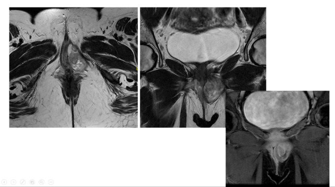 Figure-1
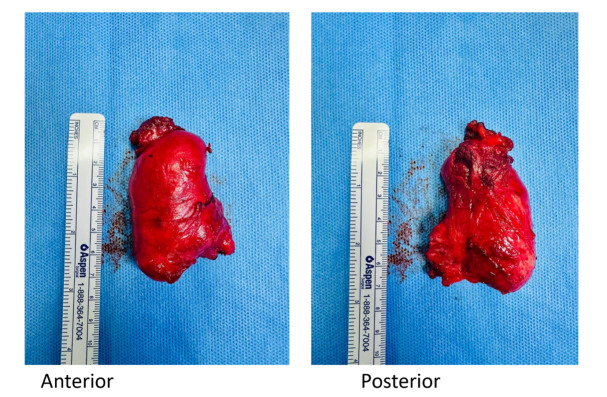 Figure-2
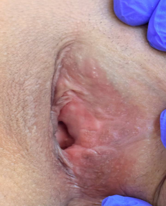 Figure-3
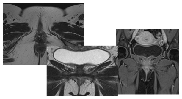 Figure-4
Peer Reviews
No public reviews on file for this paper yet. If you reviewed it on a platform where reviews are public (OpenReview, ICLR, NeurIPS, ICML), you can paste yours below so the community can read it here.
Videos
No videos yet. Explain this paper in a talk, walkthrough, or lecture? Add one.
Taxonomy
TopicsUrologic and reproductive health conditions · Testicular diseases and treatments · Urinary and Genital Oncology Studies
Introduction
Angiomyofibroblastoma (AMFB) is a rare, benign soft tissue tumor belonging to the mesenchymal tumor; it originates from blood vessels and stromal cells [1].
The woman’s genital tract, particularly the vagina and vulva, is where AMFB typically begins. The broad ligament, fallopian tube, and, less commonly, the male genital tract have also been identified as potential locations [2]. The highest prevalence rate was reported among young to middle-aged females [3].
Clinically, AFMB manifests as a slowly expanding mass; it lacks any other distinctive symptoms that set it apart from other tumors of the female genital tract [4]. Aggressive angiomyxoma, a distinct stroma myxoedematous mesenchymal tumor with a significant risk of local recurrences, must be histomorphologically distinguished from AMFB [5]. The gold standard of care is surgical removal [6]. Here, we present a case of a 36-year-old female diagnosed with vaginal AMFB, showing the diagnostic difficulties when considering this rare entity and available treatment.
Case Presentation
**
**
**
**
A 36-year-old female (P3+0) presented with a left vaginal mass noted two years prior, which had gradually increased in size. She had a history of a vulvar mass first reported in November 2023 but no significant medical or surgical history.
The initial magnetic resonance imaging (MRI) from an external facility revealed a well-defined left lateral lower vaginal lesion extending to the perineum. The radiology report suggested angiomyxoma or myxoid liposarcoma as possible differentials. The patient underwent surgical removal of the mass in July 2024 at another hospital. However, no prior imaging or clinical history was available at that time.
Histopathology from the first surgery suggested aggressive angiomyxoma based on the spindle cell tumor’s morphology. However, the diagnosis was inconclusive due to incomplete excision. An MRI in August 2024 revealed Left pelvic mass measuring 5 x 3 x 4 cm. Show bright T2 signal intensity with a swirling appearance crossing the left levator ani muscle towards the left external anal sphincter abutting the sphincter. Internal sphincter and intersphincteric fat are intact. Mass extends to the left vaginal wall with an indentation of the vaginal wall at the level of the urethra. The mass shows avid enhancement post-contrast (Figure-1).
A second surgery was planned to completely excision the mass. The 6 cm tumor, located near the urethra and puborectalis muscle, was excised with meticulous dissection (Figure-2). The postoperative site was closed using a parallel technique, ensuring optimal healing (Figure-3).
Histopathology of the second excised mass revealed a well-encapsulated stromal tumor with spindle cells embedded in a myxoid stroma. Immunohistochemical analysis supported the diagnosis of angiomyofibroblastoma. Margins were free of tumor, confirming complete resection.
Two months postoperatively, an MRI Post-resection images show complete resection with no feasible scar, residual tumor, or recurrence. There is no vaginal wall deformity. Post-contrast shows no significant abnormal enhancement (Figure-4). Incision site healing was unremarkable, and the patient reported no new symptoms.
Discussion
Fletcher et al. initially reported angiomyofibroblastoma (AMFB), a benign mesenchymal tumor, in 1992 [3]. Few instances of it occur in men, and it usually affects the external genital tract in women [7]. Women in their middle years (between the ages of 30 and 50) are generally affected by AMFB [3]. Our patient was a 36-year-old female.
Clinically, the tumor may be asymptomatic or manifest as a painless weight in the pelvis [8]. When it is found in the cervix, uterus, or urethral area, it can occasionally exhibit obstructive symptoms such as dysuria [9]. It is easily diagnosed since it is often seen in the superficial regions of the lower female genital tract. However, a tumor in the pelvis, peritoneal cavity, or ilia fossa may be discovered after it has grown considerably [10]. Regarding our case, she was symptomatic and admitted with a left vaginal mass noted two years prior, which had gradually increased in size.
Ultrasound (US) is the primary imaging technique used to evaluate vaginal masses due to its high resolution, availability, and cost-effectiveness. It can distinguish between vulvar and vaginal amyloid-beta (AMFB) and other mesenchymal tumors.[11]. AMFB is typically well-circumscribed on MRI and is often described as hypointense on T1W images, hyperintense on T2W images, and with homogenous hyperenhancement on Gd-C enhanced images. Contrast-enhanced imaging has been reported in five studies [11]. Regarding our case, the initial MRI suggested angiomyxoma or myxoid liposarcoma as possible differentials. After the first excision surgery, MRI shows bright T2 signal intensity with a swirling appearance crossing the left levator ani muscle towards the left external anal sphincter abutting the sphincter.
Malignant mesenchymal tumors, aggressive angiomyxoma, cellular angiofibroma, fibroepithelial stromal polyp, and superficial cervicovaginal myofibroblastoma are all included in the broad differential diagnosis. Usually more significant than 10 cm, aggressive angiomyxoma (AAM) is a locally infiltrative tumor of tiny, spindle- to stellate-shaped cells encased in a thick layer of a myxoid matrix with "pushing" infiltrative boundaries. Compared to angiomyofibroblastoma, the cells are often more evenly distributed and monomorphic [7]. AAM and AMFB have varied positivity for desmin and α-SMA but are immunohistochemically negative for S-100 and positive for vimentin, estrogen receptor, and progesterone receptor. On the other hand, a new molecular analysis confirms that AAM and AMFB are different types of cancer. A third of AAM patients had the HMGA2 gene arrangement reported, whereas AMFB cases do not [11]. Cellular angiofibroma, which has thicker blood arteries and lacks hormone receptor expression, is another differential diagnosis. Inflammatory myofibroblastic tumors and inflammatory fibroid polyps are further diseases that must be checked out [12][13].
Regarding our case, the radiology report suggested angiomyxoma or myxoid liposarcoma as possible differentials. Histopathology from the first surgery suggested aggressive angiomyxoma based on the spindle cell tumor’s morphology, but the diagnosis wasn’t accurate due to incomplete excision. A second surgery was planned to completely excision the mass. Histopathology of the second excised mass revealed a well-encapsulated stromal tumor with spindle cells embedded in a myxoid stroma. Immunohistochemical analysis supported the diagnosis of angiomyofibroblastoma. Margins were free of tumor, confirming complete resection.
Surgical removal is the gold standard treatment [6]. In the present case, Without any noticeable scarring, tumor recurrence, vaginal wall deformation, post-contrast enhancement, or new symptoms, the patient had a complete resection.
Conclusion
Our case is rare AMFB. AMFB may be missed when diagnosed with other masses, such as angiomyxoma or myxoid liposarcoma, so we need to do the necessary tests to diagnose and differentiate AMFB accurately from other masses. Ultrasound and MRI are initial diagnostic tools, while histopathology ensures the correct diagnosis. Surgical removal with free margins remains the appropriate treatment. Long-term follow-up is nessesary to check for poten tial recurrence, though AMFB typically carries an excellent prognosis.
Conflict of Interest
The authors declare that they have no conflict of interest.
The reference list from the paper itself. Each links out to its DOI / PubMed record.
- 1Fatusic J Hudic I Fatusic Z Mustedanagic-Mujanovic J Angiomyofibroblastoma of the vaginal portion Med Arch 2014686424510.5455/medarh.2014.68.424-425PMC 431417425648716 · doi ↗ · pubmed ↗
- 2Sherif M Mohamed I Massive angiomyofibroblastoma of the glans penis BMJ Case Rep 2019126 e 229486 e 22948610.1136/bcr-2019-229486 PMC 655741931167770 · doi ↗ · pubmed ↗
- 3Fletcher CD Tsang WY Fisher C Lee KC Chan JK Angiomyofibroblastoma of the vulva: a benign neoplasm distinct from aggressive angiomyxoma The American journal of surgical pathology 19921643738210.1097/00000478-199204000-000061314521 · doi ↗ · pubmed ↗
- 4Kwack JY Kim S Lee JS Kwon YS Case Report of Vaginal Angiomyofibroblastoma and Differential Diagnosis of Other Benign Mesenchymal Tumors in the Female Genitalia. J Clin Obstet Gynecol 2024 Mar 1343640
- 5Nielsen GP Rosenberg AE Young RH Dickersin GR Clement PB Scully RE Angiomyofibroblastoma of the vulva and vagina Modern pathology: an official journal of the United States and Canadian Academy of Pathology, Inc 199693284918685229 · pubmed ↗
- 6Sassi S Nadim C El Mohtarim Rouas L Yousfi M Lamalmi N Hassouni FE Angiomyofibroblastoma of the vulva: a case report and review of the literature Case Reports in Women’s Health 202442 e 00617 e 0061710.1016/j.crwh.2024.e 00617 PMC 1114126338827183 · doi ↗ · pubmed ↗
- 7Sims SM Stinson K Mc Lean FW Davis JD Wilkinson EJ Angiomyofibroblastoma of the vulva: a case report of a pedunculated variant and literature review Journal of Lower Genital Tract Disease 20121621495410.1097/LGT.0b 013e 318231217 b 22371044 · doi ↗ · pubmed ↗
- 8Seo JW Lee KA Yoon NR Lee JW Kim BG Bae DS Angiomyofibroblastoma of the vulva Obstetrics & Gynecology Science 201356534934910.5468/ogs.2013.56.5.349PMC 378412324328028 · doi ↗ · pubmed ↗