Post-flexible Bronchoscopy Escherichia coli Empyema: A Rare Complication or an Inflammatory Bacterial Translocation?
Ukasha Moazzam, Bakht Noor Khurshid, Cyrus Daneshvar

TL;DR
A patient with bronchiectasis developed empyema after bronchoscopy, with different bacteria found in the airway and pleural fluid.
Contribution
This case highlights the risk of post-bronchoscopy infections and challenges in antimicrobial treatment due to bacterial discordance.
Findings
Empyema occurred 10 days after bronchoscopy in a patient with bronchiectasis.
Different bacteria were isolated from bronchial washings and pleural fluid, complicating treatment decisions.
Abstract
Empyema is an unusual complication of diagnostic bronchoscopy. We report a case of a male patient in his late 70s with bronchiectasis who developed a right lower lobe empyema 10 days after undergoing flexible bronchoscopy for incidentally detected lung nodules. Bronchial washings grew Staphylococcus aureus (S. aureus) and the atypical Gram-negative bacillus Pantoea septica (P. septica), while subsequent pleural fluid culture yielded Escherichia coli (E. coli). This case highlights the risk of post-bronchoscopy infectious complications in patients with structural lung disease and illustrates how discordance between airway and pleural isolates can complicate antimicrobial decision-making.
Genes, proteins, chemicals, diseases, species, mutations and cell lines named across the full text — each resolved to its canonical identifier and authoritative record.
Click any figure to enlarge with its caption.
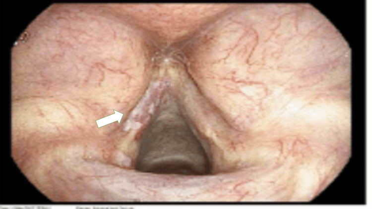 Figure 1
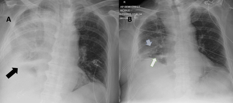 Figure 2
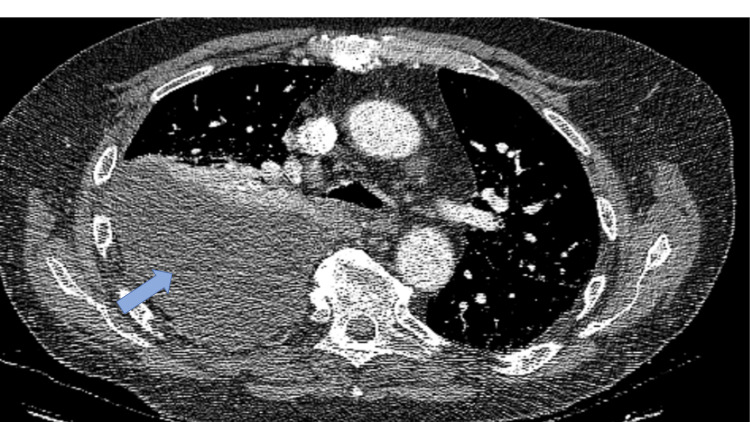 Figure 3
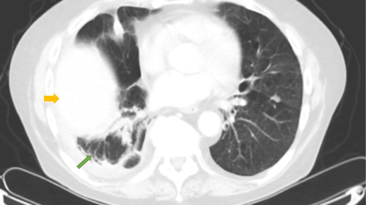 Figure 4
Peer Reviews
No public reviews on file for this paper yet. If you reviewed it on a platform where reviews are public (OpenReview, ICLR, NeurIPS, ICML), you can paste yours below so the community can read it here.
Videos
No videos yet. Explain this paper in a talk, walkthrough, or lecture? Add one.
Taxonomy
TopicsPleural and Pulmonary Diseases · Tracheal and airway disorders · Lung Cancer Diagnosis and Treatment