A Rare Case of Cutaneous Meningioma Arising on the Scalp: Diagnostic Challenges, Immunohistochemical, and Molecular Features
Kainat Memon, Richard J Digby, Azzam Ismail

TL;DR
A rare case of cutaneous meningioma on the scalp highlights the need for combining clinical and lab findings for accurate diagnosis.
Contribution
This case report emphasizes the diagnostic value of integrating histopathology, immunohistochemistry, and molecular studies for rare scalp tumors.
Findings
A scalp lesion initially thought to be an epidermoid cyst was diagnosed as cutaneous meningioma.
Combining clinical, histopathological, and molecular data is crucial for diagnosing rare extracranial meningiomas.
Cutaneous meningioma is a rare condition requiring multidisciplinary diagnostic approaches.
Abstract
The differential diagnosis for scalp lesions is broad, ranging from simple cysts to tumors. We present the case of a 49-year-old woman with a scalp lesion initially suspected to be an epidermoid cyst but later diagnosed as cutaneous meningioma on biopsy. As a rare form of extracranial meningioma, this case underscores the importance of correlating clinical findings with histopathology, such as integrating morphological features, immunohistochemical markers, and molecular studies, to establish a diagnosis in challenging cases with inconclusive clinical or radiological findings.
Genes, proteins, chemicals, diseases, species, mutations and cell lines named across the full text — each resolved to its canonical identifier and authoritative record.
Click any figure to enlarge with its caption.
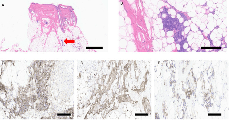 Figure 1
Peer Reviews
No public reviews on file for this paper yet. If you reviewed it on a platform where reviews are public (OpenReview, ICLR, NeurIPS, ICML), you can paste yours below so the community can read it here.
Videos
No videos yet. Explain this paper in a talk, walkthrough, or lecture? Add one.
Taxonomy
TopicsMeningioma and schwannoma management · Neurofibromatosis and Schwannoma Cases · Facial Nerve Paralysis Treatment and Research