Real-Time and Selective Detection of Pseudomonas aeruginosa in Beef Samples Using a g-C3N4-Doped Multimetallic Perovskite-Based Electrochemical Aptasensor
Sarah S. Albalawi, Naeem Akhtar, Waleed A. El-Said

TL;DR
A new electrochemical sensor detects Pseudomonas aeruginosa in beef quickly and accurately, improving food safety.
Contribution
First FeCoCuNiO-g-C3N4-based aptasensor for real-time bacterial detection in food samples.
Findings
The aptasensor achieved a detection limit of 3.03 CFU mL−1 for P. aeruginosa.
It demonstrated high selectivity and sensitivity in real beef samples.
The sensor was fabricated using a sol–gel combustion method and a graphitic pencil electrode.
Abstract
The alarming rise in foodborne illnesses, particularly those associated with microbial contamination in meat products, presents a serious challenge to global food safety. Among these microbial threats, Pseudomonas aeruginosa (P. aeruginosa) poses a critical threat due to its biofilm-forming capability and prevalence in contaminated beef, highlighting its effective real-time detection. Herein, we report the fabrication of a novel electrochemical aptasensor based on multimetal perovskite (FeCoCuNiO) doped with urea-derived graphitic carbon nitride (g-C3N4), synthesized via a sol–gel combustion method. The FeCoCuNiO-g-C3N4 nanocomposite was then coated onto a graphitic pencil electrode and functionalized with a DNA-based aptamer specific towards P. aeruginosa. The resulting aptasensor exhibited a low detection limit of 3.03 CFU mL−1 with high selectivity and sensitivity, and was…
Genes, proteins, chemicals, diseases, species, mutations and cell lines named across the full text — each resolved to its canonical identifier and authoritative record.
Click any figure to enlarge with its caption.
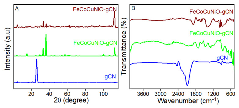 Figure 1
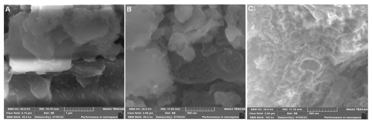 Figure 2
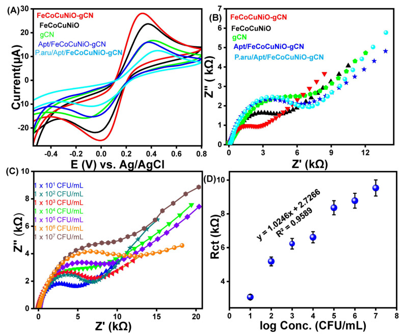 Figure 3
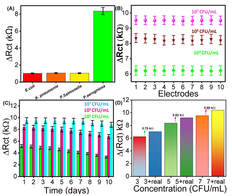 Figure 4
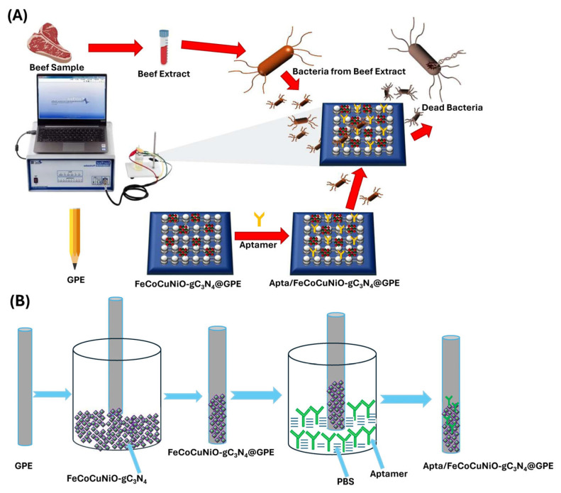 Figure 5
- —University of Jeddah
Peer Reviews
No public reviews on file for this paper yet. If you reviewed it on a platform where reviews are public (OpenReview, ICLR, NeurIPS, ICML), you can paste yours below so the community can read it here.
Videos
No videos yet. Explain this paper in a talk, walkthrough, or lecture? Add one.
Taxonomy
TopicsAdvanced Chemical Sensor Technologies · Advanced biosensing and bioanalysis techniques · Biosensors and Analytical Detection
1. Introduction
Foodborne diseases continually impact consumers, health authorities, and the food industry. Among various food products, meat, especially beef, is highly perishable, with over 20% of global meat production wasted due to microbial spoilage [1]. Fresh meat and meat products are common sites for foodborne pathogens [2]. Contamination of beef can occur through multiple routes, including cross-contamination during slaughter, unsanitary processing environments, and inadequate cold-chain practices [3]. Bacteria, the second leading cause of foodborne illnesses, pose significant threats, with approximately 3.6 million cases reported annually [4]. Of these, 1.5 million are caused by diarrheagenic Escherichia coli, Salmonella, and Pseudomonas aeruginosa (P. aeruginosa). P. aeruginosa is especially concerning because it causes gastrointestinal infections, diarrhea, abdominal pain, and nausea [5]. This highly adaptable Gram-negative bacterium threatens public health and food safety due to its inherent antibiotic resistance and ability to form biofilms rapidly [6]. This alarming adaptability highlights the urgent need for rapid, sensitive, and specific detection of P. aeruginosa at low concentrations for real-time analysis [7].
In recent years, several methods have been adopted for the detection of P. aeruginosa, including magnetic relaxation switch aptasensor [8] surface-enhanced Raman spectroscopy [9], colorimetric assays [10,11], and polymerase chain reaction techniques [12]. Although these approaches are highly sensitive and accurate, their use in bacterial detection is often limited due to long processing times, the need for specialized instruments, and complex operational procedures [13]. This highlights the need for faster, cost-effective, and user-friendly methods for the reliable diagnosis of P. aeruginosa. Electrochemical sensors have recently gained considerable attention due to their advantages, including low cost, rapid detection, ease of use, and high sensitivity [14,15]. Among these, electrochemical aptasensors stand out for their ability to provide rapid, selective, and sensitive detection of pathogens [16,17]. A key factor in improving the performance of aptasensors lies in the choice of electrode materials. An ideal electrode material should promote fast electron transfer, ensure strong and stable aptamer immobilization, and support low detection limits and high sensitivity.
In the past few decades, noble metals such as Pt [18], Pd [19], Ag [20], and Au [21]-based nanocomposites have been used for pathogen detection due to their high sensitivity and excellent stability. However, their high cost, limited abundance, and poor scalability have significantly hindered their widespread commercial applications [22]. In this regard, a diverse range of transition metal-based materials, including oxides [23], phosphides [24], nitrides [25], sulfides [26], and perovskites, have emerged as promising electrochemical sensors [27]. Among these, perovskite-type oxides (ABO_3_) have attracted considerable attention owing to their structural versatility, tunable electronic properties, and catalytic adaptability [28]. Despite these advantages, perovskites still face inherent limitations, such as relatively poor electrical conductivity and restricted exposure of active sites due to agglomeration or limited porosity [29]. To address these challenges, various conductive materials such as purines, polyaniline [30], poly (3,4-ethylene dioxythiophene) [31], urea [32], and polypyrrole have been reported [33]. Among these conductive materials, urea-derived graphitic carbon nitride (g-C_3_N_4_) has garnered significant attention due to its high electrical and thermal conductivity, good mechanical and chemical stability, high surface area, and favorable carrier transport [34,35]. Therefore, doping g-C_3_N_4_ with transition metal-based perovskite is expected to enhance its electrochemical performance and stability.
Thus, herein, we synthesized FeCoCuNiO via a sol–gel method and subsequently doped with g-C_3_N_4_ to design the electrochemical sensor (FeCoCuNiO-g-C_3_N_4_) for the sensitive detection of P. aeruginosa. To further proceed with the electrochemical applications, the as-synthesized FeCoCuNiO-g-C_3_N_4_ material was coated on a graphitic pencil electrode (GPE). To improve the selectivity of the designed sensor, the FeCoCuNiO-g-C_3_N_4_/GPE electrode was functionalized with DNA-based aptamers specific to P. aeruginosa. g-C_3_N_4_ not only enhanced the electrochemical conductivity of the sensor platform but also facilitated efficient and stable immobilization of the aptamer. Scheme 1 shows the development of g-C_3_N_4_-doped FeCoCuNiO-modified graphitic pencil electrode (FeCoCuNiO-g-C_3_N_4_/GPE)-based aptasensor and its use for P. aeruginosa detection in beef samples. The resulting aptasensor exhibited excellent electrocatalytic activity and achieved a low limit of detection (LOD) of 3.03 CFU mL^−1^. Furthermore, the FeCoCuNiO-g-C_3_N_4_/GPE-based electrochemical sensor was successfully employed for the real-time detection of P. aeruginosa in food samples. As far as we know, this is the first report describing a FeCoCuNiO-g-C_3_N_4_-based aptasensor for P. aeruginosa detection, highlighting its potential as a powerful tool for food safety measurements and environmental monitoring applications.
2. Materials and Methods
2.1. Materials
Copper(II) acetate monohydrate (Cu(CH_3_COO)2·H_2_O), ferric nitrate nonahydrate (Fe(NO_3_)3·9H_2_O), ethylenediaminetetraacetic acid (EDTA, C_10_H_16_N_2_O_8_), aqueous ammonium hydroxide solution (NH_3_, 28%), nickel(II) acetate tetrahydrate (Ni(CH_3_COO)2·4H_2_O), phosphate-buffered saline (PBS), and citric acid were all obtained from Sinopharm Chemical Reagent Co., Ltd (Shanghai, China). All the reagents were of analytical grade and used as received without further purification. The graphitic pencil electrode (GPE) with a 0.5 mm dimension and 0.0039 cm^2^ geometric surface area was purchased from the local market, Lahore, while the reference and counter electrodes were purchased from Gamry Instruments(Pennsylvania, USA). All bacterial strains, including Salmonella enterica, Klebsiella pneumoniae (K. pneumoniae), Escherichia coli (E. coli), and P. aeruginosa, were obtained from the Department of Pharmacology, Nishtar Medical University, Multan, Pakistan. P. aeruginosa aptamer sequence 5′-CCC CCG TTG CTT TCG CTT TTC CTT TCG CTT TTG TTC GTT TCG TCC CTG CTT CCT TTC TTG-3′ with Bio-RP purification was purchased from Bioneer Company, (Daejeon) South Korea, in the form of a lyophilized powder.
2.2. Synthesis of g-C3N4
Graphitic carbon nitride (g-C_3_N_4_) was synthesized via thermal polymerization of urea, following a previously reported method [36]. Briefly, 5.0 g of urea powder was finely ground to ensure uniform particle size. The obtained powder was transferred to a covered alumina crucible and subjected to calcination in a muffle furnace at 550 °C for 2 h under ambient conditions. After natural cooling to room temperature, a yellow-colored powder was collected, indicating the successful formation of g-C_3_N_4_. The product was stored in a desiccator and used for further processing.
2.3. Synthesis of Quaternary Metallic (FeCoCuNiO) Perovskite
A sol–gel combustion approach was used to synthesize the quaternary transition metal-based perovskite (FeCoCuNiO) [37]. Stoichiometric amounts of CoCl_2_·6H_2_O, Ni(CH_3_COO)2·4H_2_O, Cu(CH_3_COO)2·H_2_O, and Fe(NO_3_)3·9H_2_O were dissolved in deionized water under continuous stirring. Consequently, citric acid and EDTA were then added into the solution in a molar ratio of 2:1 (citric acid–EDTA) as complexing and chelating agents, respectively. Then the aqueous ammonium hydroxide (28%) was added to maintain the pH of the solution, and the reaction mixture was continuously stirred at 90 °C until a viscous gel formed. The obtained gel was thermally treated at 250 °C to eliminate residual moisture. Finally, the dried product was calcined at 800 °C for 10 h in air to obtain the desired crystalline FeCoCuNiO perovskite nanocomposite.
2.4. Synthesis of g-C3N4-Doped FeCoCuNiO Nanocomposite
The g-C_3_N_4_-doped quaternary metal oxide nanocomposite (FeCoCuNiO-g-C_3_N_4_) was synthesized via a thermal doping approach [38]. Briefly, 0.7 g of the previously synthesized FeCoCuNiO powder was thoroughly mixed with 5.0 g of urea using a mortar and pestle to obtain a homogeneous mixture. Pyrolysis was carried out by heating the mixture in a covered crucible at 550 °C for 2 h in a muffle furnace under an ambient air atmosphere. During this process, urea was then decomposed to form g-C_3_N_4_, which simultaneously integrated with the FeCoCuNiO phase. The obtained yellow-brownish powder was collected and stored for further application.
2.5. Fabrication of g-C3N4-Doped FeCoCuNiO Electrode
Prior to modification, the graphitic pencil electrode (GPE) was rinsed with deionized water to ensure a clean and smooth surface. A uniform suspension of the FeCoCuNiO-g-C_3_N_4_ was prepared, followed by ultrasonication for 20–30 min to achieve a stable and homogeneous mixture. Then, the GPE was suspended in an as-prepared suspension to prepare the FeCoCuNiO-g-C_3_N_4_/GPE [39]. The modified electrode (FeCoCuNiO-g-C_3_N_4_/GPE) was then rinsed with deionized water to remove unbound material and then dried before use.
2.6. Fabrication of Aptasensor
Firstly, a stock solution of aptamer (20 µL of 0.5 µM) was prepared in Tris buffer (10 mM Tris, 0.1 mM EDTA, 1 mM NaCl_2_, at pH 8.0) and then stored at 4 °C overnight to form the self-assembled monolayer. Furthermore, a series of aptamer dilutions (0.1 to 0.5 nM) was prepared in 0.1 M PBS to facilitate subsequent experimental evaluations. To immobilize the aptamer on the surface of the FeCoCuNiO-g-C_3_N_4_/GPE electrode, the fabricated FeCoCuNiO-g-C_3_N_4_/GPE electrode was incubated in aptamer solutions to enable surface functionalization and then dried overnight to obtain the aptamer immobilized FeCoCuNiO-g-C_3_N_4_/GPE (aptasensor). Finally, the fabricated aptasensor was exposed to varying concentrations of P. aeruginosa for the electrochemical detection method, including CV, for EIS measurements in 0.1 M PBS electrolyte containing 1 mM of [Fe(CN)6]^4−/3−^.
2.7. Bacterial Strains and Cultivation
The cultivation of P. aeruginosa was carried out in Luria Broth (LB) at 37 °C for 18 h under aerobic conditions, following a previously reported protocol [15]. The bacterial concentration of the resulting culture was estimated based on the McFarland Turbidity Standard [40], allowing for the approximation of colony-forming units per milliliter (CFU mL^−1^). Subsequently, serial dilutions 10^1^, 10^2^, 10^3^, 10^4^, 10^5^, 10^6^, and 10^7^ CFU mL^−1^ from the stock culture were prepared using 0.1 mol L^−1^ PBS (pH 7.4). All procedures were carried out under aseptic conditions to prevent contamination and ensure the reliability of experimental results.
2.8. Morphological, Compositional, and Electrochemical Characterizations
Structural and morphological characterizations of the synthesized nanocomposite were carried out by X-ray diffraction (XRD), Fourier transform infrared spectroscopy (FTIR), and scanning electron microscopy (SEM). Furthermore, electrochemical performance was evaluated by cyclic voltammetry (CV) and electrochemical impedance spectroscopy (EIS) using a three-electrode system potentiostat. Moreover, the instruments have been provided in the Supplementary Materials Section S1.
3. Results and Discussion
3.1. Composition and Structural Insights of Synthesized Materials
The structural and chemical properties of the synthesized materials were characterized using X-ray diffraction (XRD) and Fourier-transform infrared spectroscopy (FTIR), as illustrated in Figure 1. The XRD spectrum of g-C_3_N_4_ (Figure 1A, blue line) shows a sharp peak near 27.4° (2θ), corresponding to the (002) reflection, characteristic of interlayer stacking in g-C_3_N_4_. The peak confirms the presence of graphitic-layered structures formed by π–π stacking of conjugated planes. Multiple peaks between 20° and 80° (2θ) represent the crystalline nature of mixed-metal oxides (Figure 1A, green line). The absence of a sharp peak at 27.4° confirms no g-C_3_N_4_ contribution in this spectrum. These peaks likely correspond to NiO, CuO, Co_3_O_4_, and Fe_2_O_3_ phases, often observed in spinel or mixed oxide structures. Peaks corresponding to g-C_3_N_4_ (002) and metal oxides show the diffraction planes of (111), (200), (320), and (510), confirming the successful integration of g-C_3_N_4_ with FeCoCuNiO. Broadened peaks suggest reduced crystallite size or partial amorphization due to composite formation (Figure 1A, maroon line).
FTIR analysis further supports these findings. The spectrum of g-C_3_N_4_ shows a distinct absorption band at ~810 cm^−1^ corresponding to the breathing mode of s-triazine units, along with stretching vibrations of C–N and C=N bonds in the 1200–1650 cm^−1^ range (Figure 1B, blue line). A broad N–H stretching band near 3100–3300 cm^−1^ was also observed, arising from residual –NH or –NH_2_ groups. For FeCoCuNiO, absorption bands associated with -OH stretching (~3400 cm^−1^), symmetric/asymmetric COO^−^ stretching (~1400–1600 cm^−1^), and strong M-O vibrations below 700 cm^−1^ were clear, confirming the presence of surface hydroxyl groups, residual carboxylates [41], and metal–oxygen bonds, respectively (Figure 1B, green line). In the composite FeCoCuNiO-g-C_3_N_4_ spectrum, both g-C_3_N_4_ and metal oxide features were observed, with enhanced M-O stretching signals and slight shifts in peak positions, indicating strong interfacial interactions between the g-C_3_N_4_ and metal oxide species (Figure 1B, maroon line).
The surface morphology of the synthesized materials was examined using scanning electron microscopy (SEM), as shown in Figure 2. The FeCoCuNiO nanoparticles display a significantly different morphology of irregular-shaped, aggregated crystalline particles with relatively rough and dense surfaces, with a diameter of 210–260 nm (Figure 2A). The observed dense agglomeration suggests strong interparticle interactions, as previously supported by XRD data. The FeCoCuNiO-g-C_3_N_4_ nanocomposite reveals a successful modification of g-C_3_N_4_ over the surface of metal oxide nanoparticles, forming a heterostructured architecture (Figure 2B). Notably, the hybrid composite exhibits an increased surface roughness and well-defined interfaces between the C_3_N_4_ support and the FeCoCuNiO, with particle size ranging from 180 to 220 nm. This uniform dispersion of C_3_N_4_ over the layered oxide nanoparticles not only confirms strong physical interaction but also suggests improved electroactive sites. The improved resolution in Figure 2C provides clear evidence of the successful surface modification and uniform dispersion of g-C_3_N_4_ over the FeCoCuNiO matrix (Figure 2C).
3.2. Electrochemical Behavior of Designed Electrode
Electrochemical properties of fabricated g-C_3_N_4_, FeCoCuNiO, FeCoCuNiO-g-C_3_N_4_, Apt/FeCoCuNiO, and P. aeruginosa/Apt/FeCoCuNiO electrodes were investigated to study the electron transfer resistance by cyclic voltammetry (CV) and electrochemical impedance spectroscopy (EIS) using 0.1 M PBS electrolyte containing 1 mM of [Fe(CN)6]^4−/3−^ (Figure 3). The CV curves show that FeCoCuNiO-g-C_3_N_4_ exhibits a maximum current response (28.3 μA) compared to FeCoCuNiO (23.8 μA) and g-C_3_N_4_ (16.8 μA), indicating the superior electron transfer efficiency of FeCoCuNiO-g-C_3_N_4_ due to the synergistic effect between the multimetallic oxide and g-C_3_N_4_. In contrast, the bare FeCoCuNiO electrode showed moderate current intensity, reflecting its inherent electroactivity. The g-C_3_N_4_-modified electrode displayed relatively low current, likely due to its limited electrical conductivity. Similarly, the distinct differences were observed in the peak-to-peak separations (ΔEp) among the modified electrodes FeCoCuNiO-g-C_3_N_4_ (0.31 V), FeCoCuNiO (0.33 V), and g-C_3_N_4_ (0.35 V), reflecting their respective charge transfer kinetics and interfacial properties. Upon functionalization of the FeCoCuNiO-g-C_3_N_4_ electrode with aptamer Apt/FeCoCuNiO-g-C_3_N_4_, a noticeable decrease in the redox peak current (16.2 μA) and an increase in ΔEp (0.3 V) were observed, which is attributed to the introduction of an insulating biomolecular layer that impedes electron transfer between the electrode surface and the redox probe. Following the specific binding of P. aeruginosa to the aptamer-functionalized surface, a further suppression in current (12.6 μA) and an increase in ΔEp (0.37 V) response were recorded. This additional decline confirms the successful recognition event and suggests the formation of a compact layer that acts as a diffusion barrier, thereby restricting the accessibility of electroactive species to the electrode interface (Figure 3A). Furthermore, EIS measurements were carried out to study the charge transfer resistance (Rct) of the fabricated electrodes. The Nyquist plots of the modified electrodes have demonstrated that FeCoCuNiO-g-C_3_N_4_ exhibits the smallest semicircle, indicating the lowest Rct (2.6 kΩ) value as compared to FeCoCuNiO Rct (4.53 kΩ) and g-C_3_N_4_Rct (6.05 kΩ). The lowest Rct value of FeCoCuNiO-g-C_3_N_4_ confirms the enhanced conductivity, while FeCoCuNiO and g-C_3_N_4_ together facilitate the faster electron transfer at the electrode interface. In contrast, the Apt/FeCoCuNiO-g-C_3_N_4_ electrode shows a larger semicircle with high resistivity (4.83 kΩ), due to the presence of an aptamer layer that introduces a non-conductive barrier, hindering electron exchange between the electrode and redox species. Furthermore, the P. aeruginosa/Apt/FeCoCuNiO-g-C_3_N_4_ electrode demonstrates the largest semicircle, reflecting the highest Rct (6.05 kΩ), which confirms successful binding of the P. aeruginosa target. This biological interaction likely forms a thick insulating layer, acting as a diffusion and electron transfer barrier, thereby significantly impeding charge transport (Figure 3B).
3.3. Optimization of Biorecognition Layer
Optimization of the aptamer concentration was carried out to ensure the maximum sensor sensitivity by achieving efficient surface coverage of the electrode without influencing the target–analyte interactions. For this purpose, the EIS was conducted to determine the optimal concentration of aptamer for sensor functionalization, using five concentrations ranging from 1 to 5 nM (Figure S1). The result shows that at a low concentration of aptamer at 1 nM, the fabricated electrode shows the lowest Rct, indicating the most efficient electron transfer and favorable aptamer surface coverage. At concentrations exceeding 1 nM, a progressive increase in Rct was observed, likely due to excessive aptamer adsorption leading to surface saturation. This over-coverage may obstruct the electrode’s active sites, thereby impeding target–analyte interactions, diminishing the overall sensitivity of the fabricated electrode. Consequently, 1 nM was identified as the optimal aptamer concentration and was employed in all subsequent analyses.
3.4. Sensing and Selective Performance of the Aptasensor
The analytical performance of the fabricated Apt/FeCoCuNiO-g-C_3_N_4_ aptasensor was evaluated for the quantitative detection of P. aeruginosa across a concentration range of 1 × 10^1^ to 1 × 10^7^ CFU mL^−1^ using EIS under optimized conditions, as shown in Figure 3C. Further, the Rct was determined by fitting the EIS data to a Randles equivalent circuit model using the Zview software (Figure S2). The results show that the Rct values increase with increasing concentration of P. aeruginosa from 6.2 kΩ (1 × 10^1^ CFU mL^−1^) to 7.9 kΩ (1 × 10^2^ CFU mL^−1^), 8.8 kΩ (1 × 10^3^ CFU mL^−1^), 10.6 kΩ (1 × 10^4^ CFU mL^−1^), 11.4 kΩ (1 × 10^5^ CFU mL^−1^), 14.1 kΩ (1 × 10^6^ CFU mL^−1^), and reach a maximum of 18.3 kΩ (1 × 10^7^ CFU mL^−1^). This behavior is attributed to the effective capture of bacterial cells by the surface-immobilized aptamers, leading to the formation of a non-conductive, sterically hindering biofilm that impedes electron transfer between the redox probe and the electrode surface. To assess the sensitivity and linear range of the sensor, a calibration curve was drawn by correlating the Rct values with the logarithm of P. aeruginosa concentrations within the tested range (1.5 × 10^1^–1.5 × 10^7^ CFU mL^−1^), as shown in Figure 3D. The resulting linear regression equation was Rct = 1.0246x + 2.7266, with a correlation coefficient (R^2^) of 0.9589, demonstrating good linearity and a limit of detection (LOD) of 3.03 CFU mL^−1^. These results highlight the excellent sensitivity and linear detection range of the developed aptasensor, underscoring its potential for reliable and ultra-sensitive detection of P. aeruginosa in clinical perspectives.
To evaluate the selectivity of the fabricated aptasensor, it was challenged with different bacterial strains, including Salmonella enterica, K. pneumoniae, E. coli, and P. aeruginosa, each at a concentration of 10^5^ CFU mL^−1^ under identical conditions (Figure 4A). The results revealed a negligible Rct response to the non-target strains, whereas a noticeable increase in Rct was observed upon addition to P. aeruginosa. This distinct response underscores the high specificity of the aptamer towards P. aeruginosa, confirming the excellent selectivity of the developed aptasensor towards the targeted bacterial strain.
3.5. Stability Test of the Designed Aptasensor
Stability of the working electrode is a crucial parameter influencing the long-term reliability of electrochemical aptasensors. To assess this, EIS analysis was performed to assess the reproducibility by fabricating ten different electrodes to evaluate the ∆Rct response at a fixed concentration of P. aeruginosa (10^3^, 10^5^, and 10^7^ CFU mL^−1^) (Figure 4B). The results show a slight variation in ∆Rct across all ten electrodes at each concentration, resulting in an RSD value of 0.59, 0.684, and 0.72%. Additionally, reusability of the designed aptasensor was evaluated by performing EIS against ten consecutive measurements to evaluate the Rct of the same electrode (Figure 4C). The fabricated electrode was stored at 4 °C and evaluated daily using a fixed concentration of 10^2^, 10^5^, and 10^7^ CFU mL^−1^. The results demonstrate a decrease of 1.11, 1.32, and 1.38% with an RSD of 0.75. The findings clearly demonstrate excellent performance of the developed aptasensor for future applications.
3.6. Real Sample Analysis
The practical application of the designed aptasensor for detecting P. aeruginosa was investigated using the standard addition method in a real sample (beef) by employing EIS analysis [42]. The beef sample was prepared by following the already reported strategy (please see details in Section S2). Different concentrations of cultured P. aeruginosa (10^3^, 10^5^, and 10^7^ CFU mL^−1^) were spiked into 1 mM [Fe(CN)6]^4−^/^3−^ containing 0.1 M PBS, and the corresponding ΔRct responses were recorded. Subsequently, the same concentrations were added together with the beef sample, which resulted in slightly higher ΔRct values (0.79, 0.83, and 0.86 kΩ, respectively), as shown in Figure 4D. The results exhibited a noticeable increase in the Rct response, along with real sample recovery rates of 98% to 104% with an RSD ranging from 1.40% to 2.77%, as shown in Table S1. In addition to beef samples, the practical applicability of the as-prepared aptasensor was further validated by the detection of P. aeruginosa in tap water and raw milk. Under optimized experimental conditions, the standard addition method was employed by spiking the real samples with P. aeruginosa at concentrations of 10^3^, 10^5^, and 10^7^ CFU mL^−1^ by employing EIS measurements. The corresponding EIS responses were analyzed, and the obtained ΔRct values were used to plot the histograms (Figure S3). The results confirmed that the designed aptasensor demonstrated the robustness and reliability of the proposed sensing strategy in both tap water and raw milk. These findings underscore the biosensor’s applicability for the precise detection of P. aeruginosa in complex environments.
4. Conclusions
In summary, we reported the successful fabrication of a quaternary metal oxide nanocomposite FeCoCuNiO via the sol–gel combustion approach and subsequently doped with graphitic carbon nitride (g-C_3_N_4_). The structural and morphological study of synthesized materials was performed using XRD, FTIR, and SEM analysis, confirming the successful formation and doping of the fabricated materials. To further perform the electrochemical application, the synthesized material was then functionalized with a specific aptamer to design the aptasensor (apt/FeCoCuNiO-g-C_3_N_4_/GPE) for selective detection of P. aeruginosa. The EIS measurements demonstrated that the aptasensor exhibits sensitive detection of P. aeruginosa over a range of 1 × 10^1^–1 × 10^7^ CFU mL^−1^, with a limit of detection of 3.03 CFU mL^−1^. Furthermore, the stability tests confirm the long-term stability and reliable reproducibility of the designed aptasensor. The enhanced electrochemical performance is attributed to the synergistic interaction between g-C_3_N_4_ and FeCoCuNiO. Importantly, the achieved detection limit and wide dynamic range are in good competition with recently reported aptasensors for P. aeruginosa detection, thereby highlighting the significant potential of the designed aptasensor for the practical applicability of P. aeruginosa detection in contaminated food for food safety measurements.
The reference list from the paper itself. Each links out to its DOI / PubMed record.
- 1Akinsemolu A.A. Onyeaka H.N. Microorganisms associated with food spoilage and foodborne diseases Food Safety and Quality in the Global South Springer Berlin/Heidelberg, Germany 2024489531
- 2Li X. Zhu M. Wang S. Li W. Ren B. Qu L. Zhang X. Loop-Mediated Isothermal Amplification for Detecting Four Major Foodborne Pathogens in Meat and Meat Products Foods 202514232110.3390/foods 1413232140647072 PMC 12249438 · doi ↗ · pubmed ↗
- 3Bolohan I. Lazăr R. Mădescu B.M. Davidescu M.A. Boișteanu P.C. Microbiological Risks in the Poultry Meat Production and Processing Chain: A Systematic Review of the Literature Sci. Pap. Anim. Sci. Biotechnol.202558272284
- 4Severino N. Reyes C. Fernandez Y. Azevedo V. Francisco L.E. Ramos R.T. Maroto-Martín L.O. Franco E.F. Bacterial Foodborne Diseases in Central America and the Caribbean: A Systematic Review Microbiol. Res.2025167810.3390/microbiolres 16040078 · doi ↗
- 5El-Saadony M.T. Saad A.M. Mohammed D.M. Korma S.A. Alshahrani M.Y. Ahmed A.E. Ibrahim E.H. Salem H.M. Alkafaas S.S. Saif A.M. Medicinal plants: Bioactive compounds, biological activities, combating multidrug-resistant microorganisms, and human health benefits—A comprehensive review Front. Immunol.202516149177710.3389/fimmu.2025.149177740375989 PMC 12079674 · doi ↗ · pubmed ↗
- 6Almatroudi A. Biofilm Resilience: Molecular Mechanisms Driving Antibiotic Resistance in Clinical Contexts Biology 20251416510.3390/biology 1402016540001933 PMC 11852148 · doi ↗ · pubmed ↗
- 7Koujalagi T. Ruhal R. Mitigating Health Risks Through Environmental Tracking of Pseudomonas aeruginosa Curr. Microbiol.2024825710.1007/s 00284-024-04036-639718600 · doi ↗ · pubmed ↗
- 8Jia F. Xu L. Yan W. Wu W. Yu Q. Tian X. Dai R. Li X. A magnetic relaxation switch aptasensor for the rapid detection of Pseudomonas aeruginosa using superparamagnetic nanoparticles Microchim. Acta 20171841539154510.1007/s 00604-017-2142-2 · doi ↗