Endonephrology: Evolving Techniques of Endoscopic Ultrasound in Diagnosing Renal Lymphoma
Erika Tsuchiyose, Michael Talanian, Erik Holzwanger

TL;DR
The paper discusses a rare case where endoscopic ultrasound was used to diagnose lymphoma in the kidney.
Contribution
It presents a novel application of endoscopic ultrasound for renal tissue sampling in a complex patient case.
Findings
Endoscopic ultrasound can overcome limitations of traditional imaging for renal mass evaluation.
A case of renal lymphoma was successfully diagnosed using endoscopic ultrasound-guided tissue acquisition.
Abstract
Mucosa-associated lymphoid tissue lymphoma is a subtype of non-Hodgkin lymphoma, exceedingly rare when originating in the kidney. Most renal masses are percutaneously sampled with ultrasound or computed tomography guidance; however, limitations can include location, size, and body habitus, which may be resolved using endoscopic ultrasound–guided approaches. We present an unusual case of a 77-year-old man with history of systemic lupus erythematosus, duodenal mucosa–associated lymphoid tissue lymphoma in remission, and rectal adenocarcinoma who presented for evaluation of a right renal mass, which highlights the novelty of endoscopic ultrasound–guided renal evaluation and tissue acquisition.
Genes, proteins, chemicals, diseases, species, mutations and cell lines named across the full text — each resolved to its canonical identifier and authoritative record.
Click any figure to enlarge with its caption.
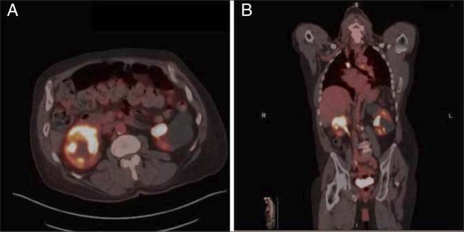 Figure 1
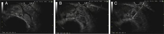 Figure 2
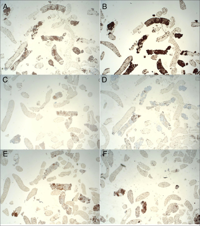 Figure 3
Peer Reviews
No public reviews on file for this paper yet. If you reviewed it on a platform where reviews are public (OpenReview, ICLR, NeurIPS, ICML), you can paste yours below so the community can read it here.
Videos
No videos yet. Explain this paper in a talk, walkthrough, or lecture? Add one.
Taxonomy
TopicsLymphoma Diagnosis and Treatment · Renal cell carcinoma treatment · Gastrointestinal Tumor Research and Treatment