A novel method for the detection of Cas9 gRNAs using a fluorophore-labeled DNA oligo
Ranmal Avinash Bandara, Zhichang Peter Zhou, Ziyan Rachel Chen, Rongqi Duan, Alan Richard Davidson, Amy P. Wong, Jim Hu

Abstract
Genes, proteins, chemicals, diseases, species, mutations and cell lines named across the full text — each resolved to its canonical identifier and authoritative record.
Click any figure to enlarge with its caption.
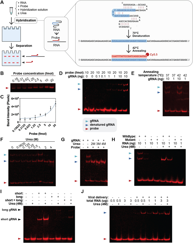 Figure 1
Peer Reviews
No public reviews on file for this paper yet. If you reviewed it on a platform where reviews are public (OpenReview, ICLR, NeurIPS, ICML), you can paste yours below so the community can read it here.
Videos
No videos yet. Explain this paper in a talk, walkthrough, or lecture? Add one.
Taxonomy
TopicsCRISPR and Genetic Engineering · RNA Interference and Gene Delivery · Advanced biosensing and bioanalysis techniques
CRISPR/Cas9 is a versatile genome editing tool that has the potential to be used to cure many genetic diseases. The system works via a guide RNA (gRNA) interacting with the Cas9 protein to form a complex that binds to a specific DNA sequence.1 The site-specific DNA binding feature of the Cas9 system can be utilized in a variety of ways to correct gene mutations or to regulate gene expression. First, the Cas9 protein can make a site-specific double-stranded break that is mainly repaired by homology-directed repair or non-homologous end-joining. Both pathways can be used for gene editing or gene insertion when proper donor DNA is present.1 Second, the Cas protein can be modified into a nickase, which creates a site-specific nick. When fused with other proteins, such as adenosine deaminase, cytosine deaminase, or reverse transcriptase, the modified Cas9 protein can be used for base editing2 or prime editing.3 While cytosine base editors and adenine base editors allow single-nucleotide changes at target sites specified by the gRNA, prime editors, with the gRNA modified to include a template sequence for reverse transcription, can offer single nucleotide substitution as well as short deletions or insertions.3 The recently improved prime editing systems include three gRNAs: an engineered pegRNA (epegRNA) to locate the target site for initiation of the prime editing, a nicking gRNA to nick the unedited DNA strand to enhance the editing efficiency, and a dead single gRNA (dsgRNA) to recruit the Cas9 protein near the editing site to enhance editing efficiency.3 In addition, the Cas9 protein can be fused with a transposase, such as in the Find and Cut-and-Transfer (FiCAT) system, to perform site-specific gene integration.4 Finally, a modified Cas9 protein can serve as a site-specific DNA binding protein to regulate gene transcription by fusing with a transcription activator or inhibitory motif. In all the above gene editing or gene regulation approaches using Cas9, proper levels of gRNA presence are essential for targeting efficiency. In the case of prime editing, even the ratio of gRNA expression is critical; the epegRNA level should be at least three-fold higher than the nicking gRNA or dsgRNA.3 Currently, there is no reliable, convenient method to precisely measure levels of gRNA expression. In this study, we developed a rapid, sensitive, cost-effective, and non-radioactive method for gRNA detection.
To design such a method, we considered using a 5′-Cy5.5-labeled DNA oligo to probe gRNAs through in-solution hybridization and detecting the hybridization products on a native polyacrylamide gel with a fluorescence-detection-capable imager, the Licor Odyssey. For the DNA oligo design, we decided to select a sequence that targets a common region of a Cas9 gRNA so that this universal probe can be used to detect gRNAs for different Cas9 target sites and for Cas9 gRNAs used for different applications, including Cas9-mediated gene integration, base editing, prime editing, and Cas9-mediated gene regulation (Fig. 1A).Figure 1. Method design, probe sensitivity test, suppressing RNA secondary structure, and enhancing the detection sensitivity with urea. (A) Diagram of experimental procedure overview. The red and blue sequences represent the probe and the probe binding region on gRNA, respectively. The red dot represents the Cy5.5 fluorophore. (B) Cy5.5 probe detection using Licor imager after native gel electrophoresis. Different amounts of indicated Cy5.5 probe were loaded onto a native polyacrylamide gel using the Blue Juice dye and run on native polyacrylamide gel for 40 min. (C) ImageJ analysis was done on gels (n = 3), as seen in Figure 1B. (D) gRNA detection using Cy5.5 probe. In vitro transcribed gRNA was hybridized in solution with 20 fmol of Cy5.5 probe. The hybridization products were separated on 10 % native polyacrylamide gel and imaged using the Licor imager. (E) Detection of gRNA in different annealing temperatures. (F, G) Effect of urea on gRNA detection. 10 ng of gRNA was incubated with 20 fmol of probe as described. Urea was added to indicated concentration and incubated. (H) Absence of detection by mutating the probe binding sequence in the gRNA. (I) Detection of gRNA in 5 μg of total RNA from mammalian cells. Short, long, and short plus long indicate total RNA from HEK293T cells transfected with plasmids expressing short, long, or short plus long gRNA, respectively. (J) Detection of gRNA in mammalian cells with viral delivery. 50 multiplicities of infection of CRISPR/Cas9 viral vector was added to 116 cells.Figure 1
We first tested the probe sensitivity (Fig. S1). To ascertain the probe sensitivity in a polyacrylamide gel similar to what we observed, different concentrations of the probe were run on a 10 % native polyacrylamide gel. As shown in Figure 1B, C, the probe was detectable from 0.16 to 20 fmol. Following confirmation of the high sensitivity, we determined whether our probe could be used to detect gRNAs. We first chose in vitro transcribed gRNA for the initial test because its high purity allows for greater specificity and accuracy as compared with gRNAs expressed from cells. To determine the minimal amount of gRNA that could be detected, we used 10, 1, and 0.1 ng of in vitro transcribed gRNA for solution hybridization (see online methods and materials). We observed that 10 ng of gRNA generated the highest signal, whereas 1 ng of gRNA showed a much lower level of signal (Fig. 1D).
To optimize the annealing temperature for hybridizing the probe and gRNA, the mixture of probe and gRNA was heated to 70 °C for 3 min and then cooled down to either 37 °C or 42 °C for 15 min. As shown in Figure 1E, the 42 °C annealing temperature yielded a stronger signal compared with 37 °C. From here, we used the 42 °C annealing temperature for the remainder of our experiments. In this test, we also observed two bands of the hybridization products due to the secondary structure of the gRNA (Fig. 1E). We envisioned that if we could eliminate the secondary structure and allow the hybridization product to run as a single band, we would gain sensitivity in detecting gRNAs in total RNA samples from mammalian cells, especially when multiple gRNAs with different sizes are present.
We used urea to disrupt the secondary structure since urea is known to disrupt hydrogen bonds and interact with nucleotides. We found that 2 M urea started to have a major effect on the gRNA secondary structure. The inclusion of 4 M urea disrupted completely the RNA secondary structure, resulting in a single band of hybridization product with a stronger fluorescent signal without separation of the probe from gRNAs (Fig. 1F, G). To ascertain the probe specificity, we designed a gRNA with the probe binding site mutated, and we showed that this RNA was not detected by the probe (Fig. 1H).
To determine whether gRNAs from total RNA isolated from mammalian cells could be detected, we transfected HEK293T cells with plasmids expressing gRNAs from the U6 promoter. We found that gRNAs could be detected in 5 μg of total RNA from cells expressing single or double gRNAs (Fig. 1I) despite different levels of expression (explained in supplementary information). We also analyzed the magnitude of urea effects on gRNA detection with the result discussed in supplementary information as well. Finally, we found that gRNAs from 116 cells transduced with helper-dependent adenoviral (HD-Ad) vector5 expressing Cas9 and gRNA could be detected with less than 1 μg of total cellular RNA, indicating the high sensitivity of the method (Fig. 1J).
In summary, these experiments demonstrated the successful development and the utility of a novel technique for gRNA detection. This method is expected to have a wide range of applications for assessing levels of gRNA expression, such as Cas9-mediated gene integration and gene editing, as well as gene regulation. Due to differences in target sites and/or in reverse transcription template sequences used in prime editing, different gRNAs may have different levels of expression. This may lead to differences in gene editing efficiency. Our method of gRNA detection can be used for assessing gRNA expression in all these cases and therefore may facilitate the development of genetic approaches for gene therapy applications.
Funding
This work was supported by the New Frontiers in Research Fund (NFRF), administered by the Social Sciences and Humanities Research Council (SSHRC) on behalf of CIHR, NSERC, and SSHRC (Grant #NFRFE-2021-00713).
CRediT authorship contribution statement
Ranmal Avinash Bandara: Data curation, Formal analysis, Methodology, Validation, Visualization, Writing – original draft, Writing – review & editing. Zhichang Peter Zhou: Methodology, Writing – review & editing. Ziyan Rachel Chen: Methodology, Writing – original draft, Writing – review & editing. Rongqi Duan: Data curation, Methodology. Alan Richard Davidson: Supervision, Writing – review & editing. Amy P. Wong: Supervision, Writing – review & editing. Jim Hu: Conceptualization, Supervision, Writing – review & editing.
Data availability
Data is available upon request.
Conflict of interests
The authors declared no conflict of interests.
The reference list from the paper itself. Each links out to its DOI / PubMed record.
- 1Ran F.A.Hsu P.D.Wright J.Agarwala V.Scott D.A.Zhang F.Genome engineering using the CRISPR-Cas 9 system Nat Protoc 8112013228123082415754810.1038/nprot.2013.143PMC 3969860 · doi ↗ · pubmed ↗
- 2Porto E.M.Komor A.C.Slaymaker I.M.Yeo G.W.Base editing: advances and therapeutic opportunities Nat Rev Drug Discov 191220208398593307793710.1038/s 41573-020-0084-6PMC 7721651 · doi ↗ · pubmed ↗
- 3Sousa A.A.Hemez C.Lei L.Systematic optimization of prime editing for the efficient functional correction of CFTR F 508del in human airway epithelial cells Nat Biomed Eng 920257213898762910.1038/s 41551-024-01233-3PMC 11754097 · doi ↗ · pubmed ↗
- 4Pallarès-MasmitjàM.IvančićD.mi R-Pedrol J.Find and cut-and-transfer (Fi CAT) mammalian genome engineering Nat Commun 12202170713486237810.1038/s 41467-021-27183-x PMC 8642419 · doi ↗ · pubmed ↗
- 5Zhou Z.P.Yang L.L.Cao H.In vitro validation of a CRISPR-mediated CFTR correction strategy for preclinical translation in pigs Hum Gene Ther 3092019110111163109926610.1089/hum.2019.074 · doi ↗ · pubmed ↗