Pyoderma Grangrenosum
Reza Aghaei, Edmund Hsu, Christopher McCoy

TL;DR
A middle-aged woman with ulcerative colitis developed painful leg ulcers diagnosed as pyoderma gangrenosum after a thorough evaluation.
Contribution
The paper presents a clinical case illustrating the characteristics and diagnosis of pyoderma gangrenosum.
Findings
Pyoderma gangrenosum ulcers were identified in a patient with ulcerative colitis.
Clinical and pathological assessments confirmed the diagnosis of pyoderma gangrenosum.
The case highlights the importance of a thorough evaluation for accurate diagnosis.
Abstract
We describe a middle-aged female with past medical history of ulcerative colitis presenting to the emergency department with bilateral painful ulcers rapidly growing on her lower legs in the prior four weeks. She was consulted by a dermatologist and after a thorough clinical and pathology assessment (as a diagnosis of exclusion), treatment for pyoderma gangrenosum was started. Pyoderma gangrenosum is a painful, chronic, ulcerative disorder often occurring in association with systemic disease. We review the clinical presentation of pyoderma gangrenosum and its complications. We describe the characteristics of ulcers with pictures from the patient. Our case illustrates the findings of pyoderma gangrenosum both clinically and pathologically.
Genes, proteins, chemicals, diseases, species, mutations and cell lines named across the full text — each resolved to its canonical identifier and authoritative record.
Click any figure to enlarge with its caption.
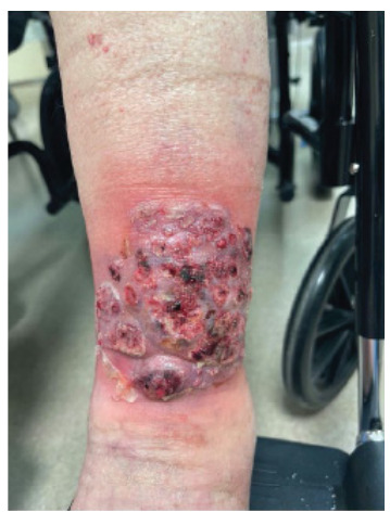 Figure 1
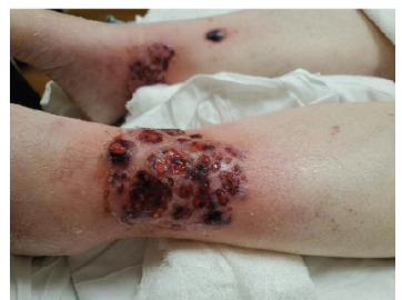 Figure 2
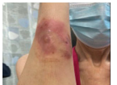 Figure 3
Peer Reviews
No public reviews on file for this paper yet. If you reviewed it on a platform where reviews are public (OpenReview, ICLR, NeurIPS, ICML), you can paste yours below so the community can read it here.
Videos
No videos yet. Explain this paper in a talk, walkthrough, or lecture? Add one.
Taxonomy
TopicsAutoimmune and Inflammatory Disorders · Autoimmune Bullous Skin Diseases · Skin Diseases and Diabetes
CASE PRESENTATION
A 51-year-old female presented with bilateral painful shin ulcers. She has a medical history of ulcerative colitis and multiple sclerosis, for which she was being treated with glatiramer acetate and mesalamine, respectively. The patient denied experiencing fever, chills, or night sweats. She also had a brown nodular painful ulcer on her right forearm, which presented four weeks prior. Her exam was significant for bilateral lower leg palm-sized cribriform ulcer, large violaceous bullous plaque on her right lower leg, and red-brown small nodular plaque on the right forearm (Images 1–?3). The patient’s clinical condition was reviewed by a dermatologist, and after a thorough clinical and pathological assessment, treatment for pyoderma gangrenosum was initiated.
DISCUSSION
Pyoderma gangrenosum (PG) is a reactive, non-infectious, inflammatory dermatosis and is classified as one of the neutrophilic dermatoses, along with Sweets syndrome and Behcets disease. The incidence of PG is approximately 0.63 per 100,000 people, with the median age of onset being 59 years.1
Although the lower legs are the most frequently affected, PG can manifest on any part of the body. The condition is often precipitated by minor trauma, a phenomenon known as “pathergy.”2
A thorough history is key with specific inquiry regarding possible pathergic response to minor or major trauma, as well as a history of pain, rapid progression, symptoms suggestive of infection or systemic disease and a detailed drug history. 3 Indeed, PG lesions are all too often misdiagnosed as simple non-healing ulcers and patients undergo debridement, which can result in catastrophic deterioration of the condition through this pathergic response. The condition predominantly affects adults, but childhood cases are rarely reported.4 More recently, Mavrakis et al have proposed new criteria based on a consensus of international experts, requiring one major and four minor criteria.5
CPC-EM CapsuleWhat do we already know about this clinical entity?Pyoderma gangrenosum is a neutrophilic dermatosis, which presents most commonly in patients with inflammatory bowel disease.What is the major impact of the image(s)?We present the classic presentation of pyoderma gangrenosum regarding images, also in a clinical course, which can assist physicians in making a diagnosis.How might this improve emergency medicine practice?Recognizing pyoderma gangrenosum classic features and presentation can help physicians increase their index of suspicion and diagnostic accuracy leading to improved outcomes.
The reference list from the paper itself. Each links out to its DOI / PubMed record.
- 1Langan SM Groves RW Card TR Incidence, mortality, and disease associations of pyoderma gangrenosum in the United Kingdom: a retrospective cohort study J Invest Dermatol 201213292166702253487910.1038/jid.2012.130 · doi ↗ · pubmed ↗
- 2Sassolas B Le Ru Y Plantin P Pyoderma gangrenosum with pathergic phenomenon in pregnancy Br J Dermatol 20001424827810.1046/j.1365-2133.2000.03444.x 10792250 · doi ↗ · pubmed ↗
- 3George C Deroide F Rustin M Pyoderma gangrenosum - a guide to diagnosis and management Clin Med (Lond)201919322483109251510.7861/clinmedicine.19-3-224PMC 6542232 · doi ↗ · pubmed ↗
- 4Graham JA Hansen KK Rabinowitz LG Pyoderma gangrenosum in infants and children Pediatr Dermatol 1994111107817084110.1111/j.1525-1470.1994.tb 00065.x · doi ↗ · pubmed ↗
- 5Maverakis E Ma C Shinkai K Diagnostic Criteria of Ulcerative Pyoderma Gangrenosum: A Delphi Consensus of International Experts JAMA Dermatol 2018154446162945046610.1001/jamadermatol.2017.5980 · doi ↗ · pubmed ↗