18F-FDG PET/CT Findings in Glandular Tularemia
Freja Gustafsson, Karl Keigo Rasmussen, Kristina Thorsteinsson, Anne-Mette Lebech, Lasse Fjordside

TL;DR
A woman with glandular tularemia showed high metabolic activity on a PET/CT scan, highlighting the challenges in diagnosing this disease.
Contribution
This case illustrates the role of 18F-FDG PET/CT in diagnosing glandular tularemia when other methods are inconclusive.
Findings
PET/CT showed high metabolic activity in the left inguinal region, suggesting abscesses.
Diagnosis was confirmed with PCR and serology for Francisella tularensis.
The case emphasizes the difficulty in diagnosing tularemia and the value of imaging.
Abstract
A 47-year-old woman presented with fever, fatigue, night sweats and inguinal glandular swelling following a tick bite. Weeks of diagnostic uncertainty followed, and a lymph node biopsy was sent to be investigated for tularemia and pathology. An 18F-FDG PET/CT scan was performed due to a suspicion of malignant lymphoma. The scan revealed high metabolic activity in the left inguinal region, which was compatible with abscesses. The diagnosis of glandular tularemia was established on a positive PCR for Francisella tularensis (F. tularensis) and positive F. tularensis serology. This case highlights the challenges of diagnosing tularemia and illustrates the role of imaging.
Genes, proteins, chemicals, diseases, species, mutations and cell lines named across the full text — each resolved to its canonical identifier and authoritative record.
Click any figure to enlarge with its caption.
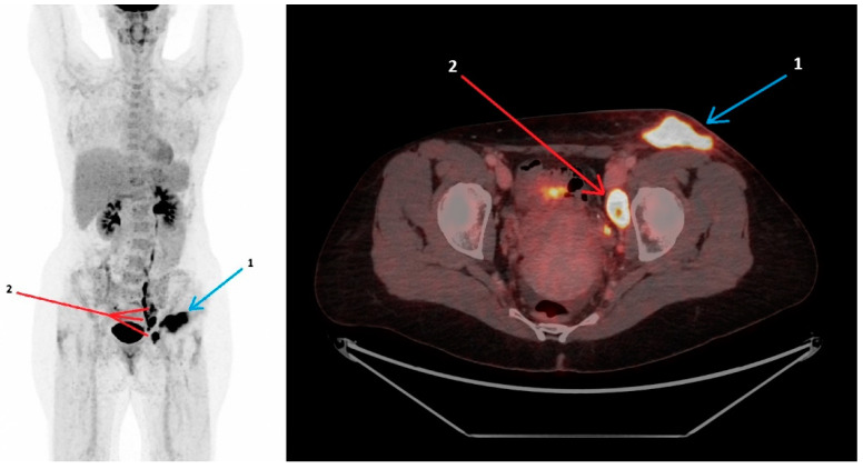 Figure 1
Peer Reviews
No public reviews on file for this paper yet. If you reviewed it on a platform where reviews are public (OpenReview, ICLR, NeurIPS, ICML), you can paste yours below so the community can read it here.
Videos
No videos yet. Explain this paper in a talk, walkthrough, or lecture? Add one.
Taxonomy
TopicsBacillus and Francisella bacterial research · Microbial infections and disease research · Yersinia bacterium, plague, ectoparasites research