A Solitary Pedunculated Nevus Lipomatosus Superficialis
Julia S Murphy, John J Fowler, Morteza Khodaee

TL;DR
A woman in her 40s had a large, irritating skin lesion on her back that was diagnosed as a rare condition called nevus lipomatosus superficialis after a biopsy.
Contribution
This case report highlights the importance of biopsy in diagnosing symptomatic pedunculated skin lesions.
Findings
A symptomatic pedunculated skin lesion was diagnosed as nevus lipomatosus superficialis via excisional biopsy.
Large pedunculated skin lesions, though often benign, should be biopsied if causing symptoms.
The case emphasizes the need for clinical evaluation and histopathological confirmation in such rare presentations.
Abstract
Large pedunculated skin lesions are a rare presentation in primary care settings. In general, these lesions are asymptomatic other than occasional irritation and discomfort. Although differential diagnosis mainly includes benign lesions, an excisional biopsy should be performed, particularly in symptomatic cases. We present the case of a woman in her 40s with an irritating large and pedunculated skin lesion on her back. Due to the symptomatic nature of the lesion, an excisional biopsy was performed, which confirmed the diagnosis of a nevus lipomatosus superficialis.
Genes, proteins, chemicals, diseases, species, mutations and cell lines named across the full text — each resolved to its canonical identifier and authoritative record.
Click any figure to enlarge with its caption.
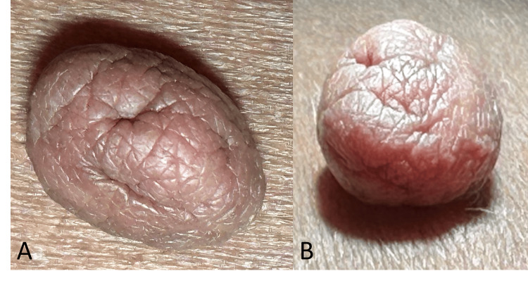 Figure 1
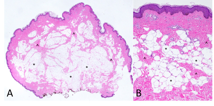 Figure 2
Peer Reviews
No public reviews on file for this paper yet. If you reviewed it on a platform where reviews are public (OpenReview, ICLR, NeurIPS, ICML), you can paste yours below so the community can read it here.
Videos
No videos yet. Explain this paper in a talk, walkthrough, or lecture? Add one.
Taxonomy
TopicsGenetic and rare skin diseases.
Introduction
Pedunculated skin lesions are attached to the body by an elongated stalk known as a peduncle. Although pedunculated skin lesions are common, the majority of them are small in size. Large pedunculated skin lesions are rare [1,2]. A well-vascularized stem is necessary for a pedunculated skin lesion to grow large [2]. These lesions typically grow slowly over several years [1-4]. While the majority of large pedunculated skin lesions are benign, there are rare instances where premalignant or malignant lesions may be present [1-5]. Benign conditions that can appear as large pedunculated cutaneous lesions include acrochordon, fibroepithelial polyps, lipoma, neurofibroma, epidermal nevus, fibrous histiocytoma, nevus lipomatosus superficialis (NLS) (lipofibroma), pyogenic granuloma, dermatofibroma, cutaneous mixed tumor, cylindroma, acquired fibrokeratoma, schwannoma, cutaneous myxoma, and cutaneous angiomyxoma [1,2]. Although extremely rare, malignant lesions presenting as pedunculated cutaneous growths may include basal cell carcinoma, melanoma, Merkel cell carcinoma, cutaneous sarcoma, and pilomatrix carcinoma [2]. While the differential diagnosis primarily involves benign lesions, an excisional biopsy should be performed, especially in cases with an unusual pattern or symptoms [1-4,6,7]. The majority of patients seek medical attention for cosmetic reasons. In this case, we present the case of a woman with a large pedunculated skin lesion on her back that had been present for several years.
Case presentation
A woman in her 40s presented with an enlarging skin lesion on her left lower back. The lesion had been growing over the last two to three years and was cosmetically bothersome to her. The lesion does not cause any pain, but it may occasionally get caught, particularly when changing clothes. On physical examination, there was a 1.4 x 1.1 x 0.8 cm, non-tender, pedunculated, flesh-colored cerebriform lesion on the left lower back (Figures 1A, 1B).
A flesh-colored, pedunculated skin lesion with cerebriform surface (A and B) on the left flank that was 1.4 × 1.1 × 0.8 cm in size, soft, mobile, and non-tender.
After discussing options with the patient, she elected to have the lesion removed. An excisional biopsy was performed, and the sample was sent for pathological evaluation. Histopathology confirmed the diagnosis of a pedunculated NLS (Figures 2A, 2B).
Histologic sections (A) demonstrate a polypoid lesion formed by lobules of adipocytes () within the reticular dermis. Bands of dermal collagen course (^) throughout the lesion (H&E, 12.5× magnification). On higher magnification (B), bands of dermal collagen (^) are intercalated among collections of mature adipocytes (), which are in close proximity to the epidermis (H&E, 100× magnification).H&E: hematoxylin and eosin
Discussion
NLS, also known as pedunculated lipofibroma, is a rare benign hamartoma characterized by the presence of adipose tissue in the dermis [3-8]. The etiology of NLS is unclear [6]. NLS affects both men and women equally, with no preference for either sex [4,6,8-11]. There have also been some case series suggesting an association with obesity and/or diabetes, but further research is necessary [5,6]. NLS has been categorized into the classic multiple form and the solitary variant [3-12]. In rare cases, NLS can ulcerate, especially in cases with irritation [4,6,8]. The classical type generally presents near birth and consists of multiple soft papulonodules and sometimes plaques [3-12]. The solitary type is more common in areas of pressure similar to acrochordons [4-8]. The solitary type is typically larger in size and appears as a single pedunculated skin lesion [4,5,7,12]. Aside from differences in their sizes, anatomical locations, and the number of lesions, both types exhibit similar clinical and histopathological features [5,6].
However, some have proposed classifying these two types as distinct entities [5,6]. Clinically, these lesions may have a smooth surface or multilobular (cerebriform) surface with prominent folds [5,6,8]. Histopathologically, NLS is characterized by mature adipose tissue within the dermis [3,5,6,8]. The recommended treatment is surgical excision, and the lesions typically do not recur [3-6,8]. Although these lesions appear benign, it is strongly recommended to send the excised samples for pathological evaluation [1,2,4,5,8,10,11].
Conclusions
Large pedunculated skin lesions are rare findings in primary care settings. These lesions are typically asymptomatic but may occasionally cause irritation or discomfort. Although the differential diagnosis often includes benign lesions, malignancy should also be considered. An excisional biopsy is recommended, especially for lesions with unusual features, those causing irritation, or those removed for cosmetic reasons.
The reference list from the paper itself. Each links out to its DOI / PubMed record.
- 1Enlarging, pedunculated skin lesion Am Fam Physician Lee JA Khodaee M 11911192852012 https://www.aafp.org/pubs/afp/issues/2012/0615/p 1191.pdf 22962900 · pubmed ↗
- 2Giant pedunculated tumors of skin Georgian Med News Wollina U Goldman A 44492020 https://www.geomednews.com/s/480918712 df 344a 4a 77508 d 4cd 7815 ab/files/uploaded/V 301_N 4_April_2020.pdf?Expires=1651816093&Signature=j WK Ihof 34sat 5mkb GRCBZCDV Syn 00cty Ca Gcn 4t Irjyy A Iyc Hb~fr Mu 7AVW 9wgh Gj D 1qxrpn LKQ 5c XN 2Pz 9ebfu S 4ZVTEN-c D 8cqg L-m K Ww Oqv Yu 2f Vr Z 618xx Ug XVP 6DW Oi 9tpky N Id 2H 7z 94K 6pw-7e CJ 9U Vr 1h Lprq H 3u 6h GGDCT 1O 0QQN 7n 9addu 1Lp 3Uuddh 0M Wzmi M 4RMM Fvngpz 5Fv Oj ZE Al 2-H Vl V Tckk Keal Rpnz E-3g D 6a E~3G-j Al X Rjgg I Mp VR 82x 6Y Fj DG~-WW Wtha J Nk JO 4feskd · pubmed ↗
- 3A rare case of a pedunculated lipoma in the perianal region: a 20-year journey Cureus Rekavari SG Mahakalkar C 016202410.7759/cureus.61304 PMC 1121283838947595 · doi ↗ · pubmed ↗
- 4Giant pedunculated lipofibroma of the thigh J Surg Case Rep Suleiman J Suleman M Amsi P Minja P Lodhia J 02023202310.1093/jscr/rjad 153PMC 1004979536998262 · doi ↗ · pubmed ↗
- 5Revisiting solitary pedunculated lipofibromas Am J Clin Pathol Adotama P Hutson SD Rieder EA Stein JA Kim RH 95495715620213412474710.1093/ajcp/aqab 075 · doi ↗ · pubmed ↗
- 6Nevus lipomatosus superficialis: a series of six cases J Cutan Pathol Abdull Gaffar B Keloth T 1191295120243785959010.1111/cup.14552 · doi ↗ · pubmed ↗
- 7Pedunculated lipofibroma J Dermatol Oztürkcan S Terzioğlu A Akyol M Altinor S Yildiz E 2882902720001082449710.1111/j.1346-8138.2000.tb 02168.x · doi ↗ · pubmed ↗
- 8Nevus lipomatosus superficialis, an unusual case report J Family Med Prim Care Singh P Anandani GM 404540471120223638769810.4103/jfmpc.jfmpc_2352_21PMC 9648317 · doi ↗ · pubmed ↗