Sequence analysis of two F1 mycobacteriophages, Deb65 and DocMcStuffins
Marcus O. Royster, Victoria Figgins, Vera Pande, Jason D. Robinson, Deeka S. Abdi, Ali Amin, Zephaniah Ansah, Ethan W. Bomersheim, Gianna Dunn, Ali A. Elfaki, Jordyn Foulk, Kate C. Ingle, Avi D. Lavu, Ved Pande, Priya T. Shan, Marie P. Smithbey, Gunnar R. Ternstrom

TL;DR
This paper analyzes the genetic sequences of two mycobacteriophages, Deb65 and DocMcStuffins, isolated from wetland soil and found to infect Mycobacterium smegmatis.
Contribution
The study identifies 41 shared genes and classifies both phages into the F1 subcluster based on gene content similarity.
Findings
Deb65 and DocMcStuffins encode 97 and 91 putative genes, respectively.
41 genes are shared between the two phages.
Both phages are assigned to the F1 subcluster of actinobacteriophages.
Abstract
Isolated from wetland soil, Deb65 and DocMcStuffins are bacteriophages with a siphoviral morphology that infect Mycobacterium smegmatis. Deb65 and DocMcStuffins encode 97 and 91 putative genes, 41 of which are shared. Based on gene content similarity to actinobacteriophages more broadly, both phages are assigned to subcluster F1.
Genes, proteins, chemicals, diseases, species, mutations and cell lines named across the full text — each resolved to its canonical identifier and authoritative record.
Click any figure to enlarge with its caption.
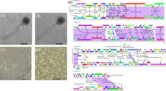 Fig 1
Fig 1| Phage | DocMcStuffins | Deb65 |
|---|---|---|
| Isolation GPS | 37°16′08.4″N | 37°16′15.1″N |
| Morphology | Siphovirus | Siphovirus |
| Capsid diameter | 50 ± 3 nm ( | 50 ± 2 nm ( |
| Tail length | ~180 ± 5 nm ( | ~190 ± 5 nm ( |
| Sequencing reads | 453,180 | 440,517 |
| Sequencing coverage, fold | 1,122 | 1,119 |
| Genome length (bp) | 58,159 | 55,767 |
| Genome end sequence | 5′ | 5′ |
| Number of open reading frames | 91 | 97 |
| GC content (%) | 62.7 | 61.6 |
| Accession number |
|
|
| SRA |
|
|
- —HHS | NIH | Eunice Kennedy Shriver National Institute of Child Health and Human Development (NICHD)
Peer Reviews
No public reviews on file for this paper yet. If you reviewed it on a platform where reviews are public (OpenReview, ICLR, NeurIPS, ICML), you can paste yours below so the community can read it here.
Videos
No videos yet. Explain this paper in a talk, walkthrough, or lecture? Add one.
Taxonomy
TopicsBacteriophages and microbial interactions · Genomics and Phylogenetic Studies · Mycobacterium research and diagnosis
ANNOUNCEMENT
Advancing our knowledge of mycobacteriophage diversity is essential for understanding the abundance, community dynamics, and evolution of Mycobacteria, an environmentally and clinically important genus (1–5). We report the sequence of two genetically distinct mycobacteriophages, Deb65 and DocMcStuffins.
Both phages were isolated from wet, silty soil samples at the College of William & Mary in Williamsburg, VA, USA, using a standard enrichment procedure (Table 1) (6). Briefly, 5 g of each soil sample was suspended in 50 mL of 7H9 media, inoculated with Mycobacterium smegmatis mc^2^ 155, and incubated in a 37°C shaker (250 rpm) for 2 days. The resulting cultures were filtered through a 0.22 µm filter, and the filtrates were plated with M. smegmatis in 7H9 top agar, yielding clear plaques for both phages, Deb65 and DocMcStuffins, after 24–48 h. Phages were then purified through three rounds of plating before being imaged by negative stain (1% uranyl acetate) transmission electron microscopy to reveal siphoviral morphologies for both phages (Fig. 1).
(a) Negative stain (uranyl acetate, 1%) transmission electron microscopy of DocMcStuffins revealing siphoviral morphology. Scale bar is 50 nm. (b) Negative stain (uranyl acetate, 1%) transmission electron microscopy of Deb65 revealing siphoviral morphology. Scale bar is 50 nm. (c) Plaque image of DocMcStuffins showing clear plaques. Scale bar is 6.125 mm. (d) Plaque image of Deb65 showing clear plaques. Scale bar is 6.125 mm. (e) Phamerator comparative alignment of Deb65 and DocMcStuffins.
Phage DNA was extracted from a lysate using the phenol-chloroform-isoamyl alcohol method and ethanol precipitated (7). DNA was prepared for sequencing using the NEB Ultra II Library Kit and sequenced using an Illumina MiSeq Sequencer (v3 reagents, single-end, 150 base read). Newbler (version 2.9) was then used to assemble the genome and Consed (version 29) to check for completeness and reveal 3′ single-stranded genome termini (8). Sequencing data, overhangs, and genome characteristics are presented in Table 1.
The genomes were annotated using DNA Master (version 5.23.6) and PECAAN (version 20221109) (9). Translational start sites were verified using the coding potential predicted by GeneMark and Glimmer (10–12), and the similarity of start sites in homologs was identified using Starterator (http://phages.wustl.edu/starterator/) and BLASTp against the Actinobacteriophage and NCBI non-redundant protein databases (6, 13). No tRNAs were identified using Aragorn version 1.2.41 (14) and tRNAscan (15).
Putative gene functions were assigned based on predictions from HHPred (using the PDB_mmCIF70, NCBI_CD, SCOPe70, and pFAM-A as databases), BLASTp, and Phamerator (Actino_draft database) for highly similar genes (16, 17). Using the gene content similarity (GCS) tool at the Actinobacteriophage database, phagesDB (https://phagesdb.org/) and clustering parameters of at least 35% GCS to actinobacteriophages, both phages are assigned to cluster F, subcluster F1 (18, 19). Default settings were used for all software.
Both phages share 41 GCS, which are primarily in the first half of the genome and include genes encoding functions in virion structure, assembly, lysis, and lysogeny, the latter consistent with the temperate lifecycle for F cluster phages (Fig. 1). Noteworthy here is that while both phages encode homologous tyrosine integrases with 90% amino acid identity (AAI), their immunity repressors only share 39% AAI. Within this genomic region, and consistent with many phages of the F1 subcluster, both Deb65 and DocMcStuffins encode anti-repressor and Cro proteins. Within the second half of the genome, where gene conservation is lower, both phages encode two glycosyltransferases that are highly conserved across the F1 subcluster. Within this region, Deb65 encodes a unique DNA methyltransferase for which no homolog exists in the Actinobacteriophage database.
The reference list from the paper itself. Each links out to its DOI / PubMed record.
- 1Jacobs-Sera D, Marinelli LJ, Bowman C, Broussard GW, Guerrero Bustamante C, Boyle MM, Petrova ZO, Dedrick RM, Pope WH, Modlin RL, Hendrix RW, Hatfull GF, Science Education Alliance Phage Hunters Advancing Genomics And Evolutionary Science Sea-Phages Program. 2012. On the nature of mycobacteriophage diversity and host preference. Edited by R. L. Modlin, R. W. Hendrix, and G. F. Hatfull. Virology (Auckl) 434:187–201. doi:10.1016/j.virol.2012.09.026PMC 351864723084079 · doi ↗ · pubmed ↗
- 2Esposito LA, Gupta S, Streiter F, Prasad A, Dennehy JJ. 2016. Evolutionary interpretations of mycobacteriophage biodiversity and host-range through the analysis of codon usage bias. Microb Genom 2:e 000079. doi:10.1099/mgen.0.00007928348827 PMC 5359403 · doi ↗ · pubmed ↗
- 3Walsh CM, Gebert MJ, Delgado-Baquerizo M, Maestre FT, Fierer N. 2019. A global survey of mycobacterial diversity in soil. Appl Environ Microbiol 85:e 01180-19. doi:10.1128/AEM.01180-1931253672 PMC 6696970 · doi ↗ · pubmed ↗
- 4Hatfull GF. 2022. Mycobacteriophages: from petri dish to patient. P Lo S Pathog 18:e 1010602. doi:10.1371/journal.ppat.101060235797343 PMC 9262239 · doi ↗ · pubmed ↗
- 5Papke RT, Doolittle WF. 2003. Phage evolution: New worlds of genomic diversity. Curr Biol 13:R 606–R 607. doi:10.1016/S 0960-9822(03)00527-X 12906814 · doi ↗ · pubmed ↗
- 6Poxleitner M, Pope W, Jacobs-Sera D, Sivanathan V, Hatfull G. 2018. Phage discovery guide. Howard Hughes Medical Institute.
- 7Sambrook J, Russell DW. 2006. Purification of nucleic aids by extraction with phenol:chloroform. Cold Spring Harb Protoc 2006. doi:10.1101/pdb.prot 445522485786 · doi ↗ · pubmed ↗
- 8Russell D. A. 2018. Sequencing, assembling, and finishing complete bacteriophage genomes. Methods Mol Biol 1681:109–125. doi:10.1007/978-1-4939-7343-9_929134591 · doi ↗ · pubmed ↗