Transcriptome of brain-like endothelial cells following coxsackievirus B3 infection
Sarah F. Hathcock, Taryn E. Keyzer, Nadine Vollmuth, Daryl W. Lam, Jon Sin, Brandon J. Kim

TL;DR
This study examines how brain-like endothelial cells respond at the genetic level when infected by coxsackievirus B3, a virus linked to meningitis.
Contribution
The study provides a new transcriptomic profile of brain-like endothelial cells infected with coxsackievirus B3.
Findings
The global transcriptome of brain-like endothelial cells was analyzed during coxsackievirus B3 infection.
The study reveals gene expression changes in endothelial cells interacting with the virus.
Findings may help understand how the virus disrupts the blood-brain barrier.
Abstract
Coxsackievirus B3 is a leading cause of viral aseptic meningitis. To gain entry to the central nervous system, it must interact with and disrupt the brain endothelial cells of the blood-brain barrier. Here, we report the global transcriptome of stem-cell-derived brain-like endothelial cells during coxsackievirus B3 infection.
Genes, proteins, chemicals, diseases, species, mutations and cell lines named across the full text — each resolved to its canonical identifier and authoritative record.
Click any figure to enlarge with its caption.
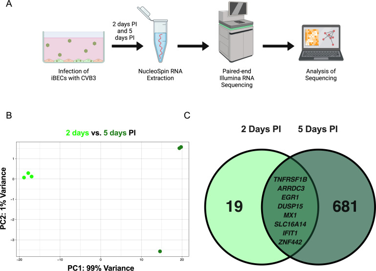 Fig 1
Fig 1- —HHS | National Institutes of Health (NIH)
- —HHS | National Institutes of Health (NIH)
Peer Reviews
No public reviews on file for this paper yet. If you reviewed it on a platform where reviews are public (OpenReview, ICLR, NeurIPS, ICML), you can paste yours below so the community can read it here.
Videos
No videos yet. Explain this paper in a talk, walkthrough, or lecture? Add one.
Taxonomy
Topicsinterferon and immune responses · RNA regulation and disease · RNA modifications and cancer
ANNOUNCEMENT
Coxsackievirus B3 (CVB3) is a pathogen in the Enterovirus genus and one of the leading causes of viral aseptic meningitis (1–4). To induce meningitis, CVB3 must access the central nervous system (CNS) by interacting with the blood-brain barrier (BBB) (5). The BBB is composed of specialized brain endothelial cells (BECs) that maintain CNS homeostasis by limiting pathogen entry into the brain parenchyma (6, 7). CVB3 can infect BECs, but their response to CVB3 is poorly understood (5).
Induced pluripotent stem-cell-derived brain-like endothelial cells (iBECs) were differentiated as described in our recent publication and others (5, 8–12). The eGFP-CVB3 construct was generated as previously described from the pMKS1 plasmid (Nancy H3 variant pH3) and an enhanced GFP (eGFP) sequence (13). The eGFP sequence resides in-frame downstream of the 5′ UTR and upstream of the viral polyprotein sequence. There is an artificial viral proteolytic cleavage site between the eGFP sequence and the viral polyprotein sequence. This allows for autocleavage of the eGFP from the viral polyprotein shortly after translation (5, 12, 13). iBECs were infected with eGFP-CVB3 or vehicle (10% fetal bovine serum (FBS) [Corning; 35010CV] in Dulbecco’s Modified Eagle Medium (DMEM) [Sigma-Aldrich; D6429]) at a multiplicity of infection (MOI) of 10 and incubated at 37°C + 5% CO_2_ for 2 or 5 days (5, 13). Viral replication was confirmed via visualization of viral eGFP and an increase of PFU at 5 days post-infection (PI) as previously described (12). RNA was isolated using the NucleoSpin RNA kit (Machery-Nagel; 740955) for one independent differentiation (n = 3). cDNA library generation and RNA-sequencing were conducted by Azenta, US, Inc using pol(A) enrichment via Oligod(T) beads and the NEBNext Ultra II RNA Library Prep Kit for Illumina (New England Biolabs, Ipswich, MA, USA). The Illumina NovaSeq 6000 platform was used to perform paired-end sequencing (2 × 150 base pairs), generating 20 million reads per sample (Fig. 1A). The quality of resulting reads was determined using FastQC version 0.11.5 (14). Adapter contamination and nucleotides with Phred quality scores under 30 were removed using TRIMGALORE version 0.4.2 (15). Reads were mapped downstream using STAR version 2.5.3a and the annotated Homo sapiens genome (GRCh38.110), and counts were quantified using featureCounts (Subread package) version 2.0.1 (16, 17). Counts were used to perform differential gene expression analysis using DESeq2 through R versions 1.34.0 and 4.1.2, respectively (18, 19). Genes were deemed differentially expressed with an adjusted P-value (Benjamini-Hochberg) of 0.05 or less and a log_2_(fold change) above 2. For the software mentioned above, default parameters were used except where otherwise noted. The methods described here are adapted from our recent work (12).
(A) Schematic of CVB3 infection of iBECs and subsequent RNA sequencing. (B) Principal component analysis of iBEC transcriptome at 2 days vs 5 days PI. (C) Venn diagram of transcripts differentially expressed at 2 days (left) vs 5 days (right) PI, with the eight commonly differentially expressed genes at both timepoints represented (middle).
The data revealed a significant difference between the transcripts of iBECs at 2 vs 5 days PI (Fig. 1B). At 2 days PI, 19 transcripts were differentially expressed, compared to 681 transcripts at 5 days PI. Of these transcripts, eight were in common between the two groups including those regulating antiviral responses (Fig. 1C) (20, 21). The use of stem-cell derived BECs to study CVB3 infection provides an advantage over other established in vitro BBB models that are not permissive to CVB3 infection (5).
The reference list from the paper itself. Each links out to its DOI / PubMed record.
- 1Sin J, Mc Intyre L, Stotland A, Feuer R, Gottlieb RA. 2017. Coxsackievirus B escapes the infected cell in ejected mitophagosomes. J Virol 91:e 01347-17. doi:10.1128/JVI.01347-1728978702 PMC 5709598 · doi ↗ · pubmed ↗
- 2Gaaloul I, Riabi S, Harrath R, Hunter T, Hamda KB, Ghzala AB, Huber S, Aouni M. 2014. Coxsackievirus B detection in cases of myocarditis, myopericarditis, pericarditis and dilated cardiomyopathy in hospitalized patients. Mol Med Rep 10:2811–2818. doi:10.3892/mmr.2014.257825241846 PMC 4227425 · doi ↗ · pubmed ↗
- 3Kaplan MH, Klein SW, Mc Phee J, Harper RG. 1983. Group B coxsackievirus infections in infants younger than three months of age: a serious childhood illness. Rev Infect Dis 5:1019–1032. doi:10.1093/clinids/5.6.10196318288 · doi ↗ · pubmed ↗
- 4Rotbart HA. 2000. Viral meningitis. Semin Neurol 20:277–292. doi:10.1055/s-2000-942711051293 · doi ↗ · pubmed ↗
- 5Mamana J, Humber GM, Espinal ER, Seo S, Vollmuth N, Sin J, Kim BJ. 2023. Coxsackievirus B 3 infects and disrupts human induced-pluripotent stem cell derived brain-like endothelial cells. Front Cell Infect Microbiol 13:1171275. doi:10.3389/fcimb.2023.117127537139492 PMC 10149843 · doi ↗ · pubmed ↗
- 6Daneman R, Prat A. 2015. The blood-brain barrier. Cold Spring Harb Perspect Biol 7:a 020412. doi:10.1101/cshperspect.a 02041225561720 PMC 4292164 · doi ↗ · pubmed ↗
- 7Abbott NJ, Patabendige AAK, Dolman DEM, Yusof SR, Begley DJ. 2010. Structure and function of the blood–brain barrier. Neurobiol Dis 37:13–25. doi:10.1016/j.nbd.2009.07.03019664713 · doi ↗ · pubmed ↗
- 8Lippmann ES, Azarin SM, Kay JE, Nessler RA, Wilson HK, Al-Ahmad A, Palecek SP, Shusta EV. 2012. Derivation of blood-brain barrier endothelial cells from human pluripotent stem cells. Nat Biotechnol 30:783–791. doi:10.1038/nbt.224722729031 PMC 3467331 · doi ↗ · pubmed ↗