Hand, Foot, and Mouth Disease in a Patient With Psoriasis: A Case Report
João A Martins, Catarina Morais, Pedro Ferreira, Andreia Baptista

TL;DR
A 48-year-old man with psoriasis developed hand, foot, and mouth disease caused by enterovirus A71, highlighting the need for awareness of this condition in adults with chronic skin diseases.
Contribution
This case report presents a rare instance of HFMD in an adult with psoriasis and no known exposure, emphasizing diagnostic considerations for similar cases.
Findings
The patient presented with painful vesicles and blisters consistent with HFMD.
Blood tests confirmed enterovirus A71 infection as the cause.
The case underscores the risk of worsening chronic skin conditions during HFMD.
Abstract
Hand, foot, and mouth disease (HFMD) is a viral infection commonly found in children but rare in adults. Patients with chronic skin conditions may worsen after an inoculation with an infectious agent with cutaneous tropism. The present case reports a 48-year-old male patient with a history of plaque psoriasis, with no identifiable epidemiological contacts, who presented at a primary health care consultation with painful vesicles, blisters, and pustules in the perioral region, elbows, hands, and soles of the feet. He was also observed in the hospital emergency department, where blood tests confirmed an enterovirus A71 infection. The patient was subsequently followed up in a dermatology consultation. This case report emphasizes the need for health professionals to consider HFMD in adults with unexplained cutaneous lesions with oral symptoms, especially when chronic dermatological diseases…
Genes, proteins, chemicals, diseases, species, mutations and cell lines named across the full text — each resolved to its canonical identifier and authoritative record.
Click any figure to enlarge with its caption.
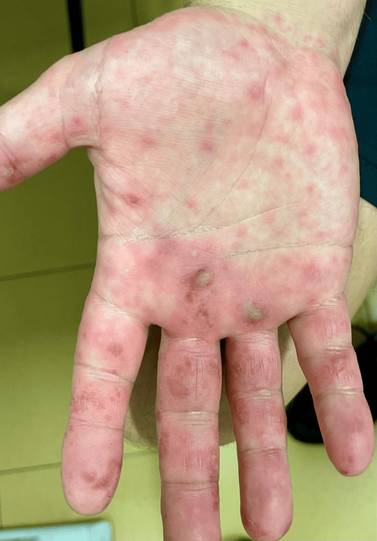 Figure 1
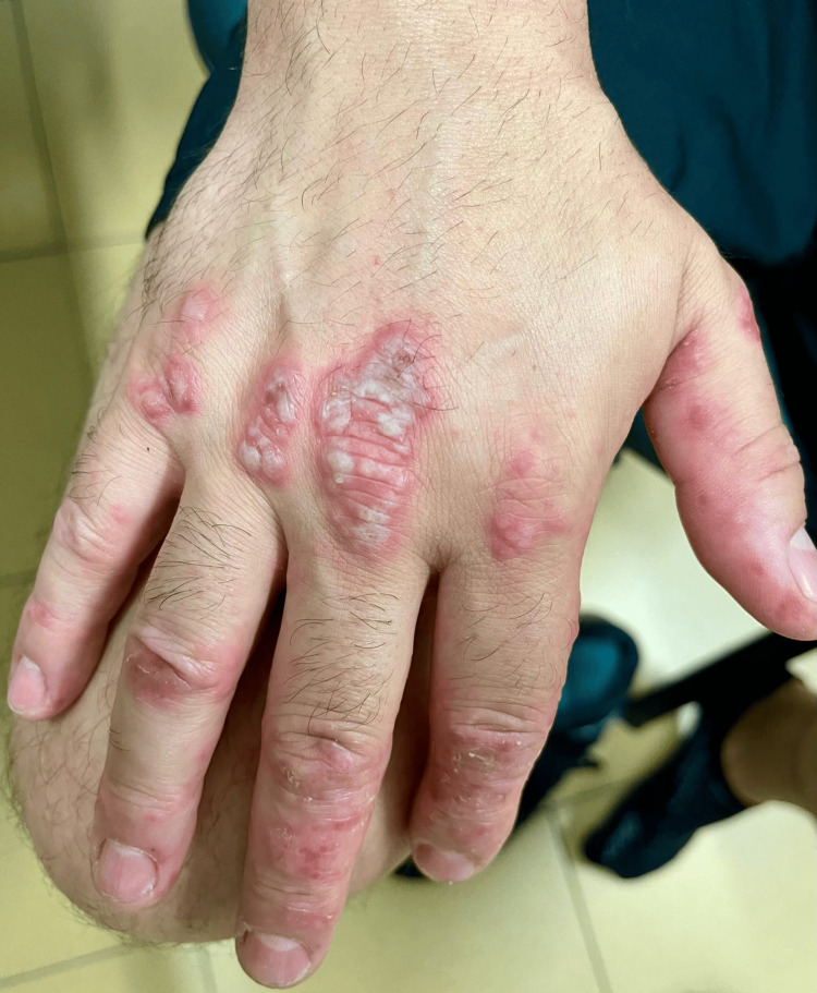 Figure 2
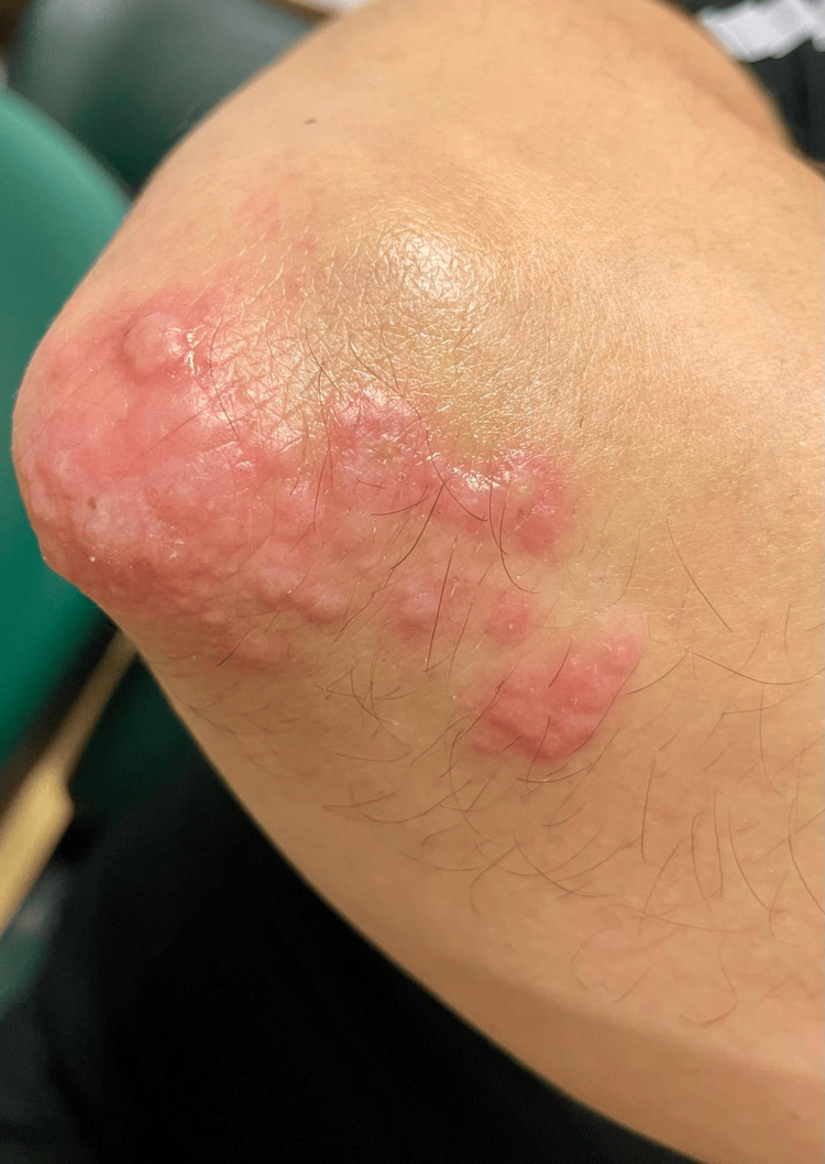 Figure 3
| Parameters | Value | Reference range |
| Hemoglobin | 15 g/dL | 13-16 g/dL |
| White blood cells | 5900/µL | 4000-11000/µL |
| Neutrophils | 3650/µL | 1500-8000/µL |
| Lymphocytes | 1540/µL | 800-4000/µL |
| C-reactive protein | 3 mg/dL | 0-0.2 mg/dL |
| Creatinin | 1.0 mg/dL | 0.7-1.2 mg/dL |
| AST | 35 UI/L | 0-40 UI/L |
| ALT | 48 UI/L | 0-50 UI/L |
| HIV 1, 2 antibodies | Negative | NA |
| HBsAg | Negative | NA |
| VDRL | Negative | NA |
| ASO | Negative | NA |
| Coxsackievirus A16 | Negative | NA |
| Enterovirus A71 | Positive | NA |
Peer Reviews
No public reviews on file for this paper yet. If you reviewed it on a platform where reviews are public (OpenReview, ICLR, NeurIPS, ICML), you can paste yours below so the community can read it here.
Videos
No videos yet. Explain this paper in a talk, walkthrough, or lecture? Add one.
Taxonomy
TopicsViral Infections and Immunology Research · Inflammatory Myopathies and Dermatomyositis · Immunodeficiency and Autoimmune Disorders
Introduction
Hand, foot, and mouth disease (HFMD) is a highly contagious viral infection, primarily affecting children under the age of 10 [1,2]. This condition is mostly caused by coxsackieviruses, though other enteroviruses can also be responsible [2]. Typical symptoms include fever, malaise, odynophagia, anorexia, and the development of painful skin lesions on the hands, feet, oral cavity, and other parts of the body [3]. Transmission occurs through direct contact with bodily secretions such as saliva, feces, or nasal secretions [2]. Although generally mild and self-limiting in children, there is a rising incidence of HFMD in adults, presenting particular diagnostic and management challenges [4,5]. Less than 1% of infections in adults are symptomatic, yet they exhibit a wide variability in symptoms, ranging from constitutional symptoms and typical skin lesions to severe presentations involving the central nervous system or the heart [6]. In patients with chronic dermatological diseases, Koebner phenomenon may occur with the worsening of their underlying condition and the need for therapeutic adjustment [7]. For these reasons, it is important for healthcare professionals to consider HFMD in the differential diagnosis of adults who present with febrile or post-febrile skin lesions, in order to avoid potential diagnostic errors, especially if there is a risk of exacerbation of a known chronic skin disease. Here, we describe a 48-year-old male patient with plaque psoriasis, previously controlled in a primary health care setting, with disease exacerbation due to HFMD.
Case presentation
A 48-year-old male, event organizer, single, without children, a smoking history of 33 pack-years, a history of plaque psoriasis for two years, regularly medicated and under control with betamethasone plus calcipotriol ointment, presented to the open clinic at a primary health care center with a one-day history of a skin rash on the hands, elbows, feet, and face, unresponsive to topical corticosteroid ointment. He also reported a prior two-week history of dry cough, odynophagia, rhinorrhea, and fever, which had spontaneously resolved. He denied risky contact with infected children or unprotected sexual relations. On physical examination, he had painful vesicles, blisters, and pustules on the perioral region, elbows, and palmoplantar regions of the hands and feet (Figures 1-3).
Vesicles, blisters and pustules on the palmar side of the right hand.
Vesicles, blisters and pustules on the dorsal side of the right hand.
Vesicles, blisters and pustules on the extensor region of the right elbow.
Given the clinical presentation, lesion location, and symptom progression, a clinical diagnosis of HFMD was made at the primary health care center, where laboratory tests were not available for diagnostic confirmation. The patient was discharged with reassurance of the clinical scenario, symptomatic measures, analgesia, and fusidic acid twice daily. After two days, he presented himself to the hospital emergency department due to worsening of the skin rash. He had new vesicles and pustules scattered on more locations, mainly on his face and ears, also painful, but with some lesions already resolving. A dermatological evaluation by a specialist was requested, who also made a clinical diagnosis of HFMD. For diagnostic confirmation and exclusion of other infections, blood tests were requested and revealed only a slight elevation of inflammatory parameters and a positive enterovirus A71 (Table 1).
He was discharged with a confirmed diagnosis of HFMD due to enterovirus A71 and advised to continue the previously prescribed treatment. Due to subsequent worsening of the plaque psoriasis, which was only responding partially to the topical therapy after the infection, the patient began follow-up in a dermatology consultation and started phototherapy with psoralen and ultraviolet A radiation. Subsequent information about the treatment efficacy is unknown because the patient moved to another location.
Discussion
This case represents an adult patient who presented with skin lesions following a respiratory illness of suggestive viral etiology (cough, odynophagia, fever, rhinorrhea), which had resolved in the previous two weeks. Several factors challenged the diagnosis in this patient. The patient had a history of plaque psoriasis and did not exhibit any extracutaneous symptoms at the time of presentation. Some lesions also had atypical locations (e.g., elbows ), possibly due to the use of topical immunosuppressive therapy. Additionally, the epidemiological source of the infection was unclear. The patient was unaware of any contact with an infected person and denied frequent contact with the pediatric population. These factors contributed to an extensive differential diagnosis, which caused further concern for the patient and the need for diagnostic confirmation in a hospital setting.
The main differential diagnoses to consider are viral rashes, bacterial rashes, and skin conditions due to systemic pathology. Among infectious causes, monkeypox, chickenpox, and syphilis are the primary considerations. For monkeypox, there were no anogenital or perioral lesions, and the lesions did not evolve into pseudo-pustules. Regarding chickenpox, there were no lesions in various stages (blisters, ulcers, scabs) or trunk lesions. In secondary syphilis, most patients have enlarged lymph nodes in multiple locations (cervical, axillary, and inguinal). Also, there is usually a widespread rash affecting the trunk, which the patient did not show.
HFMD usually presents with fever and vesicular lesions on the hands and feet and inside the mouth [3]. In adults, the symptoms can be atypical or severe, with higher fever and lesions on other parts of the body such as the arms, legs, and trunk [4]. Diagnosing atypical cases often depends on a thorough clinical history and physical examination, supported by diagnostic tests like serology or nucleic acid amplification [7]. Treatment focuses on relieving symptoms, with an emphasis on hydration and pain management [7]. The prognosis is generally good, with most cases resolving within one to two weeks and recurrences being rare. However, serious complications can occur, including painful stomatitis, pulmonary edema, myocarditis, interstitial pneumonia, and pancreatitis related to coxsackievirus [6,8]. As presented, there is also the possibility of worsening or exacerbating of chronic skin conditions, sometimes manifesting Koebner phenomenon, although there are few cases described in the literature in relation to HFMD in patients with psoriasis [7]. Patients with an underlying dermatological pathology may require more rigorous monitoring, sometimes at hospital level care, after the resolution of the infectious condition due to the need for therapeutic adjustment, whether for maintenance or crisis.
Conclusions
This case report emphasizes the need for healthcare providers to consider HFMD in adults with unexplained exanthems and oral symptoms, especially when chronic dermatologic diseases are present due to the risk of exacerbation. Atypical presentations, though rare, require accurate diagnosis for appropriate management. Further research can enhance understanding and early treatment of HFMD in adults.
The reference list from the paper itself. Each links out to its DOI / PubMed record.
- 1Atypical hand, foot and mouth disease in adults: a note on 6 cases]Ann Dermatol Venereol Flipo R Isnard C Coutard A Martres P Dumas M Blum L Begon E 8578611472020 https://www.sciencedirect.com/science/article/abs/pii/S 0151963820302532?via%3Dihub 3265479210.1016/j.annder.2020.04.025 · doi ↗ · pubmed ↗
- 2Hand, foot, and mouth disease in adults caused by Coxsackievirus B 1-B 6An Bras Dermatol Di Prinzio A Bastard DP Torre AC Mazzuoccolo LD 3213259720223527292010.1016/j.abd.2021.03.012PMC 9133263 · doi ↗ · pubmed ↗
- 3Hand, Foot, and Mouth Disease Stat Pearls [Internet] Guerra AM Orille E Waseem M Treasure Island (FL)Stat Pearls Publishing 2023 https://www.ncbi.nlm.nih.gov/books/NBK 431082/
- 4Coxsackievirus A 6 associated hand, foot and mouth disease in adults: clinical presentation and review of the literature J Clin Virol Ramirez-Fort MK Downing C Doan HQ Benoist F Oberste MS Khan F Tyring SK 3813866020142493273510.1016/j.jcv.2014.04.023 · doi ↗ · pubmed ↗
- 5Hand, foot, and mouth disease in adults Cureus Afonso C Almeida A 015202310.7759/cureus.48387 PMC 1069983038060762 · doi ↗ · pubmed ↗
- 6Hand-foot-mouth disease in an adult Cureus Gomes S Santos S Ferreira Maia I Verissimo R Carvalho T 015202310.7759/cureus.33670 PMC 992470636793831 · doi ↗ · pubmed ↗
- 7Psoriasis coxsackium JAAD Case Rep Cole DW Wang B Fullen DR Helfrich YR 22242520223567760110.1016/j.jdcr.2022.05.004PMC 9168028 · doi ↗ · pubmed ↗
- 8Severe atypical hand-foot-and-mouth disease in adults due to coxsackievirus A 6: clinical presentation and phylogenesis of CV-A 6 strains J Clin Virol Broccolo F Drago F Ciccarese G 1611020193047252010.1016/j.jcv.2018.11.003 · doi ↗ · pubmed ↗