Sequential MRI as a Diagnostic Tool for Follow‐Up of Hyaluronic Acid Dermal Filler, in a Woman Who Underwent Radiation Therapy for Oral Cancer
Gloria Bettini, Ferdinando De Negri, Roberto Amore, Sergio Rexhep Tari, Antonio Scarano

TL;DR
This paper explores how MRI can help track hyaluronic acid fillers in a cancer patient, avoiding confusion with other medical issues.
Contribution
The study highlights the MRI characteristics of hyaluronic acid fillers to aid accurate diagnosis in post-radiation therapy patients.
Findings
Hyaluronic acid appears strongly hyperintense on MRI due to its water-like signal.
The study emphasizes the importance of recognizing filler signals to avoid misdiagnosis in cancer patients.
Abstract
The present investigation aimed to inform the radiologists about the imaging features of injectable fillers in order not to confound these with true pathology or vice versa in order not to miss true pathology obscured by filler injections. The signal of hyaluronic acid closely follows the water signal because of its composition and its hydrophilic nature and appears strongly hyperintense.
Genes, proteins, chemicals, diseases, species, mutations and cell lines named across the full text — each resolved to its canonical identifier and authoritative record.
Click any figure to enlarge with its caption.
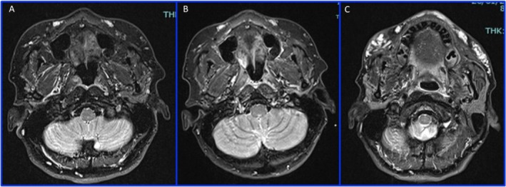 Figure 1
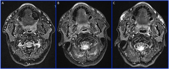 Figure 2
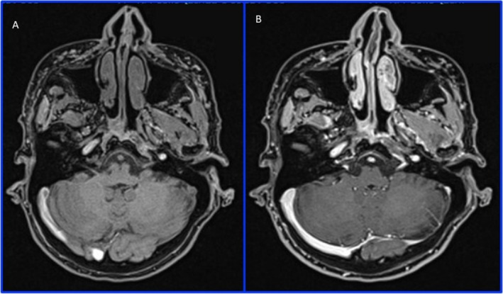 Figure 3
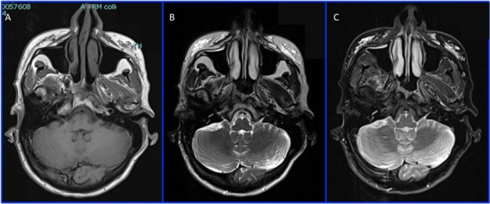 Figure 4
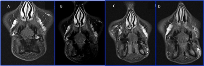 Figure 5
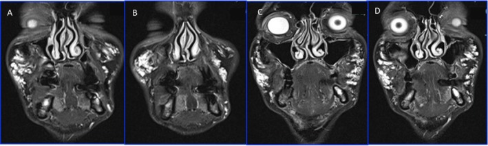 Figure 6
Peer Reviews
No public reviews on file for this paper yet. If you reviewed it on a platform where reviews are public (OpenReview, ICLR, NeurIPS, ICML), you can paste yours below so the community can read it here.
Videos
No videos yet. Explain this paper in a talk, walkthrough, or lecture? Add one.
Taxonomy
TopicsFacial Rejuvenation and Surgery Techniques · Dermatologic Treatments and Research · Body Contouring and Surgery