A guide to selecting high-performing antibodies for Protein-glutamine gamma-glutamyltransferase 2 (TGM2) for use in western blot, immunoprecipitation and immunofluorescence
Riham Ayoubi, Maryam Fotouhi, Charles Alende, Sara González Bolívar, Kathleen Southern, Carl Laflamme, Nicoletta Bianchi, Kathleen Southern, Michael A Rieger, Kathleen Southern

TL;DR
This paper evaluates commercial antibodies for TGM2 to help researchers choose reliable ones for common lab techniques.
Contribution
The study provides a standardized evaluation of TGM2 antibodies for western blot, immunoprecipitation, and immunofluorescence.
Findings
Seventeen TGM2 antibodies were tested for western blot and sixteen for immunoprecipitation and immunofluorescence.
A standardized protocol using knockout cell lines was used to assess antibody performance.
The results are shared openly to improve antibody reproducibility and guide researchers.
Abstract
Protein-glutamine gamma-glutamyltransferase 2 (TGM2) is a Ca 2+ dependent enzyme that catalyzes transglutaminase cross-linking modifications. TGM2 is involved in various diseases, either in a protective or contributory manner, making it a crucial protein to study and determine its therapeutic potential. Identifying high-performing TGM2 antibodies would facilitate these investigations. Here we have characterized seventeen TGM2 commercial antibodies for western blot and sixteen for immunoprecipitation, and immunofluorescence. The implemented standardized experimental protocol is based on comparing read-outs in knockout cell lines against their isogenic parental controls. This study is part of a larger, collaborative initiative seeking to address antibody reproducibility issues by characterizing commercially available antibodies for human proteins and publishing the results openly as a…
Genes, proteins, chemicals, diseases, species, mutations and cell lines named across the full text — each resolved to its canonical identifier and authoritative record.
Click any figure to enlarge with its caption.
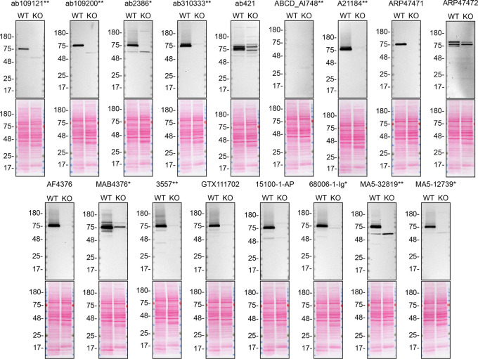 Figure 1
 Figure 2
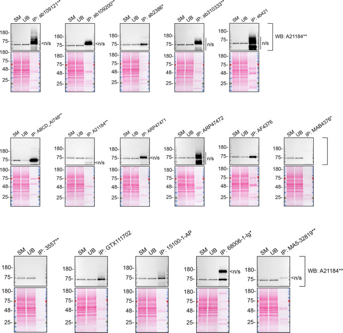 Figure 3
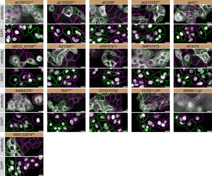 Figure 4
| Institution | Catalog number | RRID (Cellosaurus) | Cell line | Species of origin | Genotype |
|---|---|---|---|---|---|
| Abcam | ab275463 |
| A549 | Homo sapiens (Human) | WT |
| Abcam | ab261876 |
| A549 | Homo sapiens (Human) |
|
| Company | Catalog number | Lot number | RRID (Antibody Registry) | Clonality | Clone ID | Host | Concentration (μg/μL) | Vendors recommended applications |
|---|---|---|---|---|---|---|---|---|
| Abcam | ab109121** | 1058833-3 |
| Recombinant-mono | EPR2956 | rabbit | 2.79 | Wb |
| Abcam | ab109200** | 1044092-1 |
| Recombinant-mono | EP2957 | rabbit | 0.30 | Wb |
| Abcam | ab2386* | 1051063-6 |
| Monoclonal | CUB 7402 | mouse | n/a | Wb, IF |
| Abcam | ab310333** | 1056093-5 |
| Recombinant-mono | EPR28142-86 | rabbit | 0.50 | Wb, IF |
| Abcam | ab421 | 1034725-7 |
| Polyclonal | - | rabbit | n/a | Wb, IF |
| ABCD | ABCD_AI748** | 10/27/2023 |
| Recombinant-mono | 679-14-E06 | rabbit | 0.12 | Others |
| Abclonal | A21184** | 3522042510 |
| Recombinant-mono | rabbit | 1.30 | Wb, IF | |
| Aviva Systems Biology | QC18320-43546 |
| Polyclonal | - | rabbit | 0.50 | Wb | |
| Aviva Systems Biology | QC16720 |
| Polyclonal | - | rabbit | 0.50 | Wb | |
| R&D Systems (a Bio-Techne brand) | AF4376 | CFGU0119031 |
| Polyclonal | - | sheep | 0.20 | Wb |
| R&D Systems (a Bio-Techne brand) | MAB4376* | CFNO0119031 |
| Monoclonal | 716620 | mouse | 0.20 | Wb |
| Cell Signaling Technology | 3557** | 3 |
| Recombinant-mono | D11A6 | rabbit | 0.02 | Wb |
| GeneTex | GTX111702 | 44524 |
| Polyclonal | - | rabbit | 1.05 | Wb, IF |
| Proteintech | 15100-1-AP | 00081307 |
| Polyclonal | - | rabbit | 0.60 | Wb, IP, IF |
| Proteintech | 68006-1-Ig* | 10023724 |
| Monoclonal | 2D4C11 | mouse | 1.00 | Wb, IF |
| Thermo Fisher Scientific | MA5-32819** | YJ4089240 |
| Recombinant-mono | JU30-02 | rabbit | 1.00 | Wb |
| Thermo Fisher Scientific | MA5-12739* | ZA4176225 |
| Monoclonal | CUB 7402 | mouse | 0.20 | Wb, IP, IF |
- —Ontario Genomics
- —Michael J. Fox Foundation for Parkinson's Research
- —Mitacs
- —Genome Quebec
- —Genome Canada
- —Canadian Institutes of Health Research Foundation
Peer Reviews
No public reviews on file for this paper yet. If you reviewed it on a platform where reviews are public (OpenReview, ICLR, NeurIPS, ICML), you can paste yours below so the community can read it here.
Videos
No videos yet. Explain this paper in a talk, walkthrough, or lecture? Add one.
Taxonomy
TopicsErythrocyte Function and Pathophysiology · Blood properties and coagulation · Pancreatic function and diabetes
Introduction
Protein-glutamine gamma glutamyltransferase 2 (TGM2) belongs to the transglutaminase family of Ca ^2+^ dependent enzymes, regulating various cellular processes, including cell differentiation, growth and apoptosis. ^ 1 ^ ^–^ ^ 3 ^ Encoded by TGM2 gene, TGM2 protein exhibits its function through Ca ^2+^-dependent cross-linking of substrates, thereby modulating their activity. ^ 1 ^ TGM2’s catalytic activity is dependent on guanine nucleotides and Ca ^2+^ binding. ^ 4 ^ Both GTP and/or GDP act as negative regulators of TGM2, inducing a conformational change upon binding, inhibiting its cross-linking activity (closed form). Conversely, Ca ^2+^ binding prompts a conformational change to induce TGM2 activity (open form). The proteins activity is dependent on cellular location, remaining inactive intracellularly, and becoming activated upon secretion. ^ 5 ^ ^–^ ^ 9 ^
Alteration and regulation of TGM2’s activity is associated with the pathogenesis of various diseases including cancer, neurodegeneration, fibrosis, inflammatory, autoimmune disorders and liver diseases. ^ 10 ^ ^–^ ^ 13 ^ Increased TGM2 mRNA transcripts, resulting in elevated transaminase enzymatic activity, have been associated with neurodegenerative mechanism observed in Parkinson’s disease, Alzheimer’s disease and Huntington’s disease. ^ 14 ^ ^,^ ^ 15 ^ Given that α-synuclein serves as a common substrate for TGM2, elevated activity of TGM2 results in the formation of soluble aggregates as well as insoluble inclusions; distinctive features of Parkinson’s disease pathogenesis. ^ 15 ^ ^–^ ^ 18 ^ Regulating TGM2 activity using inhibitors could positively affect the human diseases in which TGM2 is implicated. ^ 19 ^ ^,^ ^ 20 ^ Identifying high-quality antibodies would accelerate TGM2 research and its potential as a pharmacological target.
This research is part of a broader collaborative initiative in which academics, funders and commercial antibody manufacturers are working together to address antibody reproducibility issues by characterizing commercial antibodies for human proteins using standardized protocols, and openly sharing the data. ^ 21 ^ ^–^ ^ 23 ^ Here, we evaluated the performance of seventeen commercially-available antibodies for TGM2 for use in western blot, and sixteen for immunoprecipitation and immunofluorescence, enabling biochemical and cellular assessment of TGM2 properties and function. The platform for antibody characterization used to carry out this study was approved by a committee of industry, academic research. It consists of first identifying appropriate human cell lines, development/contribution of equivalent knockout cell lines and finally following antibody characterization procedures on commonly used commercial antibodies. The standardized consensus antibody characterization protocols are openly available on Protocol Exchange, DOI: 10.21203/rs.3.pex-2607/v1. ^ 24 ^
Results and discussion
Our standard protocol involves comparing readouts from wild-type (WT) and knockout (KO) cells. ^ 25 ^ ^,^ ^ 26 ^ The first step is to identify a cell line(s) that expresses sufficient levels of a given protein to generate a measurable signal. To this end, we examined the DepMap transcriptomics database to identify all cell lines that express the target at levels greater than 2.5 log 2 (transcripts per million “TPM” + 1), which we have found to be a suitable cut-off (Cancer Dependency Map Portal, RRID:SCR_017655). Commercially available A549 cells expressed the TGM2 transcript at RNA levels above the average range of cancer cells analyzed. Parental and TGM2 KO A549 cells were obtained from Abcam ( Table 1).
For western blot experiments, we resolved proteins from WT and TGM2 KO cell extracts and probed them side-by-side with all antibodies in parallel on lysate and culture medium ( Figures 1 and 2).
TGM2 antibody screening by western blot on lysate.Lysates of A549 (WT and TGM2 KO) were prepared and 40 μg of protein were processed for western blot with the indicated TGM2 antibodies. The Ponceau stained transfers of each blot are presented to show equal loading of WT and KO lysates and protein transfer efficiency from the acrylamide gels to the nitrocellulose membrane. Antibody dilutions were chosen according to the recommendations of the antibody supplier. Antibody dilution used: ab109121* at 1/1000, ab109200** at 1/10000, ab2386* at 1/500, ab310333** at 1/1000, ab421 at 1/500, ABCD_AI748** at 1/10, A21184** at 1/10000, ARP47471 at 1/500, ARP47472 at 1/500, AF4376 at 1/400, MAB4376* at 1/200, 3557** at 1/500, GTX111702 at 1/500, 15100-1-AP at 1/6000, 68006-1-Ig* at 1/10000, MA5-32819** at 1/500, MA5-12739* at 1/200. Predicted band size: 77 kDa. *Monoclonal antibody, *Recombinant antibody.
TGM2 antibody screening by western blot on culture medium.A549 WT and TGM2 KO were cultured in serum free media, and 40 μg of protein from concentrated culture media were processed for western blot with the indicated TGM2 antibodies. The Ponceau stained transfers of each blot are shown. Antibody dilution used: ab109121* at 1/1000, ab109200** at 1/10000, ab2386* at 1/500, ab310333** at 1/1000, ab421 at 1/500, ABCD_AI748** at 1/10, A21184** at 1/10000, ARP47471 at 1/500, ARP47472 at 1/500, AF4376 at 1/400, MAB4376* at 1/200, 3557** at 1/500, GTX111702 at 1/500, 15100-1-AP at 1/6000, 68006-1-Ig* at 1/10000, MA5-32819** at 1/500, MA5-12739* at 1/200. Predicted band size: 77 kDa. *Monoclonal antibody, *Recombinant antibody.
As per our standard procedure, we next used the antibodies to immunoprecipitate TGM2 from A549 cell extracts. To evaluate the performance of each antibody, the TGM2 protein was detected in extracts, in each extract unbound to the antibody and corresponding immunoprecipitate ( Figure 3). To detect TGM2, a western blot was performed with an antibody successful under the conditions tested in Figure 1.
TGM2 antibody screening by immunoprecipitation on lysate.A549 lysates were prepared, and immunoprecipitation was performed using 2.0 μg of the indicated TGM2 antibodies pre-coupled to Dynabeads protein A or protein. Samples were washed and processed for western blot with the indicated TGM2 antibody. For western blot, A21184* was used at 1/10000. The Ponceau stained transfers of each blot are shown. SM=4% starting material; UB=4% unbound fraction; IP=immunoprecipitate; n/s=non-specific signal. *Monoclonal antibody, *Recombinant antibody.
For immunofluorescence, antibodies were screened using a mosaic strategy, as per our standard procedure. First, A549 WT and TGM2 KO were labelled with different fluorescent dyes in order to distinguish the two cell lines, and TGM2 antibodies were evaluated. Cells were imaged in the same field of view to reduce staining, imaging and image analysis bias ( Figure 4). Quantification of immunofluorescence intensity in hundreds of WT and KO cells was performed for each antibody tested. The images presented in Figure 4 are representative of the results of this analysis.
TGM2 antibody screening by immunofluorescence.A549 WT and TGM2 KO cells were labelled with a green or a deep-red fluorescent dye, respectively. WT and KO cells were mixed and plated to a 1:1 ratio in a 96-well plate with optically clear flat-bottom. Cells were stained with the indicated TGM2 antibodies and with the corresponding Alexa-fluor 555 coupled secondary antibody including DAPI. Acquisition of the blue (nucleus-DAPI), green (identification of WT cells), red (antibody staining) and deep-red (identification of KO cells) channels was performed. Representative images of the merged blue and red (grayscale) channels are shown. WT and KO cells are outlined with green and magenta dashed line, respectively. When the concentration was not indicated by the supplier, which was the case for all antibodies tested, except ab310333*, ABCD_AI748 and A21184**, we tested antibodies at using the dilutions listed below. At these concentrations, the signal from each antibody was in the range of detection of the microscope used. Antibody dilution used: ab109121** at 1/1000, ab109200** at 1/300, ab2386* at 1/1000, ab310333** at 1/500, ab421 at 1/1000, ABCD_AI748** at 1/1000, A21184** at 1/1000, ARP47471 at 1/500, ARP47472 at 1/250, AF4376 at 1/100, MAB4376* at 1/100, 3557** at 1/500, GTX111702 at 1/1000, 15100-1-AP at 1/300, 68006-1-Ig* at 1/500, MA5-32819** at 1/1000. Bars = 10 μm. *Monoclonal antibody, *Recombinant antibody.
In conclusion, we have screened seventeen TGM2 commercial antibodies by western blot, and sixteen by immunoprecipitation, and immunofluorescence, comparing the signal produced by the antibodies in human A549 WT and TGM2 KO cells. Several high-quality antibodies that successfully detect TGM2 under our standardized experimental protocol can be identified. Researchers who wish to study TGM2 in a different species are encouraged to select high-quality antibodies, based on the results of this study, and investigate the predicted species reactivity of the manufacturer before extending their research.
In our effort to address the antibody reliability and reproducibility challenges in scientific research, the authors recommend the antibodies that demonstrated to be underperforming under our standard procedure be removed from the commercial antibody market. Following the release of the antibody characterization, ab421 was removed from the manufacturer’s antibody catalog.
The authors do not engage in result analysis or offer explicit antibody recommendations. A limitation of this study is the use of universal protocols - any conclusions remain relevant within the confines of the experimental setup and cell line used in this study. Our primary aim is to deliver top-tier data to the scientific community, grounded in Open Science principles. This empowers experts to interpret the characterization data independently, enabling them to make informed choices regarding the most suitable antibodies for their specific experimental needs. Guidelines on how to interpret antibody characterization data found in this study are featured on the YCharOS gateway. ^ 27 ^
The underlying data for this study can be found on Zenodo, an open-access repository for which YCharOS has its own collection of antibody characterization reports. ^ 28 ^ ^,^ ^ 29 ^
Methods
The standardized protocols used to carry out this KO cell line-based antibody characterization platform was established and approved by a collaborative group of academics, industry researchers and antibody manufacturers. The detailed materials and step-by-step protocols used to characterize antibodies in western blot, immunoprecipitation and immunofluorescence are openly available on Protocol Exchange, a repository dedicated to openly sharing scientific research protocols, DOI: 10.21203/rs.3.pex-2607/v1. ^ 24 ^
Antibodies and cell line used
Cell lines used and primary antibodies tested in this study are listed in Table 1 and 2, respectively. To ensure that the cell lines and antibodies are cited properly and can be easily identified, we have included their corresponding Research Resource Identifiers, or RRID. ^ 30 ^ ^,^ ^ 31 ^
The reference list from the paper itself. Each links out to its DOI / PubMed record.
- 1Jeong EM Lee KB Kim GE : Competitive Binding of Magnesium to Calcium Binding Sites Reciprocally Regulates Transamidase and GTP Hydrolysis Activity of Transglutaminase 2. Int. J. Mol. Sci. 2020;21(3). 10.3390/ijms 21030791 31991788 PMC 7037829 · doi ↗ · pubmed ↗
- 2Kanchan K Ergülen E Király R : Identification of a specific one amino acid change in recombinant human transglutaminase 2 that regulates its activity and calcium sensitivity. Biochem. J. 2013;455(3):261–272. 10.1042/BJ 20130696 23941696 · doi ↗ · pubmed ↗
- 3Tatsukawa H Furutani Y Hitomi K : Transglutaminase 2 has opposing roles in the regulation of cellular functions as well as cell growth and death. Cell Death Dis. 2016;7(6):e 2244. 10.1038/cddis.2016.150 27253408 PMC 5143380 · doi ↗ · pubmed ↗
- 4Lorand L Graham RM : Transglutaminases: crosslinking enzymes with pleiotropic functions. Nat. Rev. Mol. Cell Biol. 2003;4(2):140–156. 10.1038/nrm 1014 12563291 · doi ↗ · pubmed ↗
- 5Liu S Cerione RA Clardy J : Structural basis for the guanine nucleotide-binding activity of tissue transglutaminase and its regulation of transamidation activity. Proc. Natl. Acad. Sci. USA. 2002;99(5):2743–2747. 10.1073/pnas.042454899 11867708 PMC 122418 · doi ↗ · pubmed ↗
- 6Achyuthan KE Greenberg CS : Identification of a guanosine triphosphate-binding site on guinea pig liver transglutaminase. Role of GTP and calcium ions in modulating activity. J. Biol. Chem. 1987;262(4):1901–1906. 10.1016/S 0021-9258(19)75724-X 2879844 · doi ↗ · pubmed ↗
- 7Singh US Erickson JW Cerione RA : Identification and biochemical characterization of an 80 kilodalton GTP-binding/transglutaminase from rabbit liver nuclei. Biochemistry. 1995;34(48):15863–15871. 10.1021/bi 00048 a 032 7495818 · doi ↗ · pubmed ↗
- 8Mian S Alaoui Sel Lawry J : The importance of the GTP-binding protein tissue transglutaminase in the regulation of cell cycle progression. FEBS Lett. 1995;370(1-2):27–31. 10.1016/0014-5793(95)00782-5 7649299 · doi ↗ · pubmed ↗