Subependymal Giant Cell Astrocytomas Without Tuberous Sclerosis: A Case Report on a Rare Medical Condition
Pranjali Nibe, Rupali Bavikar, Charusheela Gore, Gayatri Bhuibhar

TL;DR
A 22-year-old man was diagnosed with a rare brain tumor called SEGA that was not linked to a genetic condition called tuberous sclerosis.
Contribution
This case report highlights the possibility of SEGAs occurring without tuberous sclerosis in adults.
Findings
The tumor was confirmed through radiological investigations and specific protein expression.
No germline mutations in TSC1 and TSC2 were found, ruling out tuberous sclerosis.
The case emphasizes the importance of considering SEGAs in adult patients without TSC.
Abstract
Subependymal giant cell astrocytomas (SEGAs) are benign, slow-growing, noninvasive tumors frequently associated with the tuberous sclerosis complex (TSC). The tumor's location and the patient's age should be considered carefully before diagnosis. Considering SEGA as a differential diagnosis, even in adult patients without TSC, is essential. In the present case, a 22-year-old male presented with a progressive headache, dizziness, and blurring of vision. Radiological investigations confirmed the site of the tumor, and a positive expression of thyroid transcription factor 1 in the ganglion cell component, along with the absence of germline mutation in TSC1 and TSC2, led to the final diagnosis of SEGA without TSC.
Genes, proteins, chemicals, diseases, species, mutations and cell lines named across the full text — each resolved to its canonical identifier and authoritative record.
Click any figure to enlarge with its caption.
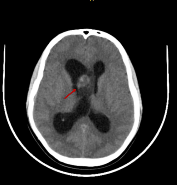 Figure 1
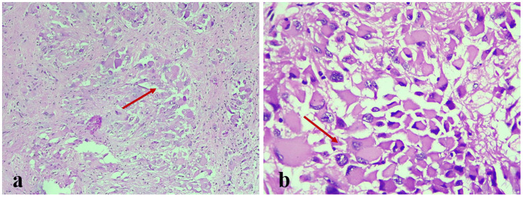 Figure 2
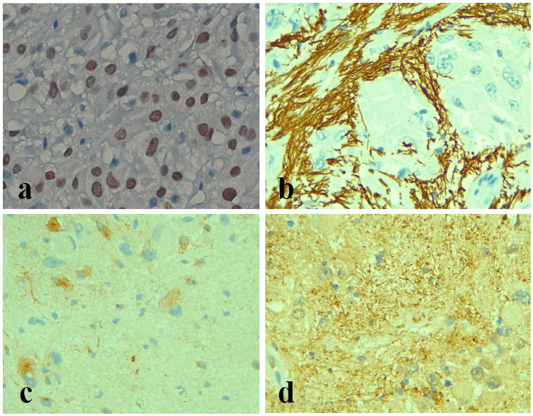 Figure 3
Peer Reviews
No public reviews on file for this paper yet. If you reviewed it on a platform where reviews are public (OpenReview, ICLR, NeurIPS, ICML), you can paste yours below so the community can read it here.
Videos
No videos yet. Explain this paper in a talk, walkthrough, or lecture? Add one.
Taxonomy
TopicsTuberous Sclerosis Complex Research · Neurofibromatosis and Schwannoma Cases · Medical Imaging and Pathology Studies