Case Report: Activated B-cell-diffuse large B-cell lymphoma
Padmashri Kalmegh, Alka Hande, Madhuri Gawande, Swati Patil, Archana Sonone, Aayushi Pakhale, Soundarya Ravi, Padmashri Kalmegh, Junn-Liang Chang, Padmashri Kalmegh, Padmashri Kalmegh

TL;DR
This case report describes a rare oropharyngeal lymphoma diagnosed as activated B-cell-diffuse large B-cell lymphoma in a female patient.
Contribution
The report highlights the clinical, histological, and immunohistochemical features of a rare lymphoma case to aid in early diagnosis.
Findings
The patient presented with a palatal swelling and ulceroproliferative lesion.
Diagnosis of activated B-cell-diffuse large B-cell lymphoma was confirmed via punch biopsy and immunohistochemistry.
The case emphasizes the importance of correlating clinical and pathological findings for accurate diagnosis.
Abstract
Lymphomas of the oral and oropharyngeal regions are rather uncommon, and diagnosis can be challenging and confusing due to the multiple histological subgroups. Lymphomas are the third most common type of tumor in the head and neck region and are brought on by the lymphoreticular system. The two forms of lymphoma are Hodgkin’s lymphoma and non-Hodgkin’s lymphoma (NHL). Herein, we present a case report of oropharyngeal lymphoma. The female patient reported with a complaint of swelling over the palatal region for two to three months. An ulceroproliferative lesion was evident over the posterior palatal region. We diagnosed reactive lymphadenitis based on an incisional biopsy. To confirm the diagnosis and rule out other conditions, a punch biopsy followed by immunohistochemical studies were done. Features suggestive of activated B-cell-diffuse large B-cell lymphoma were confirmed. Among…
Genes, proteins, chemicals, diseases, species, mutations and cell lines named across the full text — each resolved to its canonical identifier and authoritative record.
Click any figure to enlarge with its caption.
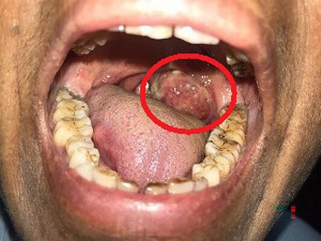 Figure 1
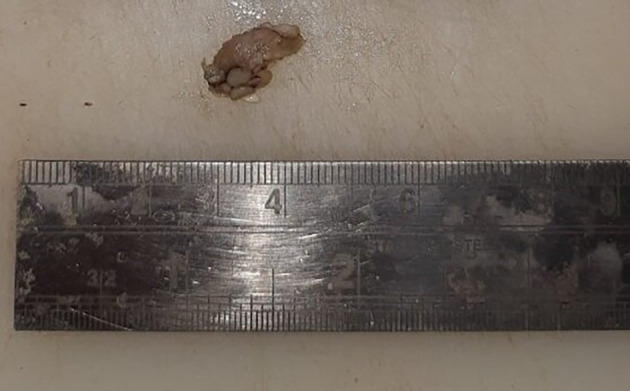 Figure 2
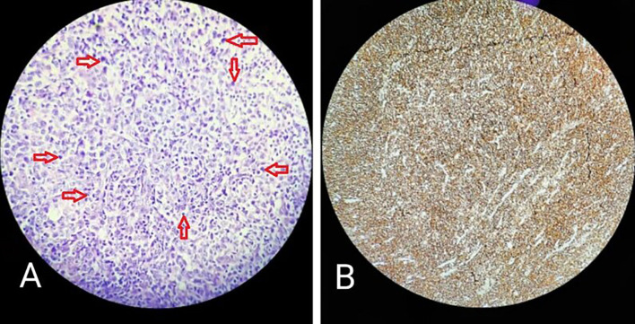 Figure 3
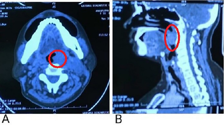 Figure 4
Peer Reviews
No public reviews on file for this paper yet. If you reviewed it on a platform where reviews are public (OpenReview, ICLR, NeurIPS, ICML), you can paste yours below so the community can read it here.
Videos
No videos yet. Explain this paper in a talk, walkthrough, or lecture? Add one.
Taxonomy
TopicsLymphoma Diagnosis and Treatment · Viral-associated cancers and disorders · Chronic Lymphocytic Leukemia Research
Introduction
Samuel Wilks coined the term “Hodgkin’s disease,” which is now known as “Hodgkin lymphoma,” in recognition of the earlier report by Thomas Hodgkin from Guy’s hospital in London. ^ 1 ^ Lymphomas are a diverse group of lymphoid malignancies with a range of clinical behaviour patterns and therapeutic responses. The histologic type, clinical variables, and more recently, molecular traits all affect the prognosis. ^ 2 ^ In terms of prevalence, lymphomas rank ninth among males and tenth among women worldwide. Lymphomas are the second most prevalent type of cancer in the head and neck region, and they rank third after squamous cell carcinoma and salivary gland cancers in the oral cavity. Even though lymphomas account for 3% to 5% of all reported cases in the head and neck region, they are the most prevalent non-epithelial malignant tumors in the same region. ^ 3 ^ Typically, no underlying cause of lymphoma is found in any specific occurrence. Nonetheless, other environmental, viral, and genetic risk factors for lymphoma have been discovered. Certain lymphomas have been linked to a number of infectious agents, including Helicobacter pylori, Borrelia burgdorferi, Chlamydia psittaci, and Campylobacter jejuni. ^ 4 ^ We adhere to the CARE reporting guidelines to report this Case report. ^ 5 ^
Case report
A 63-year-old female, presented to the outdoor patient department of our institution in January 2023, with a complaint of a non-healing ulcer over the posterior palatal region for two to three months. She described no symptoms before two to three months but later observed swelling over the posterolateral region of the soft palate. Initially, it was 2 × 1 cm in size and increased to the current size of 4 × 3 cm approximately. She described difficulty in mastication and swallowing. A change in consistency and quantity of saliva was reported by the patient. No history of trauma was present. Intraorally, a single ulceroproliferative lesion present over the anterior faucial pillar was observed ( Figure 1, marked with a red circle). The lesion was oval, reddish pink in color, with ill-defined borders, soft to hard in consistency, nonmobile, and had an irregular surface. Uvula deviated to the left side. Lymph nodes in the left submandibular region were palpable, of size 1 × 2 cm, non-tender, and firm in texture. In December 2022, an incisional biopsy was taken with a histopathological diagnosis of reactive lymphadenitis. This was followed by a punch biopsy ( Figure 2) in January 2023, with histopathological features suggestive of a lymphoproliferative lesion. No extraoral extension of the growth was evident.
Clinical photo of the patient showing swelling on the posterolateral portion of the soft palate.
Punch biopsy specimen (2 × 1.7 cm).
Histological features
Haematoxylin and Eosin-stained sections showed predominantly a diffuse infiltrate of large-sized atypical B cell population comprising of scant cytoplasm. The nucleus was double the size of the nucleus of a normal lymphocyte, vesicular chromatin, and prominent nucleoli ( Figure 3A, marked with red arrows). Complete effacement of normal lymphoid tissue architecture was present.
A: Diffuse infiltration of large lymphocytes (marked by red arrow); B: CD45 positive.
Immunohistochemical findings
Immunohistochemical features with cytokeratin staining were negative, CD45 was strongly positive with an intensity score of 3, proportionate scores of 4 ( Figure 3B), and CD20 positive. On the basis of histopathological and immunohistochemical features, the lesion was suggestive of an activated B-cell (ABC) subset of diffuse large B-cell lymphoma (DLBCL).
Radiographic features
A contrast-enhanced computerized tomography (CECT) presented evidence of a soft tissue density lesion with few non-enhancing necrotic areas noted in the left tonsillar fossa measuring 2.2 × 2 × 2.7 cm ( Figure 4A, marked with a red circle) with the following extensions: a. Anteromedially extending into the oropharynx causing partial obstruction with non-visualization of uvula suggestive of involvement abutting the base of the tongue and vallecula ( Figure 4B, marked with a red circle). b. Posteroinferiorly involves the posterior pharyngeal wall. c. Laterally involving the peritonsillar space including the pharygobasillar fascia, styloglossus and stylopharyngeus, and the posterior belly of the digastric.
A: Non-enhancing necrotic areas noted in the left tonsillar fossa; B: Anteromedial extension abutting to the base of the tongue.
Treatment
DLBCL becomes less fatal and may even be cured with combination chemotherapy, which only successfully treats less than 50% of patients. The type of therapy will either be curative, where survival is the goal, or palliative, where the goal is to improve the patient’s quality of life, depending on the patient’s status. ^ 4 ^ Rituximab, cyclophosphamide, doxorubicin, vincristine, and prednisone (R-CHOP) is the standard treatment for DLBCL patients. Approximately 60–70% of DLBCL patients who continue this regimen are free of illness. ^ 6 ^
In the present case, chemotherapy (cyclophosphamide, hydroxy doxorubicin, oncovin, and prednisone) was used to treat the patient. The patient continued receiving CHOP chemotherapy, and her overall health is improving. A four-month follow-up showed regression of the swelling. The patient is still on treatment.
Discussion
Malignant lymphomas are a category of neoplasms with variable degrees of malignancy that are derived from the lymphoid tissue’s fundamental cells, the lymphocyte, and histiocytes at any stage of development. ^ 7 ^ The classification of lymphomas originating from these normal lymphoid populations is challenging since the physiologic immunological roles of lymphocytes vary depending on lineage and degree of development. NHL and Hodgkin lymphoma (HL) are the two main categories into which lymphomas are frequently categorized. NHLs are a variety of lymphoproliferative malignancies that have a far higher propensity to disseminate to extranodal locations than Hodgkin’s lymphoma. ^ 2 ^ NHL, a class of neoplasms, occurs due to the uncontrolled proliferation of B-, T-, or Natural Killer (NK) cells. T-cell or NK cell-derived illnesses account for 10 to 15 percent of all NHL cases. ^ 8 ^ DLBCL, which accounts for 30–40% of cases in various parts of the world, is the most common NHL. It is most commonly seen in the sixth to seventh decade of life. ^ 9 ^ DLBCL is a diverse entity in terms of clinical presentation, genetic advances, therapeutic options, and prognosis. There was a significant advancement when gene expression profiling (GEP) was applied to further detect this variety and provide a rationale for categorizing patients in the study of DLBCL. DLBCL patients are divided into subtypes named germinal center B-cell-like (GCB), ABC, and 10–15% remain unclassified. The ABC subtype is derived from B cells that are undergoing plasma cell development. Patients with the ABC subtype often have the worst prognosis than those with the GCB subtype. ^ 10 ^ However, there is morphological and clinical heterogeneity in DLBCL. However, patients with GCB DLBCL still do considerably better than those with ABC DLBCL. ^ 11 ^ Expression of CD20 (cluster of differentiation antigens) in lymphoma cells is essential for both making an accurate diagnosis and creating a strategy for biological therapeutic therapy. CD20 antigen is expressed on the surface of neoplastic cells in the majority of B-cell lymphomas, albeit the degree of CD20 expression varies depending on the type of lymphoma and the degree of lymphoma B-cell differentiation. Hence, it is assumed that CD20 expression is high in DLBCL and hairy cell leukemia cells, and low in B-cell chronic lymphocytic leukemia (CLL). ^ 12 ^ Particularly, ABC DLBCLs express X-box binding protein-1 (XBP-1), a significant modulator of the secretory phenotype of plasma cells. ^ 13 ^ About 25% of ABC DLBCL samples taken from patients have BLIMP1-inactivating mutations in the PRDM1 gene. ^ 14 ^ By inhibiting the expression of the majority of mature B-cell differentiation genes, BLIMP1 promotes plasmacytic differentiation. ^ 15 ^ Consistent activation of the nuclear factor-kappa B (NF-κB) signaling pathway, which stimulates cell survival and proliferation while inhibiting apoptosis, is another pathogenic characteristic of ABC DLBCLs. ^ 16 ^ In order to ensure the most effective use of investigational treatments or currently accessible, frequently expensive therapeutic agents, pathologists must also evaluate the markers required to support their usage. Additionally, pathologists must prioritize sufficient and viable tumor tissue for high-throughput molecular analysis. ^ 17 ^ ^–^ ^ 21 ^
According to the location and histologic type, clinical presentation differs substantially. Biopsy in conjunction with immunological investigations of biopsy tissue is the only accurate way to identify and classify these lesions. ^ 22 ^ No single antibody has been useful for subdividing DLBCL or determining prognosis, according to research by Meyer et al. Because of this, many antibody combinations or methods have been created. ^ 23 ^ For the differential diagnosis of 40 cases of oral NHL, Van der Waal et al. used the following antibodies: L26 (CD20, a pan B-cell marker), CD 79a (the immunoglobulin anchoring molecule, so a B-cell marker), CD3, and UCHL 1 (CD45RO), both pan T-cell markers, BerH2 (CD30), and Mib1 (staining primarily B cells). ^ 24 ^ Forty cases of oral cavity NHL were studied by Kemp et al. for sex, age, location, clinical presentation, and WHO histological subtype. They found that 98% of the lymphomas in their investigation had a B cell lineage, and the majority of these were histologically subtyped as diffuse large B cell lymphomas (58%). Our study is in accordance with Kemp et al., who used an immunohistochemical panel that included CD3, CD5, CD20, CD45RO, CD79a, leukocyte common antigen, Bcl-2, CD10 and CD23 to validate the lineage and help characterize the subtype. According to them, molecular testing is typically useful in determining genetic infiltration and seems to be a useful supplement to diagnosis. ^ 25 ^ In nonimmunocompromised patients, diffuse large B-cell lymphoma in the head and neck region was the most prevalent form observed, according to Hicks and Flaitz, 2001. ^ 26 ^ Hashimoto and Kurihara (1982), reviewed pathological characteristics of oral NHL and according to his nine cases and review of the literature, he concluded that B-cell lymphoma is the most common histotype in oral NHL. ^ 27 ^ Therefore, in order to diagnose a rare condition, this case report emphasizes the value of early biopsies of oral lesions and the use of a definitive marker among other available immunohistochemical markers.
Limitations
The study has limitations due to the patient’s ongoing treatment and the relatively short follow-up time.
Conclusions
Oral NHL is a very uncommon disorder that may cause localized swelling, inflammation, or discomfort in the vestibule, gingiva, or posterior hard palate. DLBCL is a genetically heterogeneous disease with equally different clinical manifestations. Hence, one can determine the malignant nature and prognosis of various kinds of lymphomas by correlating the histological characteristics of these entities. Pathologists should evaluate immunophenotypic and genetic markers that are useful for determining prognosis. In order for the patient to receive therapy at an early stage and have a favorable prognosis, a thorough evaluation of the patient and appropriate investigations are needed for a precise diagnosis.
Consent
Written informed consent for publication of their clinical details and clinical images was obtained from the patient.
The reference list from the paper itself. Each links out to its DOI / PubMed record.
- 1Wang HW Balakrishna JP Pittaluga S : Diagnosis of Hodgkin lymphoma in the modern era. Br. J. Haematol. 2019 Jan;184(1):45–59. 10.1111/bjh.15614 30407610 PMC 6310079 · doi ↗ · pubmed ↗
- 2Jiang M Bennani NN Feldman AL : Lymphoma classification update: T-cell lymphomas, Hodgkin lymphomas, and histiocytic/dendritic cell neoplasms. Expert. Rev. Hematol. 2017 Mar 4;10(3):239–249. 10.1080/17474086.2017.1281122 28133975 PMC 5514564 · doi ↗ · pubmed ↗
- 3Arruda JA Ade Schuch LF Conte Neto N : Oral and oropharyngeal lymphomas: A multi-institutional collaborative study. J. Oral Pathol. Med. 2021 Jul 1;50(6):603–612. 10.1111/jop.13211 34091952 · doi ↗ · pubmed ↗
- 4Mugnaini EN Ghosh N : Lymphoma. Prim. Care. 2016 Dec 1;43(4):661–675. 10.1016/j.pop.2016.07.012 27866584 · doi ↗ · pubmed ↗
- 5Padmashri K Hande A Gawande M : Activated B-cell-Diffuse Large B-Cell Lymphoma: A Case Report.[Dataset]. Zenodo. 2023. 10.5281/zenodo.7918599 · doi ↗
- 6Cao X Ye Q Orlowski RZ : Waldenström macroglobulinemia with extramedullary involvement at initial diagnosis portends a poorer prognosis. J. Hematol. Oncol. 2015 Dec;8:1–1. 10.1186/s 13045-015-0172-y 26104577 PMC 4487966 · doi ↗ · pubmed ↗
- 7Singh R Shaik S Negi BS : Non-Hodgkin’s lymphoma: a review. J. Fam. Med. Prim. Care. 2020 Apr;9(4):1834–1840. 10.4103/jfmpc.jfmpc_1037_19 32670927 PMC 7346945 · doi ↗ · pubmed ↗
- 8Hans CP Weisenburger DD Greiner TC : Confirmation of the molecular classification of diffuse large B-cell lymphoma by immunohistochemistry using a tissue microarray. Blood. 2004 Jan 1;103(1):275–282. 10.1182/blood-2003-05-1545 14504078 · doi ↗ · pubmed ↗