Ophthalmomyiasis Case Caused by Two Blow Fly (Diptera: Calliphoridae) Species in North America
Taylor B. Parker, Kelly A. Meiklejohn, Gregory A. Dahlem, Ralph C. Eagle, Marius J. Heersink

TL;DR
A rare case of eye infection caused by two types of blow flies was reported in North America, highlighting unusual larval behavior.
Contribution
This is the first documented case of ophthalmomyiasis caused by two blow fly species in a single individual.
Findings
DNA analysis identified Lucilia coeruleiviridis and Phormia regina as the two blow fly species involved.
Morphological examination confirmed only Lucilia coeruleiviridis.
This case suggests competitive behavior among blow fly species in unusual ecological conditions.
Abstract
Ophthalmomyiasis is the result of fly larvae feeding on the tissues of the eye. Commonly associated with poor hygiene and open wounds, this condition is rare and often stigmatized. Treatment can be straightforward, and full recovery is common. Identifying the species responsible for ophthalmomyiasis is important for the medical, forensic, and entomological communities. Here, we present a case of ophthalmomyiasis where 30–40 blow fly (Diptera: Calliphoridae) larvae were removed from the eye of a human male. A representative subsample of five larvae was used for taxonomic identification via two approaches (a) DNA analysis, via sequencing of the complete mitochondrial genome (mtGenome) and comparison of the mtGenome and mitochondrial COI barcode region to GenBank, and (b) morphology, examination of the posterior spiracles using microscopy, and comparison to published larval descriptions of…
Genes, proteins, chemicals, diseases, species, mutations and cell lines named across the full text — each resolved to its canonical identifier and authoritative record.
Click any figure to enlarge with its caption.
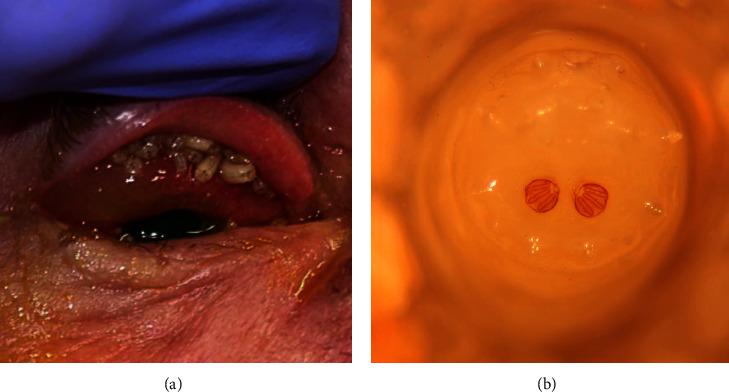 Figure 1
- —National Science Foundation
- —College of Veterinary Medicine, Kansas State University
- —Carolinas
Peer Reviews
No public reviews on file for this paper yet. If you reviewed it on a platform where reviews are public (OpenReview, ICLR, NeurIPS, ICML), you can paste yours below so the community can read it here.
Videos
No videos yet. Explain this paper in a talk, walkthrough, or lecture? Add one.
Taxonomy
TopicsForensic Entomology and Diptera Studies · Yersinia bacterium, plague, ectoparasites research · Dermatological diseases and infestations
1. Introduction
Myiasis is a type of parasitism that results from dipterous larvae developing in live vertebrate tissue. Larvae across many species within the fly family Calliphoridae—commonly known as blow flies—are known for having diverse feeding habits, ranging from necrophages feeding on decayed matter (including human bodies), to obligate parasites feeding exclusively on live tissue, and facultative parasites which can feed on live or dead animal matter (depending on environmental conditions). For species reported to cause obligate parasitism, adult flies will commonly lay their eggs in moist areas of tissue, such as wounds or entrances to the body, and the larvae will feed on the tissue to continue through development.
Ophthalmomyiasis is an uncommon and underdocumented phenomenon due to surrounding stigmas. This condition specifically refers to the infestation of (peri-)ocular tissue by fly larvae. This is most commonly known to occur with the larvae of the human botfly (Dermatobia hominis) (e.g., [1, 2]) and the sheep nasal botfly (Oestrus ovis) (e.g., [3, 4]) but has been documented in other fly genera (e.g., Lucilia (Calliphoridae) and Sarcophaga (Sarcophagidae)) [4]. Most patients present with ophthalmomyiasis externa, referring to larvae infestation of the external structures of the eye, including the cornea and conjunctiva. Some patients may progress to ophthalmomyiasis interna, when larvae penetrate the eye, although this is rare [5]. The condition is most commonly observed in (sub)tropical regions and is associated with poor hygiene and close animal contact. Other common risk factors for ophthalmomyiasis include ocular wounds, advanced age, debilitation, and poor access to nutrition and care. Given the rarity and health impacts of ophthalmomyiasis, when cases are encountered in practice, it is important to identify the species present.
Morphological identification is the gold standard method of taxonomic identification of a range of organisms including insects. Although reliable and low-cost, there are two major hurdles to the routine identification of blow fly larvae using morphology: (a) there are a limited number of experts globally and (b) the majority of species lack distinguishing characteristics visible using a standard light microscope. Given this, it can be difficult to reliably identify blow fly larvae, especially at the species level. DNA analysis can be used as a supplemental or alternative method for taxonomic identification. Typically, regions from the mitochondrial genome (mtGenome) are targeted to permit DNA-based species identifications in insects, including blow flies, given (a) the high copy number of the mtGenome means recovery of mitochondrial DNA (mtDNA) is likely even in highly compromised samples, (b) the mtGenome shows accelerated rates of evolution which can be harnessed to permit discrimination even among closely related taxa, and (c) there are sufficient mitochondrial sequences available in public databases (e.g., GenBank [6] and BOLD [7]) to allow comparisons of unknowns [8].
In this paper, we document a case of ophthalmomyiasis and outline the DNA-based and morphological approaches implemented to identify the blow fly species present.
2. Materials and Methods
2.1. Case Report
A 59-year-old Caucasian male was referred to a tertiary-care center in Philadelphia, PA, for right eye pain and redness with mild blurry vision. The patient complained of “bugs crawling on my face” and eye redness for approximately one week. Of note, the patient had previously been treated for clinical alcoholism and was unhoused at the time of presentation.
On first presentation, multiple white larvae were noted to crawl out of the patient's right eye when the eye was manipulated. The patient's vision was measured to be 20/30 in the right eye and 20/20 in the left eye by Snellen acuity. External examination showed 1-2+ injection and chemosis. On upper eyelid eversion, approximately 30 to 40 larvae were noted to emerge from the patient's upper fornix (Figure 1(a)). Lower forniceal exam was normal. With fluorescein staining, most of the superior conjunctiva and approximately 80% of the cornea were found to have an epithelial defect without corneal stromal infiltrate or thinning. Pupil examination, extraocular motility, intraocular pressure, and anterior segment examination were normal. Dilated fundoscopic examination was normal, without evidence of intraocular infestation by larvae.
The patient was then given topical anesthesia, and all visible larvae were removed bedside with blunt tip forceps. Approximately 40 living larvae were collected in specimen cups. Upper and lower fornices were swept with a cotton-tip applicator multiple times to ensure no larvae remained. The patient was then treated with aggressive applications of antibiotic ointment every 3 hours to promote reepithelialization and to suffocate any remaining larvae. The patient remained inpatient for 3 days, and each day was examined with improving clinical exam and without evidence of further larvae infection. On the last day of admission before signing out of the hospital against medical advice, the patient's epithelium had completely healed, conjunctival injection and chemosis had resolved, and the patient's vision returned to 20/20 by Snellen acuity. The patient did not return for outpatient follow-up examination.
2.2. Identification of Larvae
2.2.1. DNA Analysis
Five larvae were selected for DNA analysis that represented diversity with regard to size and developmental stage (e.g., second- or third-instar; Table 1). Using a sterile scalpel, the anterior third of each larva was dissected and used as an input for DNA isolation using the DNeasy Blood and Tissue Kit (Qiagen, Hilden, Germany) according to the manufacturer's instructions [9]. The quantity of total genomic DNA recovered from each larva was determined using the Qubit 3 Fluorometer (Invitrogen, Waltham, MA, USA) and the Qubit dsDNA kit (Invitrogen). The mtGenome of each sample was sequenced to permit taxonomic identification and briefly involved the following steps: (1) DNA libraries were constructed from isolated total genomic DNA using half reactions of the KAPA Hyper Prep Kit (Roche, Basel, Switzerland) and KAPA Illumina compatible unique dual-indexed adapter kit (Roche) following the manufacturer's recommendations [10, 11], (2) mtDNA was enriched using a custom hybridization capture panel designed off Calliphora vicina (Calliphoridae; GenBank accession NC_019639.1; Daicel Arbor Biosciences, Ann Arbor, MI, USA) following the “High Sensitivity” protocol in the Hybridization Capture for Targeted NGS manual [12] without modification, (3) libraries enriched for the mtGenome were sequenced on an Illumina MiniSeq (Illumina, San Diego, CA, USA) using a Mid Output kit (2 X 150 paired end sequencing), and (4) raw sequence reads were imported into the CLC Genomics Workbench (Qiagen) and the mtGenome for each sample was assembled using a modified pipeline from Molto et al. [13] using reference mtGenomes from various blow fly species. Published primers [14] were used to locate the cytochrome oxidase subunit I (COI) barcode region from each mtGenome alignment, given this region is often used for species identification in insects. Both the mtGenome and COI barcode regions from each sample were individually searched against GenBank using the default parameters of the Standard Nucleotide Basic Local Alignment Search Tool (blastn). To interpret the blastn results and determine the appropriate level of taxonomic assignment, the Organization of Scientific Area Committees proposed standard 2021-S-0006 Standard for the Use of GenBank for Taxonomic Assignment of Wildlife [15] was consulted. The remaining posterior two-thirds of each larva were subjected to morphological examination for species identification.
2.2.2. Morphology
The larval remains were identified using published figures of third instar larvae of Calliphoridae [16], a pictorial key to fly larvae of public health importance [17], and personal notes from blow fly rearings by G. Dahlem. Only the posterior portion of the larvae was available for morphological study, as the anterior portion was destroyed for the DNA analysis. This meant that structures such as the cephalopharyngeal skeleton and the prothoracic spiracles were not available for examination. Features of the posterior spiracular pit and posterior spiracles were available, and taxonomic identifications were based on these larval features. Morphological identification was completed in a blinded fashion; details of DNA analysis were not provided to prevent biasing of the morphological results.
3. Results
For each sample, a complete mtGenome (excluding the variable control region) was recovered totaling >14,900 base pairs in length (Table 1). When utilizing the mtGenomes to search GenBank, three larvae were taxonomically identified at the species level as Lucilia coeruleiviridis (Calliphoridae; ID # 2, 4, 5; Table 1) and two as Phormia regina (Calliphoridae; ID # 1 and 3; Table 1). The percentage of identical bases for each mtGenome generated in this study when compared to the sequences contained within GenBank was ≥99%. These assignments were further verified by phylogenetic placement, whereby the mtGenomes from the five larvae specimens in this study clustered with reference mtGenomes from L. coeruleiviridis and P. regina downloaded from GenBank (results not shown). When utilizing only the 658-bp COI barcode region for taxonomic assignment, sample ID #1 and 3 were also assigned to the species level based on the blastn results as P. regina (Table 1). However, genus-level assignment to Lucilia was only possible for larvae ID # 2, 4, and 5; there are multiple Lucilia species whose COI barcode region sequences are 100% identical, including L. coeruleiviridis, L. mexicana, and L. eximia, making species level assignment using only this region not possible.
Morphological identification was completed on the posterior two-thirds of each of the five larvae subjected to DNA analysis (Table 1). The specimen representing sample ID #1 was a second instar larva, and no figures were available to identify that specimen to the species level; thus, no taxonomic identification was rendered for this sample (Table 1). The remaining samples were all third instar larvae, which could be compared to published illustrations. Morphologically, samples ID #2, 4, and 5 (Table 1) corresponded to L. coeruleiviridis using the structure of the posterior spiracles (Figure 1(b)). Sample ID #3 (Table 1) was initially identified as L. coeruleiviridis because it showed a complete peritreme. Published figures of P. regina posterior spiracles indicate an incomplete peritreme, not enclosing the button. The specimen of sample ID #3 was more lightly sclerotized, which may indicate that it recently molted to the third instar and may account for the discrepancy with respect to the peritreme. A published photo of a third instar P. regina larva [18] seems to show a complete peritreme, which is different from what has been illustrated in other publications. Considering this variability, we could not confidently identify sample ID #3 to the species level, and thus, it was only classified as Calliphoridae (Table 1).
4. Discussion
Healthy individuals who have average hygiene are not likely to experience myiasis. Ophthalmomyiasis is an uncommon form of myiasis and, to our knowledge, finding two blow fly (Calliphoridae) species in the same individual has not been previously reported in the scientific literature. Moreover, Phormia and Lucilia species are from separate Calliphoridae subfamilies, Chrysomyinae and Luciliinae, respectively. Neither P. regina nor Lucilia sp. prefers living tissue over decaying tissue for larval growth development [19]. However, P. regina and L. coeruleiviridis are competitors, reported to fill the same niche in the same environment [20].
Our results highlighted that the COI barcode region did not permit species-level resolution among closely related Lucilia species. Given it has been previously reported that there is not enough genetic variability to distinguish between L. coeruleiviridis and L. mexicana using only the COI barcode region [21], reduced taxonomic resolution with this region was not unexpected in this study. Notably, L. coeruleiviridis is most commonly found in the eastern part of the United States (where the patient in this study resided), whereas closely related L. mexicana and L. eximia are mostly found in the western United States and Central/South America, respectively [22–24]. These observations suggest, in concordance with our mtGenome results, that the three Lucilia larvae are most likely L. coeruleiviridis.
The morphological identification of the blow fly larvae, which is difficult for even an experienced entomologist, gave consistent results for the three samples we found belonging to the Lucilia genus. However, the two samples identified as P. regina using DNA analysis could not be confirmed using morphological identification as they were especially small, one being a second instar and the other may have recently molted to third instar. In future ophthalmomyiasis cases, it would be beneficial to perform the morphological identification before destructive DNA analysis. This would ensure that all informative features of the larva useful for taxonomic assignment could be examined. Despite this, we concluded that it was beneficial to present the results of the morphological identification of the posterior two-thirds of each of the five larvae to support our DNA analysis results, due to the rarity of finding two species in the same case.
5. Conclusion
This is the first reported instance in which two different genera of blow flies were found in the same human ophthalmomyiasis case. Using mtGenome sequences, we were able to taxonomically assign the larvae to either P. regina or L. coeruleiviridis; these results were largely confirmed via COI analysis and morphology. Ocular myiasis is rare but preventable and treatable. Education of both patients and providers around the world is necessary to decrease the number of myiasis cases. Identifying the myiasis agent furthers forensic and entomological research, as well as informs medical professionals on species that could cause harm to patients.
The reference list from the paper itself. Each links out to its DOI / PubMed record.
- 1Emborsky M. E. Faden H. Ophthalmomyiasis in a child The Pediatric Infectious Disease Journal 2002211828310.1097/00006454-200201000-000242-s 2.0-003614297811791112 · doi ↗ · pubmed ↗
- 2Bangsgaard R. Holst B. Krogh E. Heegaard S. Palpebral myiasis in a Danish traveler caused by the human bot-fly (Dermatobia hominis) Acta Ophthalmologica Scandinavica 200078448748910.1034/j.1600-0420.2000.078004487.x 2-s 2.0-003386574210990060 · doi ↗ · pubmed ↗
- 3Jordan V. Mowatt L. Ophthalmomyiasis externa due to sheep nasal botfly in rural Jamaica Tropical Doctor 2019491484910.1177/00494755188096032-s 2.0-8505956870130419775 · doi ↗ · pubmed ↗
- 4Singh G. Chauhan S. Sood A. Thakur K. Vidi, vini, vinci: external ophthalmomyiasis infection that occurred, and was diagnosed and treated in a single day: a rare case report Oman Journal of Ophthalmology 20092313013210.4103/0974-620X.5731320927210 PMC 2903918 · doi ↗ · pubmed ↗
- 5Martinez-Rojano H. Huerta H. Sámano R. Chico-Barba G. Mier-Cabrera J. Plascencia-Nieto E. S. Ophthalmomyiasis externa and importance of risk factors, clinical manifestations, and diagnosis: review of the medical literature Diseases 2023114 p. 18010.3390/diseases 1104018038131986 PMC 10743177 · doi ↗ · pubmed ↗
- 6NCBI Gen Bank overview 2022 https://www.ncbi.nlm.nih.gov/genbank/
- 7Ratnasingham S. Hebert P. D. N. bold: the Barcode of Life Data System (http://www.barcodinglife.org) Molecular Ecology Notes 20077335536410.1111/j.1471-8286.2007.01678.x 2-s 2.0-3424814994318784790 PMC 1890991 · doi ↗ · pubmed ↗
- 8Meiklejohn K. A. Burnham-Curtis M. K. Straughan D. J. Giles J. Moore M. K. Current methods, future directions and considerations of DNA-based taxonomic identification in wildlife forensics Forensic Science International: Animals and Environments 2021110003010.1016/j.fsiae.2021.100030 · doi ↗