Complex presentation of a left Fronto-zygomatic Dermoid cyst; a case report
Laith A Ayasa, Sara Rahhal, Alaa Khaled Najjar, Mohammed Aliwaiai, Asad Aldarawish, Izzeddin Bakri

TL;DR
A 50-year-old woman with persistent headaches was found to have a rare dermoid cyst in her craniofacial region, successfully removed through surgery.
Contribution
This case report highlights the importance of considering dermoid cysts in the differential diagnosis of craniofacial lesions.
Findings
A dermoid cyst was successfully excised via craniotomy in a patient with refractory headaches.
Histopathological analysis confirmed the diagnosis of a fronto-zygomatic dermoid cyst.
The case emphasizes the variable clinical presentation of dermoid cysts.
Abstract
We present a case of craniofacial dermoid cyst in a 50-year-old female. The patient's complaint was persistent refractory headaches with no other significant neurological symptoms. Diagnostic imaging revealed the presence of a lesion in the left fronto-zygomatic region. Surgical intervention involved a craniotomy that led to a successful excision of the dermoid cyst. The diagnosis was subsequently confirmed by histopathological analysis. This case underscored the importance of considering DC as a potential diagnosis for any craniofacial lesion, given their diverse presentations and associated complications.
Genes, proteins, chemicals, diseases, species, mutations and cell lines named across the full text — each resolved to its canonical identifier and authoritative record.
Click any figure to enlarge with its caption.
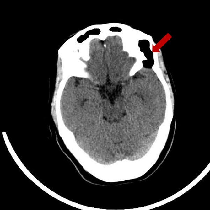 Figure 1
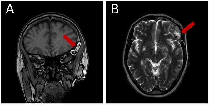 Figure 2
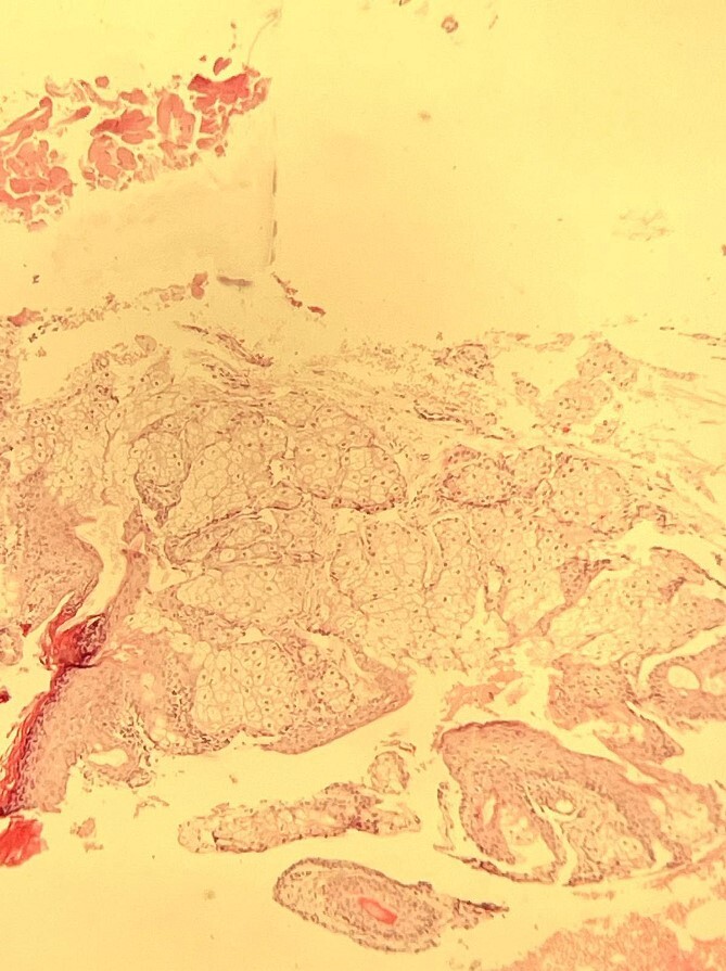 Figure 3
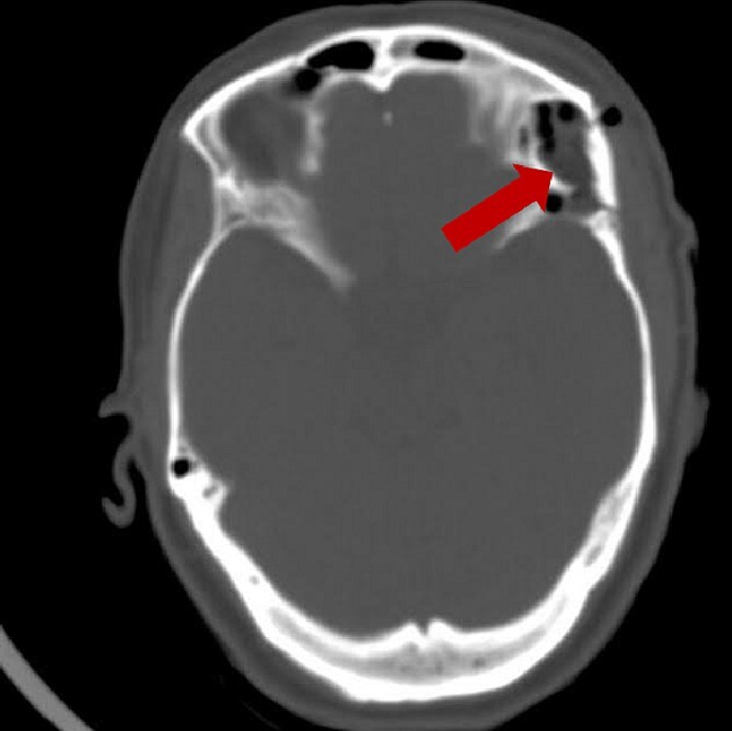 Figure 4
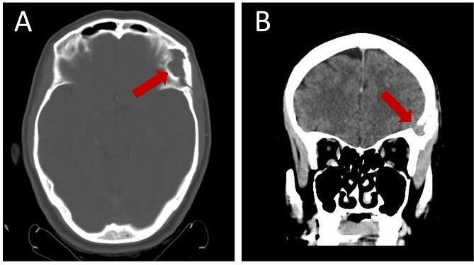 Figure 5
Peer Reviews
No public reviews on file for this paper yet. If you reviewed it on a platform where reviews are public (OpenReview, ICLR, NeurIPS, ICML), you can paste yours below so the community can read it here.
Videos
No videos yet. Explain this paper in a talk, walkthrough, or lecture? Add one.
Taxonomy
TopicsTeratomas and Epidermoid Cysts · Fetal and Pediatric Neurological Disorders · Spinal Dysraphism and Malformations