Transient activation of retinopathy of prematurity secondary to erythrocyte suspension transfusion
Alparslan Şahin

TL;DR
This case report shows that giving red blood cell transfusions to premature infants can temporarily worsen a serious eye condition, but it often resolves on its own.
Contribution
The novelty lies in demonstrating that ROP progression after erythrocyte suspension transfusion can be transient and self-resolving in some cases.
Findings
Two premature infants developed type 1 ROP after erythrocyte suspension transfusion.
Clinical findings resolved within a few days without intervention.
Observation for 24-48 hours is suggested before deciding on treatment.
Abstract
Retinopathy of prematurity (ROP) is a serious retinal vascular disorder that needs prompt diagnosis, and treatment to prevent undesired visual outcomes. Due to its shorter period of disease progression, it is important to be hasty in treating ROP. Erythrocyte suspension (ES) aggravates the progression of ROP. However, this progression may be transient as in the present case reports. This case report aimed to present two cases that developed type 1 ROP after erythrocyte suspension transfusion. Clinical findings of the patients were resolved within a few days without any intervention. Premature infants receiving ES treatment can be observed for 24-48 hours, and the treatment can be planned after determining the persistence of the plus sign. Abbreviations: ES = Erythrocyte suspension, ROP = Retinopathy of prematurity, NICU = neonatal intensive care unit
Genes, proteins, chemicals, diseases, species, mutations and cell lines named across the full text — each resolved to its canonical identifier and authoritative record.
Click any figure to enlarge with its caption.
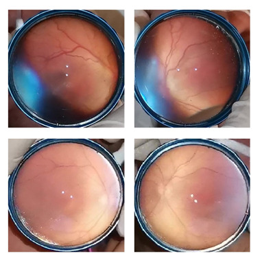 Fig. 1
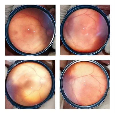 Fig. 2
Peer Reviews
No public reviews on file for this paper yet. If you reviewed it on a platform where reviews are public (OpenReview, ICLR, NeurIPS, ICML), you can paste yours below so the community can read it here.
Videos
No videos yet. Explain this paper in a talk, walkthrough, or lecture? Add one.
Taxonomy
TopicsRetinopathy of Prematurity Studies · Neonatal Health and Biochemistry · Neonatal Respiratory Health Research
Introduction
Retinopathy of prematurity (ROP) is a serious retinal vascular disorder that may cause blindness. Several risk factors are defined for the progression of ROP. Erythrocyte suspension (ES) transfusion is a well-defined risk factor for ROP progression. Ninety percent of premature infants will get at least one ES transfusion [1]. Zhu et al. reported that ES transfusion is an independent risk factor for ROP progression, particularly in extremely preterm infants [2]. It has been reported that the incidence of ROP was 1.68 times higher in the ES transfusion group than in those of the non-transfused group of premature infants [3].
Prompt diagnosis and treatment are important to prevent devastating visual outcomes. Although it is necessary to be hasty in treating the disease, in some cases the delay in treatment may result in favor of the patient, as in the two case reports mentioned below. This report presented two cases with the diagnosis of type 1 ROP after ES transfusion that resolved within a few days after diagnosis without any surgical intervention.
Case reports
Case 1
A 26-week-old infant with a birthweight of 790 grams was hospitalized in the neonatal intensive care unit (NICU). The first ocular examination of the patient was performed on the 28th day of age. It revealed bilateral mild vitreous haze, but no plus disease, and zone I immature retina without any stage of ROP. Consequent two examinations revealed the same findings other than resolving vitreous haze. Unfortunately, the fourth examination revealed a significant plus disease with stage 1 disease in zone I. Hence, bilateral intravitreal injection of bevacizumab was recommended. However, the treatment of the patient was delayed because of waiting for the legal guardians’ consent and lack of drug availability. Two days after diagnosis, a bilateral injection of bevacizumab was scheduled. Before injection, the retinal examination was performed. It was observed that the bilateral plus sign significantly disappeared with stable retinal disease as stage 1 in zone I. Because of the detailed examination of the patient’s medical history, it was observed that the patient was given an ES transfusion a day before the appearance of the plus disease. Treatment was cancelled. Consequently, the follow-up examinations did not show progression of ROP. Retinal vascularization was completed without any intervention (Fig. 1).
The fundus images of case 1. Note the plus sign just a day after ES transfusion (Top). Two days after this examination the plus sign recovered without any intervention (Bottom)
Case 2
A 27-week-old infant with a birthweight of 1000 grams was followed in the NICU. The first two examinations did not reveal type 1 ROP (bilateral zone I immature retinal vascularization without plus sign). The third examination revealed bilateral zone I stage 1 disease with plus disease. The patient had received an ES transfusion two days before this examination. Hence, a closer follow-up was recommended. Two days later, the examination revealed complete recovery of plus sign with zone I stage 1 disease. The patient was followed up closely. The ROP was regressed and complete retinal vascularization was observed without any complication (Fig. 2).
The fundus images of case 2. The plus sign 2 days after ES transfusion (Top). Plus sign disappeared two days after the previous examination (Bottom)
Discussion
There are well-defined risk factors for the development of ROP [4]. Transfusion of ES is one of the reported risk factors [2]. ES transfusion is usually needed if the infants’ vital hemodynamic findings are impaired. Almost all extreme premature infants will get at least one ES transfusion [5]. ES transfusion primarily affects ROP through two mechanisms [6]. Because of their immature antioxidant system and high transferrin saturation, preterm newborns are particularly susceptible to the oxidative stress induced by free iron overload. Secondly, HbA, adult hemoglobin, has a lower affinity for oxygen than that of HbF. Therefore, it may lead to more release of oxygen to the tissues due to its lower affinity.
The present case reports indicated that ES transfusion aggravated the plus sign that needs treatment in infants with ROP. Besides, waiting for a few days after ES transfusion might prevent unnecessary treatments in these infants. It should be kept in mind that ES transfusion may cause transient acceleration of the clinical findings of ROP due to ES transfusion, especially plus sign, and the infants should be followed closely. It can be interpreted that a few days after blood transfusion, the plus sign may improve as these babies stabilize hemodynamically.
Conclusion
In the case of ROP progression, factors that worsen clinical findings should be questioned before treatment. Patients receiving ES treatment can be observed for 24-48 hours, and the treatment can be planned after determining the persistence of the plus sign. However, close follow-up is warranted after ES transfusion.
Conflict of Interest Statement
The author declares no conflict of interest.
Informed Consent and Human and Animal Rights Statement
Informed consent has been obtained from all individuals included in this study. Minor patient consent to publish the case and images were gathered.
Authorization for the use of human subjects
Ethical approval: The research related to human use complies with all the relevant national regulations and institutional policies, is by the tenets of the Helsinki Declaration, and has been approved by the Ethical Committee of Bower Hospital, Diyarbakir, Turkey (2023/0901, 15.09.2023).
Acknowledgments
None.
Sources of Funding
The author received no financial support for the research, authorship, and/or publication of this article.
Disclosures
None.
The reference list from the paper itself. Each links out to its DOI / PubMed record.
- 1Strauss RG Anaemia of prematurity: pathophysiology and treatment Blood Rev 2010242212252081736610.1016/j.blre.2010.08.001PMC 2981681 · doi ↗ · pubmed ↗
- 2Zhu Z Hua X Yu Y Zhu P Hong K Ke Y Effect of red blood cell transfusion on the development of retinopathy of prematurity: A systematic review and meta-analysis P Lo S One 202015 e 02342663251258210.1371/journal.pone.0234266 PMC 7279893 · doi ↗ · pubmed ↗
- 3Ludwig CA Chen TA Hernandez-Boussard T Moshfeghi AA Moshfeghi DM The epidemiology of retinopathy of prematurity in the United States Ophthalmic Surgery Lasers and Imaging Retina 20174855356210.3928/23258160-20170630-0628728176 · doi ↗ · pubmed ↗
- 4Kim SJ Port AD Swan R Campbell JP Chan RVP Chiang MF Retinopathy of prematurity: a review of risk factors and their clinical significance Surv Ophthalmol 2018636186372967961710.1016/j.survophthal.2018.04.002PMC 6089661 · doi ↗ · pubmed ↗
- 5Maier RJ Sonntag J Walka MM Changing practices of red blood cell transfusions in infants with birth weights less than 1000 g J Pediatr 20001362202241065782910.1016/s 0022-3476(00)70105-3 · doi ↗ · pubmed ↗
- 6Stutchfield CJ Jain A Odd D Williams C Markham R Foetal haemoglobin, blood transfusion, and retinopathy of prematurity in very preterm infants: A pilot prospective cohort study Eye 201731145114552854865110.1038/eye.2017.76PMC 5639193 · doi ↗ · pubmed ↗