Targeted Six-Week Intensive Physiotherapy for a Case of Tuberculous Meningitis With a Syndrome of Inappropriate Antidiuretic Hormone Secretion
Arjavi A Pakhan, Raghuveer Raghumahanti

TL;DR
This case report shows how a six-week intensive physiotherapy program improved the quality of life and functional abilities of a patient with severe tuberculous meningitis.
Contribution
The study demonstrates the effectiveness of early and targeted physiotherapy in managing complications of tuberculous meningitis.
Findings
The patient showed significant improvement in muscle strength and independence in daily activities after six weeks of physiotherapy.
Early physiotherapy intervention helped maintain joint integrity and improve postural strength in a TBM patient.
The program enhanced the patient's ability to perform activities of daily living and swallow independently.
Abstract
Tuberculous meningitis (TBM) is a severe form of extrapulmonary tuberculosis (TB) characterized by the invasion of Mycobacterium tuberculosis into the meninges surrounding the brain and spinal cord. It triggers an intense inflammatory response, leading to neurological complications if not promptly and adequately managed. TBM often precipitates muscle weakness, neurological deficits, respiratory challenges, swallowing difficulties, joint contractures, and pain. Physiotherapy intervention is essential in treating these problems by personalized treatment strategies and treatment plans to enhance muscle strength, motor control, coordination, and overall mobility. This case report aims to highlight the significant role of physiotherapy in improving the quality of life (QOL) and functional abilities of patients with TBM. The current case report reviews the case of a 73-year-old male who…
Genes, proteins, chemicals, diseases, species, mutations and cell lines named across the full text — each resolved to its canonical identifier and authoritative record.
Click any figure to enlarge with its caption.
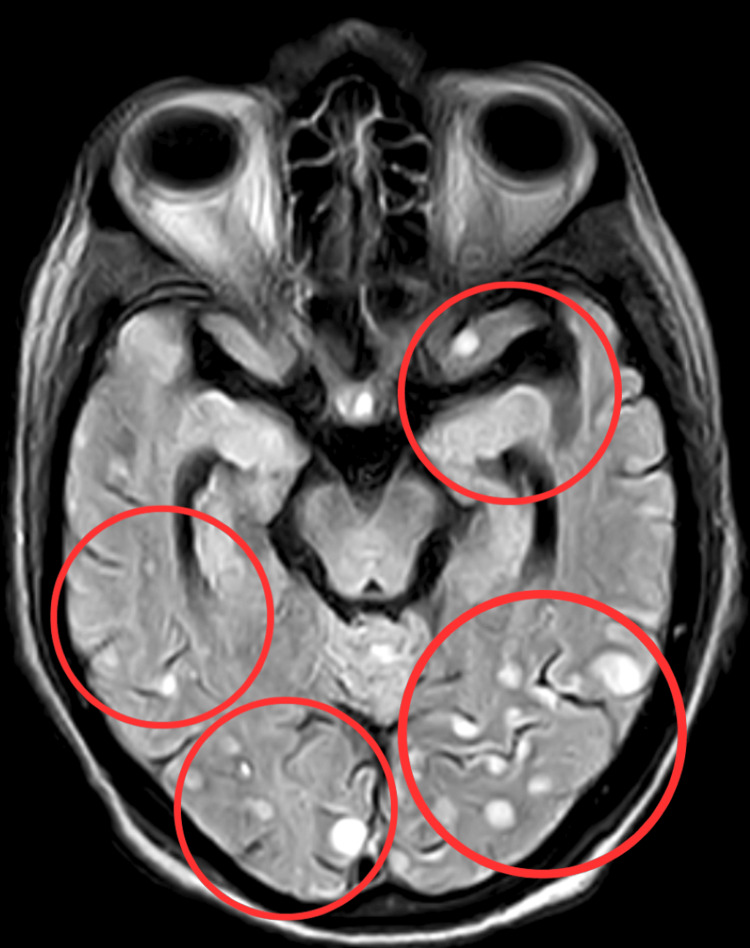 Figure 1
 Figure 2
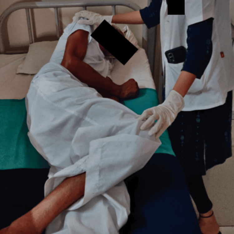 Figure 3
| MMT | Pre-treatment | Post-treatment | ||||
| Muscles | Right | Left | Right | Left | ||
| Shoulder | ||||||
| Flexors | 3/5 | 3/5 | 4/5 | 4/5 | ||
| Extensors | 3/5 | 3/5 | 4/5 | 4/5 | ||
| Abductors | 3/5 | 3/5 | 4/5 | 4/5 | ||
| Adductors | 3/5 | 3/5 | 4/5 | 4/5 | ||
| Elbow | ||||||
| Flexors | 3/5 | 3/5 | 4/5 | 4/5 | ||
| Extensors | 3/5 | 3/5 | 4/5 | 4/5 | ||
| Wrist | ||||||
| Flexors | 3/5 | 3/5 | 4/5 | 4/5 | ||
| Extensors | 3/5 | 3/5 | 4/5 | 4/5 | ||
| Hip | ||||||
| Flexors | 2/5 | 2/5 | 3/5 | 3/5 | ||
| Extensors | 2/5 | 2/5 | 3/5 | 3/5 | ||
| Abductors | 2/5 | 2/5 | 3/5 | 3/5 | ||
| Knee | ||||||
| Extensors | 2/5 | 2/5 | 3/5 | 3/5 | ||
| Flexors | 2/5 | 2/5 | 3/5 | 3/5 | ||
| Ankle | ||||||
| Dorsiflexors | 2/5 | 2/5 | 3/5 | 3/5 | ||
| Plantarflexors | 2/5 | 2/5 | 3/5 | 3/5 | ||
| Problems faced by the patient | Goals | Intervention | Rationale | ||
| 0-2 weeks | 2-4 weeks | 4-6 weeks | |||
| Respiratory complications | To prevent respiratory complications | Initiate deep breathing exercises, targeting the diaphragmatic and thoracic regions, involving inhalation and exhalation for three sets of 10 breaths. In week 2, further introduce longer breath-hold durations to enhance respiratory endurance | Additional deep breathing techniques (diaphragmatic and pursed lip breathing) to further enhance respiratory capacity | Incentive spirometry, to further enhance respiratory capacity | Increase the oxygen level. Help restore the diaphragmatic function of muscle |
| Other secondary complications | To prevent bed sores, contracture, and circulatory problems | Repositioning every two hours to prevent pressure ulcers and DVT. Employed supportive pillows and sandbags. In week 2, continue these practices while educating the patient on self-repositioning techniques to enhance engagement and maintain optimal limb elevation | Continue emphasizing proper positioning every two hours to prevent pressure ulcers and DVT. Reinforce the importance of limb elevation and educate the patient on self-repositioning techniques | Deep breathing exercises and effective coughing techniques. Regular practice of these techniques is emphasized to prevent chest complications | To prevent DVT, contracture, and circulatory complications |
| Muscle weakness | To improve muscle strength | Initiated isometric exercises for key muscle groups (biceps, triceps, deltoid, quadriceps) at a light to moderate intensity. Perform 1-2 sets of 10-15 repetitions. In week 2, maintain the current exercises with the same resistance level or consider progressing to bodyweight squats or step-ups for the lower limb muscles | Maintain current strength training exercises with a focus on the lower limb muscles. Perform two sets of 10-15 repetitions. In week 3, consider introducing bodyweight squats or step-ups for the lower limbs. Exercises are conducted at a light to moderate intensity | Progress the lower limb strength training exercises by introducing resistance bands or light dumbbells. Focus on exercises that target the quadriceps, hamstrings, glutes, and calf muscles. Perform 2-3 sets of 10-15 repetitions at a light to moderate intensity | Enhance the QOL. Enhance mobility pain management |
| Difficulty in movement | To maintain joint integrity | Passive and active assisted movements for bilateral upper and lower limbs, consisting of 10 repetitions for each movement to enhance joint ROM. In week 2, maintain these exercises while gradually increasing the number of repetitions to challenge the patient's mobility | Continue mobility with joint exercises while considering more advanced procedures, such as pendulum swings and joint mobilization techniques | Continue with joint mobility exercises and introduce advanced techniques like contract-relax stretching and PNF to further enhance the ROM | Improve muscle strength and prevent joint stiffness |
| Difficulty in transfer | To initiate bed mobility and to make patient transfer | Mobility training-bed rolling, bedside sitting, and wheelchair mobility exercises aimed at promoting functional independence | Continue with mobility training and make it more challenging to help the patient become more independent and safer in everyday tasks | Maintain the current mobility exercises and introduce more challenging activities that simulate daily tasks, including step-ups and controlled walking on even surfaces | Maintain their independence in ADLs and improve mobility |
| Poor balance and coordination | To balance independently | Balance training with a 30-minute elevation of the bed head to challenge balance and stability. In week 2, maintain this regimen while incorporating more advanced balance exercises, such as single-leg balance and proprioception activities | Maintain the 30-minute bed head elevation and incorporate more challenging balance exercises, such as tandem stance and proprioception activities, to further enhance stability and coordination | Continue with balance exercises and advance to dynamic balance activities, like walking on uneven surfaces or using balance boards, to further enhance stability and coordination | Improve stability, to reduce the risk of fall |
| Sitting difficulty | To sit independently | Assess the patient's sitting difficulty and initiate bedside sitting with maximum support for five minutes. In week 2, continue with bedside sitting while progressively reducing support, aiming for increased independence | The duration to build the patient's sitting endurance. In week 3, work on achieving independent sitting without any support | Focus on achieving independent sitting without support. Gradually increase the duration of sitting exercises to enhance endurance and overall functional independence | Sitting independently without any support |
| Difficulty in swallowing | To improve swallowing | Changing food textures, optimizing posture during meals, and using adaptive tools | Based on patient feedback and needs, optimize food textures, posture during meals, and adaptive tools to enhance the swallowing process | Incorporate more complex food textures and optimize mealtime posture | Enhancement of nutritional status. To prevent aspiration. To prevent choking |
| Outcome measures | Pre-treatment | Post-treatment |
| Functional Outcome Swallowing Score | 3/4 | 1/4 |
| Barthel Index of Activities of Daily Living Scale | 0/0 | 40/100 |
| Expanded ICU Mobility Scale and Intensity Classification | 0/6 | 4/6 |
| Quality of Life Scale | 57/112 | 69/112 |
Peer Reviews
No public reviews on file for this paper yet. If you reviewed it on a platform where reviews are public (OpenReview, ICLR, NeurIPS, ICML), you can paste yours below so the community can read it here.
Videos
No videos yet. Explain this paper in a talk, walkthrough, or lecture? Add one.
Taxonomy
TopicsInfectious Diseases and Tuberculosis · Orthopedic Infections and Treatments · Hematological disorders and diagnostics
Introduction
Tuberculous meningitis (TBM), miliary tuberculosis (TB), and multiple tuberculomas represent distinct yet interconnected clinical entities, each stemming from the insidious Mycobacterium tuberculosis infection. These TB manifestations reflect this pathogen's remarkable ability to disseminate through the bloodstream and lymphatic system, causing diverse clinical presentations with intricate diagnostic and therapeutic challenges [1]. TBM is one of the most common and serious forms of TB in India. India has the highest burden of TB in the world, accounting for over 25% of global cases. Syndrome of inappropriate antidiuretic hormone secretion (SIADH) is a known complication of TBM, occurring in 5-30% of cases. Mycobacterium tuberculosis infiltrates the meninges around the brain and spinal cord, resulting in a severe type of extrapulmonary TB known as TBM. This invasion triggers an intense inflammatory response, leading to neurological complications if not promptly and adequately managed [2]. TBM is notorious for its subtle initial symptoms, which often include fever, headache, and altered mental status, progressing to more severe manifestations such as cranial nerve deficits and focal neurological signs [3]. Early diagnosis and initiation of anti-tubercular therapy are pivotal for favorable outcomes, as delays can result in significant morbidity and mortality [4].
Hematogenous spread tuberculous bacilli, which spread widely throughout the body and produce multiple tiny granulomas in different organs, are the hallmarks of miliary TB. Numerous organ systems can be impacted by these minute granulomas, often known as "miliary" lesions, which cause the condition's varied clinical presentations [5]. Individuals may have systemic signs that resemble other infectious or malignant processes, such as fever, weight loss, and sweats at night [6]. Because of its complex clinical picture, miliary TB is often difficult to diagnose. On the other hand, multiple tuberculomas, which typically affect the brain and lungs, are localized accumulations of Mycobacterium tuberculosis within tissues or organs [7]. These tuberculomas may cause localized neurological impairments or pulmonary symptoms, depending on where they are anatomically located [8]. Sometimes, TBM or miliary TB coexists with multiple tuberculomas, complicating the clinical presentation and diagnostic assessment [9,10].
The ensuing cascade of events disrupts the blood-brain barrier, forms caseating granulomas, and releases pro-inflammatory cytokines, ultimately leading to neurological damage [8]. TBM is diagnosed through a combination of clinical assessment and laboratory tests. Lumbar puncture is used to analyze the cerebrospinal fluid (CSF) for biochemical signs of TB infection such as low glucose, elevated proteins, and the presence of TB bacteria. Neuroimaging like computed tomography (CT) or magnetic resonance imaging (MRI) scans can reveal basilar meningeal inflammation indicative of TB infection. Microbiological tests like GeneXpert on the CSF can provide rapid evidence of TB bacteria. Evaluating for accompanying pulmonary TB through chest imaging and sputum evaluation is important for diagnosing and treating TBM. Assessing for exposure history or immunological evidence of latent TB is critical for determining if the meningeal TB represents primary infection or reactivation of old infection. Clinical findings like fevers, mental status changes, and hydrocephalus also aid diagnosis [11]. Physiotherapy is crucial in the multidisciplinary approach to treating patients with TBM [12]. TBM often inflicts severe neurological dysfunction in patients, causing muscle weakness, sensory disturbances, and cognitive impairments [13]. In the comprehensive management of such complications, physiotherapy plays a crucial component in rehabilitation, focusing on physical aspects to restore functional independence and overall quality of life (QOL) [14]. Physiotherapy uses various methods to treat patients with motor deficits, including passive and active range of motion (ROM) exercises [15]. This is important because it prevents complications like joint stiffness and muscle contractures, which are frequently seen in individuals who have been immobilized. Physiotherapy aids in the restoration of mobility and functional independence by progressively increasing the ROM [5].
Moreover, physiotherapists focus on enhancing gait and balance, which are problems that individuals with neurological dysfunction often face. TBM survivors often experience challenges in walking and maintaining proper posture, making them susceptible to falls and related injuries [16]. Through specialized training and exercises, physiotherapy is pivotal in helping patients achieve optimal balance and gait patterns, ultimately enhancing their overall mobility. Sensory deficits represent another neurological issue encountered by TBM patients. These deficits can significantly diminish patients' QOL. Physiotherapists employ sensory re-education techniques to assist patients in regaining sensory perception, such as touch and proprioception. These methods are precious in reinstating normal function and reducing discomfort associated with sensory deficits [17]. The main aim is to highlight the significant role of physiotherapy in improving QOL and functional abilities in a 73-year-old male diagnosed with TBM. A six-week targeted intensive rehabilitation program was initiated based on the patient's impairments.
Case presentation
Patient information
A 73-year-old male with right-hand dominance was admitted to the neurology intensive care unit (ICU) with complaints of generalized weakness and difficulty in swallowing. The patient had a history of recurrent fever occurring every month for the past six months and visited a private clinic where paracetamol was prescribed to him. In September, the patient's condition worsened as he started experiencing severe headaches, recurrent fever, chills, generalized weakness, and body pain. Subsequently, the patient was admitted for further evaluation.
Clinical findings
The first examination was done following the patient's and family's consent. The patient was conscious, cooperative, and well-oriented to time, place, and person. The patient's attitude of the limb was in supine lying, and the upper limb exhibited slight shoulder abduction with elbow flexion and a neutral wrist position while in a supine position. The lower limb showed external hip joint rotation, extended knee, and ankle plantarflexion. The Glasgow Coma Scale (GCS) was E4V5M6 on higher mental function evaluation. The patient was connected to external medical appliances, including a Foley catheter and a Ryles tube. On the sensory examination, the sensory functions in the upper extremities were intact, while superficial reflexes, including fine touch and pin-prick, were absent in the lower extremities. On motor examination, the muscle tone of the upper and lower extremities was normal. According to the Oxford grading system, manual muscle testing (MMT) showed 3/5 in the upper limb, which is full ROM against gravity without resistance; in the lower limb, it was 2/5, which is full ROM in gravity eliminated, shown in Table 1. Kernig's sign was positive. High-resolution CT (HRCT) of the thorax reveals diffusely scattered miliary nodules in bilateral lung parenchyma with a patch area of consolidation in the left lower lobe and mediastinal lymphadenopathy-miliary TB. MRI of the brain reveals evidence of variable-sized round to oval lesions noted in the bilateral cerebral hemisphere and bilateral cerebellar hemisphere. The lesions appear hypointense on TI WI and hyperintense on T2 WI/FLAIR, showing blooming on SWI and no restriction on DWI suggestive of (s/o) partial calcification. There is the prominence of sulco-gyral space, ventricular system, Sylvian fissure, and cerebellar folia s/o age-related atrophic changes, as shown in Figure 1.
MRI of the brainThe red circles show variable-sized round to oval lesions noted in the bilateral cerebral hemisphere and bilateral cerebellar hemisphereMRI: magnetic resonance imaging
Therapeutic intervention
A targeted six-week intensive physiotherapy (TIP-6) intervention was started as soon as the patient was in ICU. Table 2 shows the treatment protocol given to the patient for six weeks. Figure 2 and Figure 3 show the patient being rehabilitated.
Passive ROM exerciseROM: range of motion
Rolling facilitation
Outcome measures
The TIP-6 was carried out for three weeks following the progression outcome measures, which showed improvement after the physiotherapy intervention. Outcome measures are mentioned in Table 3.
Discussion
In TBM, the inflammatory response often leads to muscle weakness and neurological deficits. Physiotherapy interventions play a pivotal role in improving muscle strength and mobility. By engaging in targeted exercises such as isometric muscle training and bodyweight squats, the patient regains muscular strength and re-establishes neural pathways. This neurological reconnection, facilitated by physiotherapy, aids in reversing muscle atrophy and promoting coordinated muscle function, ultimately countering the muscle weakness seen in TBM. Physiotherapy effectively addresses respiratory problems that may occur due to reduced lung function and weaker respiratory muscles in patients with TBM. Exercises that involve deep breathing, incentive spirometry, and other breathing methods aid in increasing lung capacity and regaining the function of the diaphragmatic muscle. Physiotherapy enhances the muscles that control breathing, increases endurance, and improves oxygen exchange. As a result, this intervention improves the patient's respiratory health in general, helping to improve oxygenation and avoiding problems such as pneumonia. Physiotherapy can help with balance and coordination problems, mobility challenges, and transfer difficulties. The exercises promote neural plasticity, allowing the patient to regain functional independence and improve overall mobility. Additionally, balance training fosters neural adaptations, enhancing stability and coordination, thus reducing the risk of falls. Physiotherapy's modifications to food textures and mealtime posture, combined with adaptive tools, help the patient improve their swallowing ability. By optimizing these aspects, physiotherapy ensures that the patient receives proper nutrition and reduces the risk of aspiration. Physiologically, these interventions enhance the coordination of swallowing muscles and reduce the chances of food or liquids entering the airway, ultimately preventing choking and improving the patient's overall nutritional status.
As demonstrated by critical measurements, targeted intensive physiotherapy intervention made a significant difference in the patient's health. Initially, at 0/100, the Barthel Index increased to 40/100 after treatment, showing a substantial improvement in the patient's ability to do daily tasks without assistance. Furthermore, the Functional Outcome Swallowing Score (FOSS) improved from 3/4 to 1/4, indicating substantial progress in the patient's ability to swallow safely without the risk of aspiration. These results highlight the comprehensive impact of physiotherapy on the patient's overall well-being, encompassing gains in muscle strength, mobility, respiratory health, and ADLs. The results of this case report highlight the importance of incorporating rehabilitation programs into the management of TBM patients. Early initiation of rehabilitation, even while patients are in the ICU, can lead to improved outcomes and reduced disability.
The results align with similar studies on rehabilitating TBM patients. Suisan and Thohari presented two severe TBM cases who received a multimodal sensory stimulation program incorporating positioning, ROM exercises, and tactile and auditory stimuli during their ICU stay. After two weeks, both patients showed remarkable progress with GCS scores improving from E1V1M1 to E4V5M6, allowing transfer from ICU to high-dependency unit. The intensive early rehabilitation likely contributed to their neurological and functional improvements [18].
In a study by Wang et al., they discussed a case of an immunocompetent patient who had a brain abscess caused by TB. This patient developed problems like Gerstmann's syndrome and right-sided apraxia. However, following TB treatment and rehabilitation, the patient made a full recovery, resumed their regular activities, and even went back to work. This case highlights the importance of intensive rehabilitation, precise surgery (stereotactic surgery), and effective TB treatment when dealing with such conditions. However, after a meticulously thought-out six-week rehabilitation program, notable progress was observed, including the patient's ability to sit independently without assistance [7].
Conclusions
In this case, the patient had TBM with multiple tuberculoma with miliary TB with SIADH, and physical therapy was started when the patient was in the ICU. TIP-6 interventions improved the patient's abilities and independence while preventing respiratory and secondary complications. Total muscle strength had all improved significantly, as shown in the above scales. Physiotherapy helps the patient to enhance his ADLs and QOL.
The reference list from the paper itself. Each links out to its DOI / PubMed record.
- 1Tuberculous meningitis Stat Pearls [Internet] Slane VH Unakal CG Treasure Island (FL)Stat Pearls Publishing 2022 http://www.ncbi.nlm.nih.gov/books/NBK 541015/31082059 · pubmed ↗
- 2Protective effect of BCG against tuberculous meningitis and miliary tuberculosis: a meta-analysis Int J Epidemiol Rodrigues LC Diwan VK Wheeler JG 11541158221993814429910.1093/ije/22.6.1154 · doi ↗ · pubmed ↗
- 3False-positive tuberculin skin tests: what is the absolute effect of BCG and non-tuberculous mycobacteria?Int J Tuberc Lung Dis Farhat M Greenaway C Pai M Menzies D 11921204102006 https://pubmed.ncbi.nlm.nih.gov/17131776/17131776 · pubmed ↗
- 4Presentation and outcome of tuberculous meningitis in a high HIV prevalence setting P Lo S One Marais S Pepper DJ Schutz C Wilkinson RJ Meintjes G 06201110.1371/journal.pone.0020077 PMC 309827221625509 · doi ↗ · pubmed ↗
- 5Pediatric and adult meningeal, parenchymal, and spinal tuberculosis: a neuroimaging review J Neuroimaging Mertiri L Freiling JT Desai NK Kralik SF Huisman TA 202310.1111/jon.1317738073450 · doi ↗ · pubmed ↗
- 6Tuberculous meningitis with pulmonary miliary tuberculosis: a clinicoradiological study Neurol India Kalita J Misra UK Ranjan P 194196522004 https://pubmed.ncbi.nlm.nih.gov/15269468/15269468 · pubmed ↗
- 7Treatment outcomes of tuberculous meningitis in adults: a systematic review and meta-analysis BMC Pulm Med Wang MG Luo L Zhang Y Liu X Liu L He JQ 2001920193169459910.1186/s 12890-019-0966-8PMC 6833188 · doi ↗ · pubmed ↗
- 8Pathophysiology and prognosis in Vietnamese adults with tuberculous meningitis J Infect Dis Thwaites GE Simmons CP Than Ha Quyen N 1105111518820031455187910.1086/378642 · doi ↗ · pubmed ↗