Ichthyosis Skin Changes in a Patient With Hereditary Hemochromatosis
Neha Arora, Kaycee Nguyen, Andrew Hudson, Lindsay Bicknell

TL;DR
A patient with hereditary hemochromatosis developed ichthyosis-like skin changes, highlighting the need for physicians to recognize dermatologic symptoms of this condition.
Contribution
This case report highlights the under-recognized association between ichthyosis and hereditary hemochromatosis.
Findings
A 56-year-old male with HH presented with brown plate-like scales consistent with ichthyosis vulgaris.
Ichthyosis in HH is not well documented, despite the high prevalence of skin manifestations in this disease.
Physicians should be aware of dermatologic signs of hereditary hemochromatosis for early recognition.
Abstract
Hereditary hemochromatosis (HH) is characterized by elevated iron absorption in the body, leading to iron accumulation with subsequent dysfunction and end-organ damage. While the progression of the disease can result in arthralgias, hepatomegaly, cardiomyopathies, and diabetes, over a third of HH patients present with cutaneous manifestations. We present the case of a 56-year-old male with HH who presented to dermatology with a rash and diffuse scaling. The patient exhibited brown plate-like scales clinically consistent with diffuse ichthyosis vulgaris. While ichthyosis has been seen in patients with idiopathic hemochromatosis, its association with HH is not well reported. Due to the high prevalence of cutaneous involvement in hereditary hemochromatosis, physicians should familiarize themselves with ichthyosis and the other dermatologic manifestations of this disease.
Genes, proteins, chemicals, diseases, species, mutations and cell lines named across the full text — each resolved to its canonical identifier and authoritative record.
Click any figure to enlarge with its caption.
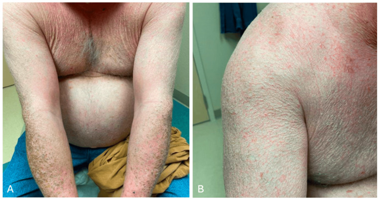 Figure 1
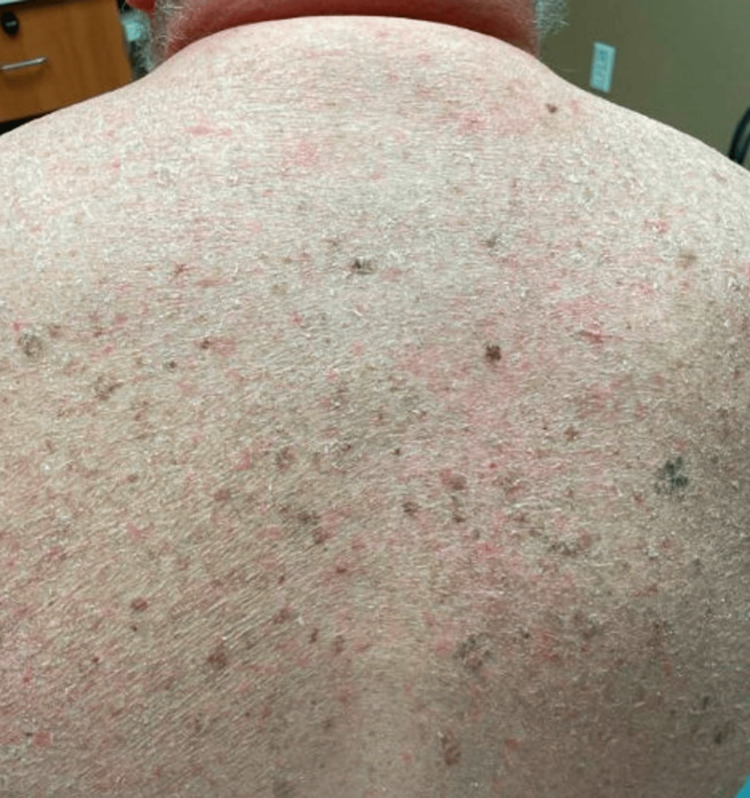 Figure 2
Peer Reviews
No public reviews on file for this paper yet. If you reviewed it on a platform where reviews are public (OpenReview, ICLR, NeurIPS, ICML), you can paste yours below so the community can read it here.
Videos
No videos yet. Explain this paper in a talk, walkthrough, or lecture? Add one.
Taxonomy
TopicsHealth, Education, and Physical Culture
Introduction
Hereditary hemochromatosis (HH) is an autosomal recessive genetic disorder characterized by an imbalance in iron homeostasis. It is the most common inherited single-gene disorder in the American Caucasian population, with at least one in 10 people carrying the mutation [1]. HH is characterized by increased iron absorption and is primarily caused by a mutation in the HFE gene [1]. This mutation impairs the function of hepcidin, leading to pathologic increases in iron storage, which can deposit in tissues throughout the body and lead to systemic symptoms [1]. Since females naturally lose a portion of the excess iron through menstruation, pregnancy, and lactation, the affected males usually develop the disease manifestations earlier than females [2]. The clinical presentation of HH can be vague, and patients often report fatigue and arthralgias as early symptoms. However, the complications associated with this condition include hepatomegaly leading to cirrhosis, cardiomyopathies and dysrhythmias, diabetes, and symmetric arthropathies [2]. With early detection and treatment, the progression of the disease to the liver, heart, and endocrine glands occurs less frequently now, although dermatologic manifestations are still commonly reported.
Case presentation
A 56-year-old male with hereditary hemochromatosis presented with a chronic rash with diffuse scaling throughout his arms (Figure 1), back (Figure 2), scalp, and ears. The patient’s arms exhibited plate-like, or “fish-like,” brown scales resembling diffuse ichthyosis vulgaris. The patient reported dryness and scaliness since childhood, and prior therapy included over-the-counter emollients with minimal relief.
Brown scales present along the patient’s arms and forearms
Diffuse scaling throughout the patient’s back
The patient had been previously found to have compound heterozygosity for mutation in the HFE gene, confirming a diagnosis of HH. The patient’s laboratory results showed elevated hemoglobin, glycosylated hemoglobin (HbA1c), ferritin, total iron-binding capacity, and transferrin. The patient reported undergoing therapeutic phlebotomy for the management of HH, which did not impact the severity or distribution of the skin findings.
The patient was instructed to use urea cream and emollients containing lactic acid in all affected areas. Further genetic testing of family members was deferred at the time per patient preference and given its low likelihood of changing management. The patient reported improvement in dryness and scale six months following the initiation of treatment.
Discussion
Approximately one-third of HH patients present with skin hyperpigmentation, which can be generalized but is more commonly observed on exposed skin [2]. The hyperpigmentation can appear gray due to dermal iron accumulation (described as “slate-gray skin”) or brown from increased melanin production [3,4]. Less frequently, the mucous membranes and conjunctiva may also be affected [3]. While skin hyperpigmentation is the most prevalent cutaneous finding of HH, various other dermatologic manifestations have been reported.
Ichthyosis describes a group of skin conditions characterized by impaired skin barrier integrity leading to transepidermal water loss and epidermal hyperproliferation [5]. This subsequently results in the appearance of fish-like scales and skin flaking. Ichthyosis disorders can be inherited or acquired in the setting of malignancy, infection, and inflammatory or autoimmune disorders [5].
Although ichthyosis in HH patients is not often discussed, cases of patients with idiopathic hemochromatosis presenting with ichthyosis-like skin changes have been documented [6]. Clinical presentation ranges from xerosis to generalized ichthyosis vulgaris, with the most commonly affected areas being the forearms, wrists, and feet [6]. Although not the case for our patient, typically, ichthyotic changes worsen with exacerbations of hemochromatosis but are alleviated with phlebotomy [6]. Additional dermatologic manifestations can include skin atrophy with hyperkeratosis predominantly in the pretibial region, palmar erythema, spider angiomas, lipodystrophy, and alopecia [6]. The loss of body hair and koilonychia may occur, but these changes are often not reversible even after treatment [6]. Our patient did not present with any of the additional cutaneous findings listed above.
The first-line treatment for ichthyosis includes creams and ointments containing agents such as salt, urea, or glycerol to increase the skin’s water-binding capacity [7]. Topical keratolytic formulations with alpha-hydroxy acids, salicylic acid, and high-dose urea can also be used in combination with retinoids to promote skin cell turnover and prevent hyperproliferation [7]. Phlebotomy can be considered in patients with ichthyosis refractory to standard treatments [7].
Interestingly, the patient’s intermittent phlebotomy for the management of HH had not improved his ichthyosis. The treatment plan included first-line agents including urea cream for improved hydration and emollients containing lactic acid for its keratolytic properties. After six months, the patient reported improvement in ichthyosis findings with treatment.
Conclusions
Hereditary hemochromatosis (HH) can present with a wide variety of symptoms including musculoskeletal findings, endocrine dysregulation, liver disease, and skin hyperpigmentation. Here, we present the case of a 56-year-old male with hereditary hemochromatosis who exhibits diffuse ichthyosis vulgaris. Given the high prevalence and potential clinical significance of skin manifestations in HH, physicians should familiarize themselves with the varied cutaneous presentations of hereditary hemochromatosis, including ichthyosis vulgaris.
The reference list from the paper itself. Each links out to its DOI / PubMed record.
- 1Recognition and management of hereditary hemochromatosis Am Fam Physician Brandhagen DJ Fairbanks VF Baldus W 853860652002 https://pubmed.ncbi.nlm.nih.gov/11898957/11898957 · pubmed ↗
- 2ACG clinical guideline: hereditary hemochromatosis Am J Gastroenterol Kowdley KV Brown KE Ahn J Sundaram V 1202121811420193133535910.14309/ajg.0000000000000315 · doi ↗ · pubmed ↗
- 3Cutaneous manifestations of chronic liver disease Clin Liver Dis Patel AD Katz K Gordon KB 3513602420203262027610.1016/j.cld.2020.04.003 · doi ↗ · pubmed ↗
- 4Skin manifestations of liver diseases Ann Hepatol Koulaouzidis A Bhat S Moschos J 18118462007 https://pubmed.ncbi.nlm.nih.gov/17786146/17786146 · pubmed ↗
- 5Recent advances in understanding ichthyosis pathogenesis F 1000 Res Marukian NV Choate KA 5201610.12688/f 1000 research.8584.1PMC 492673427408699 · doi ↗ · pubmed ↗
- 6Cutaneous manifestations of idiopathic hemochromatosis: study of 100 cases Arch Dermatol Chevrant-Breton J Simon M Bourel M Ferrand B 1611651131977836692 · pubmed ↗
- 7Management of ichthyosis: a brief review Skin Therapy Lett Limmer AL Nwannunu CE Patel RR Mui UN Tyring SK 57252020 https://pubmed.ncbi.nlm.nih.gov/32023022/32023022 · pubmed ↗