Sarcomatoid hepatocellular carcinoma mimics hepatic abscess on contrast‐enhanced ultrasound
Haibo Luo, Xiaoling Leng, Guiwu Chen, Wenqin Liu, Yanhua Xie

TL;DR
A rare liver cancer called sarcomatoid hepatocellular carcinoma can look like a liver abscess in imaging tests, making it hard to diagnose correctly.
Contribution
This case highlights how SHCC can mimic a hepatic abscess using contrast-enhanced ultrasound and CT scans.
Findings
SHCC was misidentified as a hepatic abscess using imaging techniques.
The diagnosis was confirmed only after pathological examination.
SHCC has both epithelial and mesenchymal tumor features.
Abstract
Sarcomatoid hepatocellular carcinoma (SHCC) is a rare subtype of hepatocellular carcinoma characterized by abdominal pain or persistent fever with an inflammatory reaction. Here, we report a case of SHCC mimicking hepatic abscess described by not only ultrasonography but also computer tomography. SHCC is a rare subtype of hepatocellular carcinoma characterized by epithelial and mesenchymal tumor features with sarcomatoid morphology. Here, we report a case of SHCC described by ultrasonography and computer tomography as well as confirmed by pathological examination. A 71‐year‐old male patient with a mass located in the liver was described by imaging, including routine abdominal ultrasound, contrast‐enhanced ultrasound, plain computer tomography, and enhanced computer tomography. A diagnosis of sarcomatoid hepatocellular carcinoma (SHCC) was confirmed by pathological examination.
Genes, proteins, chemicals, diseases, species, mutations and cell lines named across the full text — each resolved to its canonical identifier and authoritative record.
Click any figure to enlarge with its caption.
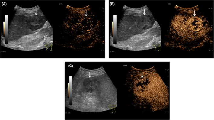 Figure 1
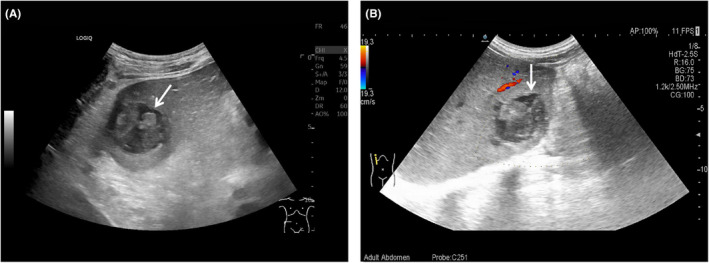 Figure 2
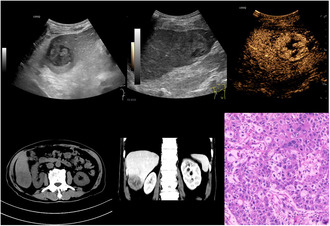 Figure 3
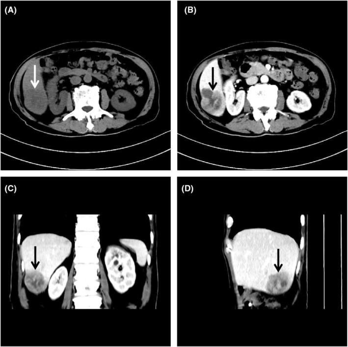 Figure 4
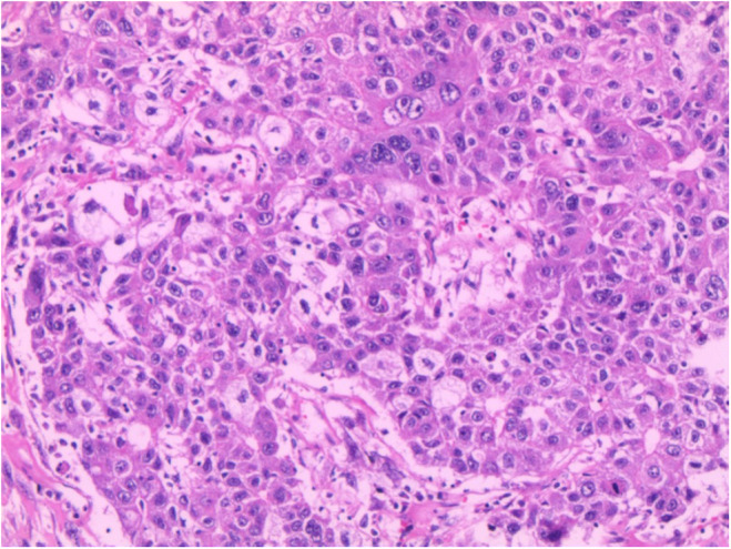 Figure 5
Peer Reviews
No public reviews on file for this paper yet. If you reviewed it on a platform where reviews are public (OpenReview, ICLR, NeurIPS, ICML), you can paste yours below so the community can read it here.
Videos
No videos yet. Explain this paper in a talk, walkthrough, or lecture? Add one.
Taxonomy
TopicsCholangiocarcinoma and Gallbladder Cancer Studies · Peptidase Inhibition and Analysis · Neuroendocrine Tumor Research Advances
Sarcomatoid hepatocellular carcinoma (SHCC), is a rare subtype of hepatocellular carcinoma characterized by epithelial and mesenchymal tumor features with sarcomatoid morphology, whose incidence is only 5% of hepatocellular carcinoma.1 Compared with hepatocellular carcinoma, SHCC has more rapid tumor growth and poorer disease progression. However, SHCC is easily misdiagnosed and delays the optimal treatment due to the clinical and imaging features being similar to hepatic abscesses. Here, we present a case image, including routine abdominal ultrasound, contrast‐enhanced ultrasound, plain computer tomography, and enhanced computer tomography, of SHCC that mimics hepatic abscesses.
A 71‐year‐old male patient presented to our hospital with a fever of unknown origin for 1 month, whose temperature is approximately 38°C. Physical examination revealed a right epigastrium slight tenderness. Laboratory tests revealed hypersensitive C‐reactive protein of >5.00 mg/L, routine C‐reactive protein of >200.00 mg/L, white blood cell of 21.36 × 10^9^ /L, neutrophils of 18.43 × 10^9^ /L, activated partial thromboplastin time of 45.30 s, fibrinogen quantification of 8.21 g/L, hepatitis B virus surface antigen of <0.01 IU/mL, hepatitis B virus surface antigen–antibody of 32.12 mIU/mL, hepatitis B virus e antigen of 0.439 S/CO, hepatitis B virus e antigen–antibody of 1.59 S/CO, and hepatitis B virus core antigen–antibody of 6.41 S/CO, alpha feto protein of 50.98 ng/mL.
Initially, routine abdominal ultrasound explored a hypoechoic mass located in the right hepatic lobe with irregular anechoic areas internally (Figure 1). Additionally, a contrast‐enhanced ultrasound indicated a mass with rapidly high peripheral enhancement during the arterial phase and with low peripheral enhancement during the portal venous phase without internal enhancement throughout all the phases (Figure 2). Furthermore, the mass had low density with internal cystic degeneration on plain computer tomography and obvious peripheral enhancement with no internal enhancement on enhanced computer tomography (Figure 3). Ultimately, the patient underwent a surgical mass excision, and an SHCC diagnosis was confirmed (Figure 4). No recurrence or metastasis was found during the 5‐month postoperative follow‐up.
According to the previous literature, the patient of SHCC has abdominal pain or persistent fever with an inflammatory reaction due to infection and necrosis in malignancy. Routine abdominal ultrasound and plain computer tomography reveal the mass is diffuse liquefactive necrosis with or without metastasis or portal and hepatic vein thrombosis.2 Contrast‐enhanced ultrasound and enhanced computer tomography observe the mass with irregular, thick, circular, and high enhancement.3 However, the evidence of SHCC remains too deficient to differentiate it from hepatic abscesses, and thus further research is necessary.
AUTHOR CONTRIBUTIONS
Haibo Luo: Writing – original draft. Xiaoling Leng: Supervision. Guiwu Chen: Writing – review and editing. Wenqin Liu: Formal analysis; visualization. Yanhua Xie: Formal analysis; visualization.
FUNDING INFORMATION
No funding was received for this study.
CONFLICT OF INTEREST STATEMENT
The authors declare no conflicts of interest.
CONSENT
Written informed consent was obtained from the patient to publish this report in accordance with the journal's patient consent policy.
The reference list from the paper itself. Each links out to its DOI / PubMed record.
- 1Sadiq AM , Mjemmas MG , Sadiq AM , Nkya GZ . Sarcomatoid hepatocellular carcinoma in a young African female. SAGE Open Med Case Rep. 2021;9:2050313 X 211052452.10.1177/2050313 X 211052452 PMC 850420834646567 · doi ↗ · pubmed ↗
- 2Wang CW , Ye XH . Sarcomatoid intrahepatic cholangiocarcinoma: a case report. Adv Ultrasound Diag Ther. 2023;7(1):38‐41.
- 3Yang Z , Lv K , Zhao YN , Pan M , Zhang C , Wei S . Sarcomatoid hepatocellular carcinoma mimicking hepatic abscess: a case report. Medicine (Baltimore). 2020;99(39):e 22489.32991488 10.1097/MD.0000000000022489 PMC 7523807 · doi ↗ · pubmed ↗