3D-printable portable open-source platform for low-cost lens-less holographic cellular imaging
Stephan Amann, Max von Witzleben, and Stefan Breuer

TL;DR
This paper introduces a low-cost, 3D-printable, open-source digital holographic microscope that uses off-the-shelf components to enable high-resolution cellular imaging in resource-limited settings.
Contribution
It provides a fully open-source, portable, and affordable lens-less holographic imaging platform with demonstrated micron-scale resolution using simple, off-the-shelf parts and 3D-printed housing.
Findings
Achieved 1.55 μm resolution with laser source
Demonstrated imaging of micro-spheres, red blood cells, and plant cells
Validated imaging capability with standardized test target
Abstract
Digital holographic microscopy is an emerging potentially low-cost alternative to conventional light microscopy for micro-object imaging on earth, underwater and in space. Immediate access to micron-scale objects however requires a well-balanced system design and sophisticated reconstruction algorithms, that are commercially available, however not accessible cost-efficiently. Here, we present an open-source implementation of a lens-less digital inline holographic microscope platform, based on off-the-shelf optical, electronic and mechanical components, costing less than $ 190. It employs a Blu-Ray semiconductor-laser-pickup or a light-emitting-diode, a pinhole, a 3D-printed housing consisting of 3 parts and a single-board portable computer and camera with an open-source implementation of the Fresnel-Kirchhoff routine. We demonstrate 1.55 {\mu}m spatial resolution by laser-pickup and…
Click any figure to enlarge with its caption.
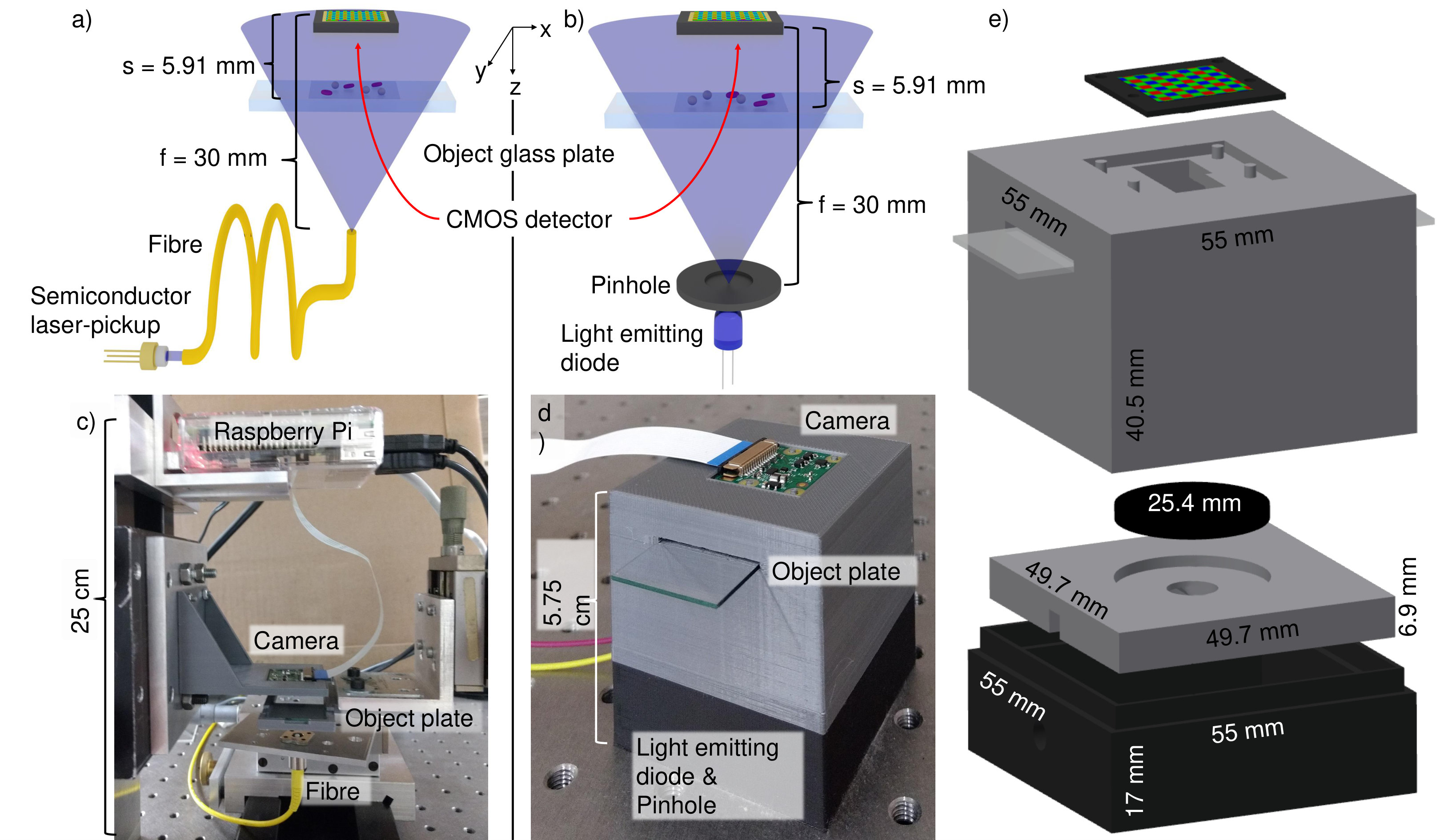 Figure 1
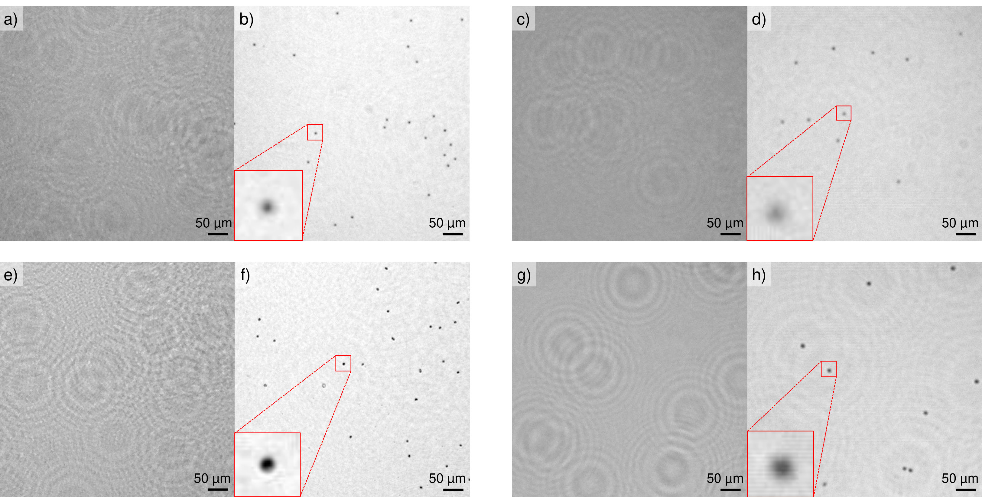 Figure 2
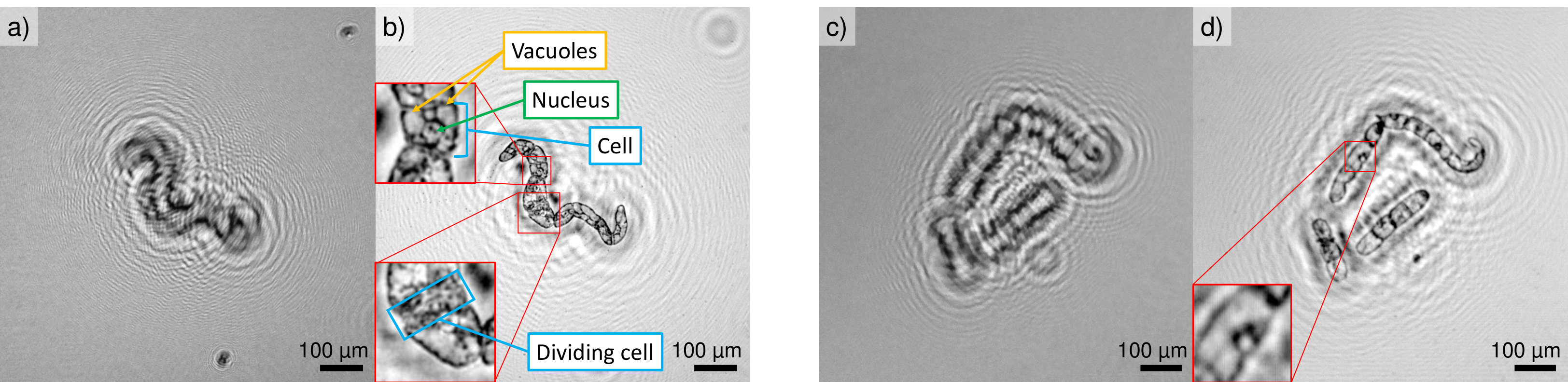 Figure 3
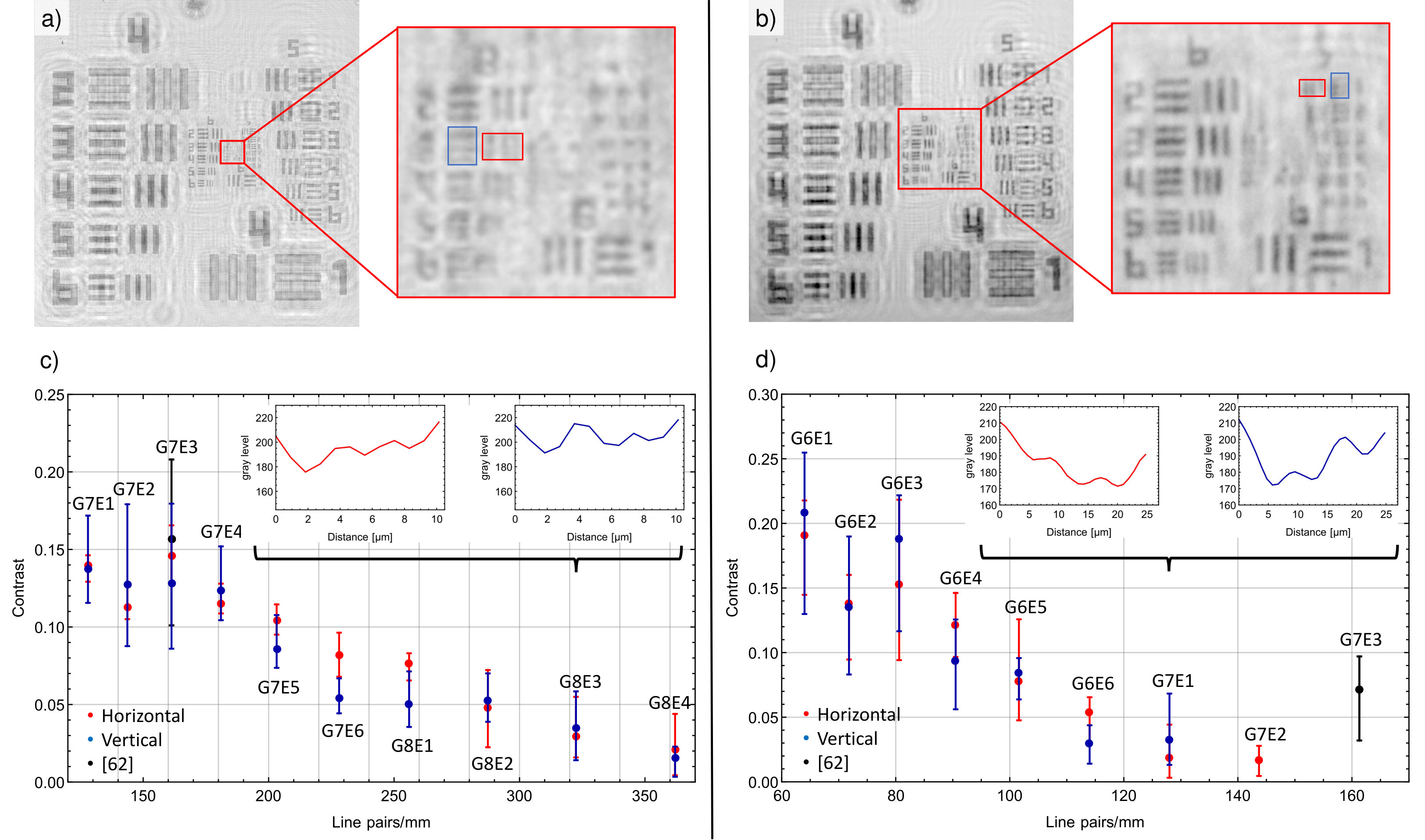 Figure 4
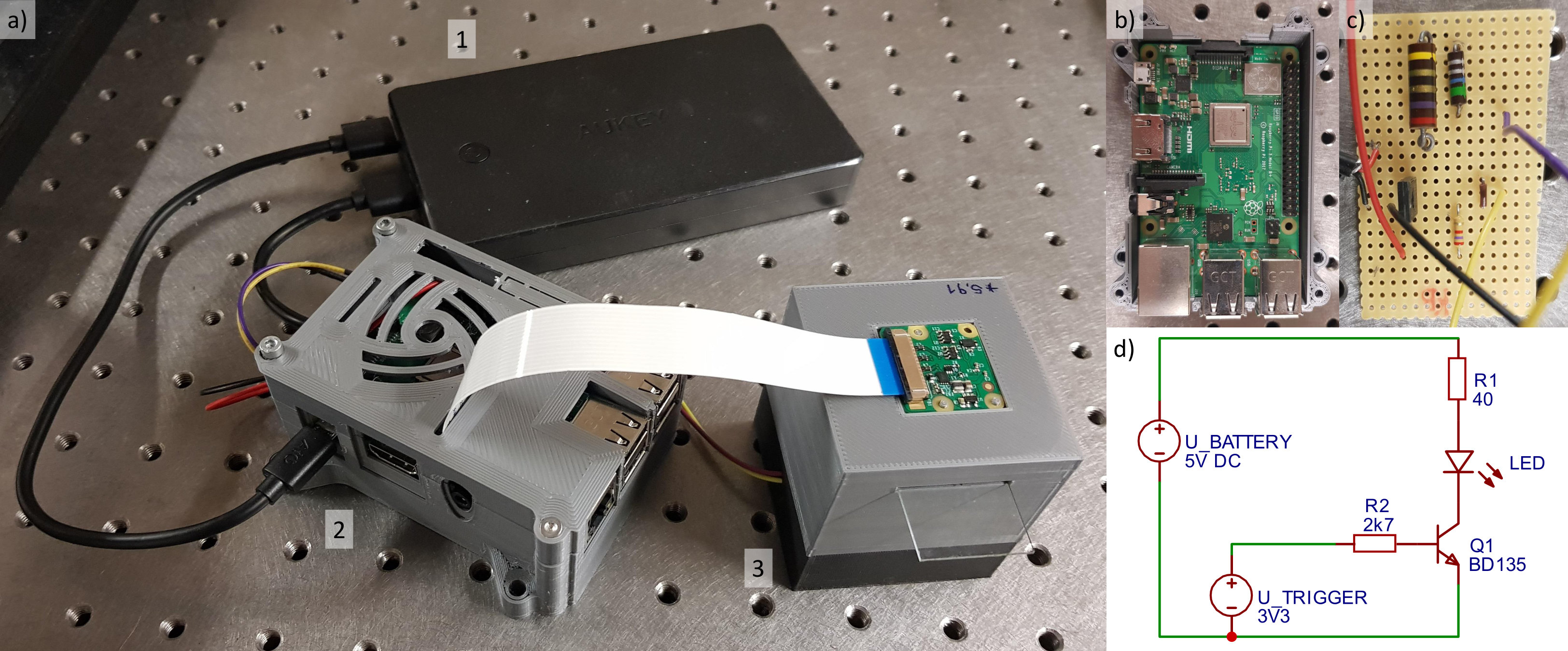 Figure 5
Peer Reviews
No public reviews on file for this paper yet. If you reviewed it on a platform where reviews are public (OpenReview, ICLR, NeurIPS, ICML), you can paste yours below so the community can read it here.
Videos
No videos yet. Explain this paper in a talk, walkthrough, or lecture? Add one.
3D-printable portable open-source platform for low-cost lens-less holographic cellular imaging
Stephan Amann1, Max von Witzleben1, Stefan Breuer1
1Institute for Applied Physics, Technische Universität Darmstadt, Schlossgartenstraße 7, 64289 Darmstadt, Germany
Digital holographic microscopy is an emerging potentially low-cost alternative to conventional light microscopy for micro-object imaging on earth, underwater and in space. Immediate access to micron-scale objects however requires a well-balanced system design and sophisticated reconstruction algorithms, that are commercially available, however not accessible cost-efficiently. Here, we present an open-source implementation of a lens-less digital inline holographic microscope platform, based on off-the-shelf optical, electronic and mechanical components, costing less than \$$190. It employs a Blu-Ray semiconductor-laser-pickup or a light-emitting-diode, a pinhole, a 3D-printed housing consisting of 3 parts and a single-board portable computer and camera with an open-source implementation of the Fresnel-Kirchhoff routine. We demonstrate 1.55,\upmu\upmu,\upmu,\upmu,\upmu$m sizes. The imaging capability is validated by imaging-contrast quantification involving a standardized test target. The presented 3D-printable portable open-source platform represents a fully-open design, low-cost modular and versatile imaging-solution for use in high- and low-resource areas of the world.
Introduction
Digital inline holography (DIHM) is an imaging modality within the fast evolving field of imaging microscopy research since a few decades [1] and is based on Gabor’s holographic principle [2]. By wide-field illumination of semi-transparent nano- to micron-sized objects with either a coherent or incoherent point light source, an interference pattern forming the hologram is created within a detection plane. It consists of the scattered part of the beam, object beam, and the unscattered (transmitted) part, the reference beam. The hologram contains amplitude and phase information of the imaged objects and allows for a numerical reconstruction of the object’s light field. To date, digital holography has been pioneered across a broad range of the electromagnetic spectrum: Terahertz[3] and infrared [4] holography bear the attractive potential to penetrate opaque media, whereas UV [5, 6] and X-ray [7, 8] illumination enable imaging on nanometer scale. Electron holography today is a mature research field and widely in use, allowing for molecular imaging [9, 10]. Digital holography within the visible wavelength regime has attracted considerable attention where a broad spectrum of light sources has been employed, ranging from gas [11, 12], solid state [13] and semiconductor [1, 14, 15, 16] lasers over light emitting diodes (LEDs) [17, 18, 19, 20] to halogen lamps [21]. Recently, ultra-broadband digital holography with sunlight illumination has been demonstrated, employing a new reconstruction algorithm [22]. In digital off-axis holography (DOAH), reference and object beam are spatially separated and recombine at an angle within the detector plane. Such experiment can already be compact, combining for example a collimation lens and two gradient-index lenses forming the point-light source and which enables a highly stable off-axis digital holographic system [23]. This helps reducing twin images, typically a challenge in holography set-ups [24]. During reconstruction a conjugate, out of focus image of the object is obtained in addition to the reconstructed object. This can lead to residual fringes, decreased contrast and an over all reduced image quality. In DOAH, object and twin image can be separated in the Fourier domain of the hologram. DOAH set-ups require a rather large number of optical components including mirrors, beam splitters and collimation lenses. Complementary to DOAH, DIHM constitutes a compact holography implementation that avoids to spatially separate the reference beam from the object beam. DIHM setups typically comprise of coherent or incoherent point light source, a 2D digital detector array and, positioned in between, the micron-scale object sample to be imaged. It has been demonstrated in the ultraviolet[5], visible[13], near-infrared[25] up to the mid-infrared[26] wavelength regime. In DIHM, only a negligible influence of the twin image on the quality of microscopic object analysis has been demonstrated [1, 27], as the twin image is defocused across the whole detector array when the distance between detector and object is large as compared to the object size. Moreover, for DIHM several approaches to numerically remove the twin image from the reconstructed image have been demonstrated [28, 29, 30]. Ideal light sources are semiconductor photonic emitters such as laser diodes (LDs) and LEDs thanks to their compactness, high electro-optical efficiencies, comparable low price and availability. Partially coherent light sources can even enhance the result in DIHM as coherent speckle noise is reduced [27] while being less susceptible to mechanical vibrations. Considering the generally less complex implementation, easy replacement, lower price, high reliability and reduced safety issues, LEDs appear as ideal DIHM light sources for student and early researcher education. To ensure homogeneous illumination, the LED or LD light can be coupled into an optical fibre ensuring a Gaussian beam profile at the fibre’s end [19, 31, 20, 15, 32]. In addition, more compact or complex experimental set-ups can be realized thanks to the mechanical fibre flexibility [31]. Both charge-coupled device (CCD) and complementary metal-oxide-semiconductor (CMOS) cameras are commonly used as detectors, with an increasing number of CMOS chips due to recent advances in terms of sensitivity and reduced pixel size. Two parallel tracks of current DIHM research can be identified, focusing on two different detection schemes: On-chip microscopy on the one hand, where the sample under investigation is located close to the CMOS detector. Its advantage is the large field-of-view which corresponds to the whole detector area. Several on-chip microscopes employing low-coherent LED illumination [17, 31, 33] have been demonstrated making them already cost-efficient and easy to operate. In combination with several pixel super-resolution approaches using multi-height imaging [19], wavelength scanning [20], sub-pixel shifting[34] or flowing samples [35], sub-micron spatial resolution is possible. Sub cellular imaging of malaria infected blood cells [36], red blood cell (RBC) imaging with a cell phone camera [18] and colour imaging using DIHM [31] have been reported. On the other hand, fringe magnification technique is studied where the sample is located near the point source [1, 23, 15]. Here, the maximum lateral resolution is accompanied by a decreased field-of-view (see equation (5)).
DIHM research has already reached the stage of commercialization where for example several DIHM implementations exist including lens-less inline and DOAH schemes [37] delivering lateral resolutions of m. A submersible holographic microscope for remote in-vivo oceanic microscopy has been reported [38] and is now commercially available [39]. Moreover, lens-based DOAH has recently been employed in in-line industrial production control [40] and for imaging of semiconductor structures [41]. In life-sciences, DIHM has the advantage of working label-free, which enables non-invasive, in-vivo study of biological samples, for example RBCs [42, 43, 44], parasited mouse RBCs [45], sperm cells [15], diarrhea parasites [17], and several aquatic organisms [38]. Moreover, as information about the whole sample volume is encoded in one single hologram, processing speed is substantially increased as compared to microscopic scanning techniques such as confocal or fluorescence microscopy. Thus, large sample volumes can be analyzed efficiently, such as cell cultures of human cancer cells [46, 47] as well as microplastic pollution in marine environments [48]. Furthermore, by acquiring and analyzing sequences of images, DIHM enables long term cell evolution studies [32] and micro-particle tracking [49]. Although differently complex, portable and low-cost DIHM solutions have recently been reported [50, 18, 15, 51], critically important details neccessary for their realization in a laboratory are mostly disclosed. Such important details include details on employed of light sources, distances between objects and light source or detector, methods of reconstruction, non obvious limitations or design or construction files thus making it challenging to set up a DIHM without too much prior expertise and avoids access to low-cost micron spatial resolution imaging. In this work first, we present two experimental DIHM platforms employing an LED and a LD as illumination sources and operate the both by a portable single-board computer and camera. The LD has been disassembled from a standard commercially available Blu-ray disk drive. The housing and all mechanical mounts are 3D printed. [52]. Second, we describe the implemented open-source hologram reconstruction based on HoloPy [53] and Fiji plugin [54]. All employed code is open-source accessible aiming at triggering further developments and sharing between research laboratories, diagnostic labs and science education. Third, we demonstrate microscopic 2D-imaging of polystyrene micro-spheres (PMSs) and mature human RBCs imaging with micron spatial resolution. Fourths, we perform 2D-imaging of larger tobacco BY-2 cells (TBY2s). Fifths, we quantify the achieved spatial resolution by optical contrast analysis of an united states air force (USAF) microscopic imaging test target. Sixths, we summarize our efforts in developing a 3D-printable open-source platform for cellular imaging and provide a brief outlook on our activities.
Laboratory and 3D printed opto-mechanical DIHM setups
Both developed DIHM implementations, described in the following, aim at achieving and validating the maximum achievable spatial resolution when an temporally and spatially coherent source as well as a temporally and spatially incoherent semiconductor light source at equivalent emission wavelength are considered. First, a LD-based lens-less DIHM platform is developed that aims at demonstrating single-digit micrometer spatial resolution cellular imaging by a temporally coherent source and is schematically depicted in Fig. 1 (a). It employs a nm emitting LD in a laser-pickup which has been dismounted from a commercially available standard Blu-ray disc drive. The laser emission is coupled into a standard single-mode fibre. The diverging fundamental Gaussian mode beam is directed towards the object glass plate at a distance of mm from the fibre exit facet. The CMOS camera is positioned at a distance of mm. Second, an LED-based lens-less DIHM platform depicted schematically in Fig. 1 (c) is designed and constructed by 3D printable parts, with the over all system costs amounting to less than \,190. Compared to Fig. [1](#Sx1.F1) (a), in Fig. [1](#Sx1.F1) (b) a 430 nm emitting high-power LED is employed as a temporally and spatially incoherent semiconductor light source. A fraction of the emitted light is passed through a high-precision pinhole where 1.1 \upmuz=5.91 mm). For live cell imaging, such ultra-low optical power is of critical importance, as cell damage by light exposure needs to be minimised. To construct the LED-based platform in Fig. [1](#Sx1.F1) b), first three mechanical parts in Fig. [1](#Sx1.F1) c) are 3D-printed, see section “Methods”, and assembled as sketched in Fig. [1](#Sx1.F1) d) and e) where also specific dimensions of the platform are depicted. For both experiments, equal spatial separations between emission facet, microscope glass plate carrying the micro-objects under investigation and CMOS detector are chosen. We chose a distance of 30 mm between light source and detector in order to reach a compact experimental set-up, while still maintaining illumination of the hole detector area. The position of the object emerges from resolution optimization, see section “Resolution”. A Raspberry Pi single-board portable computer and a Raspberry Pi CMOS detector camera with a pixel size of (1.12\times 1.12),\upmu28,125,\delta\delta_{\textnormal{Laser}}=0.87,\upmu\delta_{\textnormal{LED}}=0.92,\upmu\upmuf=$30 mm between fibre facet and CMOS detector are expected for the LD and LED light source, respectively.
Image reconstruction
Following the hologram acquisition, information retrieval of the cellular objects deposited on the object glass plate is performed numerically. The propagation of light fields is completely described by diffraction theory. Hence it is possible to reconstruct amplitude and phase information of the objects from their interference patterns generated on the camera. At the object location the pattern is focused and reveals the shape and morphology of micro-objects. Numerically, arbitrary planes can be re-focused in retrospect yielding access to volumes with a large number of objects in different heights in z-direction which can be studied with acquiring a single image. The propagation of a wave front towards the detector is described by the Fresnel-Kirchhoff diffraction integral
[TABLE]
where denotes the wave field on the detector, is the incident wave, the transmission function of the object, and and are two points in the object plane respectively detector plane[55]. The amplitude and phase distribution of the object can be obtained via an inverse integral
[TABLE]
with the reference wave and the intensity distribution of the hologram in the detector plane . For the reconstruction, an open-source plugin for the open-source microscope software Fiji, see “Methods” section, that implements the angular spectrum estimation for small distances in the order of micrometers up to several centimeters, is employed. The amplitude distribution in the reconstructed plane is calculated using two Fourier transforms
[TABLE]
where is the height of the reconstructed plane, the wave vector, the index of refraction, the number of pixels and the pixel size of the CMOS detector. and denote Fourier Transform and the inverse Fourier Transform. The formalism described above is implemented in two open-source reconstruction software packages, see “Methods” section. In the following, we elaborate and identify two easy to implement reconstruction softwares on a standard desktop computer or potentially also on a mobile phone. HoloPy, a software package for python, allows for hologram reconstruction, but also hologram simulation and scattering calculations. The algorithm to reconstruct point source holograms is based on Fresnel-Kirchhoff diffraction[1]. It considers a background subtracted hologram, experimental parameters including distances and light source wavelengths and then reconstructs the hologram using two Fourier transforms. Alternatively, hologram fitting is provided by HoloPy where the position of a scatterer is simulated in order to produce the same interference pattern instead of image back-propagation [56]. For the case of a known number of scatterers, this method can be recommended as it allows to reconstruct spherical or cylindrical object shapes. It is less practicable, however, when arbitrarily shaped micro-objects are of interest, as for example folded RBCs. For the latter case, an open-source plugin [57] for Fiji, see “Methods” section, is a possible solution with a user-friendly graphical-user-interface, implemented in Java. Phase, amplitude and intensity distribution of micro-objects can be reconstructed at arbitrary heights in z-direction.
Micro particle and cellular imaging
In the following, we first capture and image standardized PMSs of diameter m by both the LD and LED-based lens-less DIHM platform and reconstruct the resulting object properties by the Fiji plugin. Second, we investigate anonymized mature RBCs as micro-objects in the same manner. Third, we image cell suspension culture TBY2s. The recorded holograms and subsequently reconstructed object planes for multiple PMSs and RBCs are depicted in Fig. 2. Laser-based DIHM hologram (a) and reconstruction (b) is presented next to LED-based DIHM hologram (c) and reconstruction results (d). Both insets depict an isolated PMS or RBC enlarged to five times its original size. It becomes evident that the LD-based platform provides sharper images where a larger number of interference fringes can be captured per object. These fringes overlap within the hologram resulting in a hologram with more grainy texture as compared to the LED-based results in Fig. 2 (a,c). In contrast, the LED-based reconstructed image is considerably more washed out resulting in comparably extended objects. Accordingly, for human RBCs imaged by the LD-based platform, the oval disk shape can clearly be resolved as depicted in Fig. 2 (e,f). Several RBCs appear to be tilted in their spatial position, resulting in an elliptical shape. This is in stark contrast to the results obtained by the LED-based platform where information retrieval, for example on the cell morphology, are scarce. However, by the LED-based platform, individual cells can clearly been distinguished, thus exemplifying its potential for individual cell counting or tracking.
In order to validate both platform’s imaging capabilities also for extended cellular objects, fast growing plant tobacco TBY2s have been prepared and imaged. TBY2s are employed in various fields of plant biology as a model material and are ideally suited for cellular and molecular analyses[58]. Corresponding results are depicted in Fig. 3. The recorded holograms and reconstructed object planes for an isolated TBY2 are depicted for LD-based DIHM hologram (a) and reconstruction (b) is presented whereas the hologram, obtained by the LED-based DIHM, is depicted in (c) and the corresponding reconstruction in (d). Both platforms allow to successfully access individual cell segments with a length of m as well as internal structures including cell nuclei and vacuoles. In Fig. 3b), a dividing cell undergoing mitosis can be observed. For LED illumination, individual vacuoles are not distinguishable. This is not surprising, as the vacuole membrane thickness is around one order of magnitude smaller than the cell wall thickness of (7-10) nm for plant vacuoles[59] as compared to (71-87) nm for tobacco leaf cells walls [60]. However it is possible to make out the nuclei of several cells. Interestingly, TBY2s infer a more complex interference pattern as compared to both PMSs and RBCs, indicating a stronger absorption and thus increased hologram contrast. We found that Fiji revealed a substantially faster reconstruction as compared to HoloPy. The reconstruction of 10 planes of a digital hologram by the Fiji plugin demands 30 seconds computational time on a regular consumer PC as compared to several minutes by HoloPy. Towards larger volumes, the reconstruction time can theoretically be improved by performing computations on a graphics processing unit [11] as demonstrated for live imaging [61]. In the following section, we aim to quantify the theoretical lateral resolution as well as the spatial resolution experimentally achieved by both the LD-based and LED-based DIHM platforms.
Resolution
In DIHM, the lateral resolution is bounded by the optical assembly numerical aperture () and the illumination wavelength [1]:
[TABLE]
suggesting a shorter wavelength for a higher lateral resolution. For a digital holographic microscope with a pixel number , with pixel size , an illumination wavelength and a distance between object and detector plane, this translates to
[TABLE]
As described in [62], a maximum lateral resolution can be achieved at a distance
[TABLE]
between object and detector, with the distance between light source and detector. This originates from the consideration, that a higher resolution is possible, the closer the object is placed to the detector, however at the same time the interference fringes move closer. Here, denotes the distance at which different interference rings are still resolved by different camera pixels. The axial resolution of the system can be calculated according to
[TABLE]
By equation (6), the theoretical resolution for both developed platforms can be estimated. For m, , mm, nm and nm, respectively, lateral resolutions of m and m are theoretically possible by the selected, optimum platform design. To evaluate the experimentally achieved resolution, a 1951 USAF microscopic imaging test target on a glass microscopic slide serves as a reference object consisting of groups of horizontal and vertical lines with decreasing spatial frequency. The resulting reconstructed amplitude images acquired with the LED (a) and LD (b) setup are depicted in Fig. 4. With LED illumination, element 1 of group 7 is the last resolvable element corresponding to a resolution of 128 line pairs/mm and a line width of 3.91 m. For LD illumination, element 3 of group 8 is still resolvable, leading to a resolution of 322.5 line pairs/mm and a line width of 1.55 m. This is mostly a result of the higher temporal and spatial resolution of the laser in comparison to the used LED, as well as the smaller wavelength. The resolving power with LD illumination is thus significantly higher.
Finally, to quantify the achieved image contrast and thus evaluate the capability of the developed DIHM setups, in the following the intensity of a single USAF element is averaged along its axis and plotted (see red and blue rectangle in Fig. 4a and b). Then, the contrast of consecutive extreme points is calculated by . Each element consists of 5 dark lines which leads to 4 contrast values. The average contrast is then displayed in dependence on the resolution in line pairs/mm, with the error bars ranging from the minimum to the maximum value. The resulting image contrast of different elements of the respective USAF image is depicted in Fig. 4c) and d). Results obtained by both light sources indicate decreasing contrast values with increasing line pairs. The maximum resolvable elements are as shown by the the intensity profile plots for horizontal and vertical elements 3 of group 8 in the inset of Fig. 4c). For LED, the maximum resolvable element is element 1 of group 7 as depicted in the inset of Fig. 4d). We compared our contrast values to reported values [63], where an unspecified laser or a LED with a central wavelength of 470 nm (25 m pinhole) was used for image acquisition. The reported value for laser illumination coincides with our results. With LED illumination, the reported value reachs a higher resolution, group 7 element 3 is still resolvable. This could be due to a higher signal-to-noise ratio of the employed camera, the smaller bandwidth of only 10 nm as well as the use of advanced reconstruction algorithms. The achieved spatial resolution of both setups is suited to image and detect individual micro-particles and cellular objects including ensembles of microscopic biological samples. For the LD-based DIHM, sharper interference pattern and a higher number of interference fringes could be captured. Thus, more object information is retrieved yielding a crisper reconstructed image including more details. We attribute this to the LD’s higher second-order temporal coherence as well as spatial coherence. The LED’s spatial coherence could be increased by reducing the pinhole diameter which would require increased LED biasing currents which then require efficient cooling of the LED and thereby increasing the physical dimensions of the setup. Our DIHMs allow to image cellular objects with dimensions smaller than m as well as microscopy of larger objects with a spatial resolution of m. To ensure maximum possible resolution, a LD-based DIHMs is recommended, whereas for applications where high temporal stability, compact set-up, ruggedness and reduced costs a required, LED-based DIHMs is recommended.
Conclusion
A 3D printable platform for lens-less holographic cellular imaging with open accessible software solutions has been developed delivering spatial resolutions of m by LD or m by LED illumination. A 405 nm Blu-ray semiconductor laser-pickup coupled to an optical fibre and a 430 nm high power LED in combination with a m pinhole have been successfully employed as DIHM light sources. Despite its lower degree of temporal coherence, the LED proved to be of advance in terms of implementation, price and lower safety concerns. A single-board portable Raspberry Pi computer and camera operate the light sources as well as perform the image acquisition. By an open-source software implementation of the Fresnel-Kirchhoff algorithm, we imaged and succesfully reconstructed m PMS and human RBCs with a diameter of about m, as well as TBY2s with an individual size of about m. Less than W of optical power were sufficient for holographic imaging microscopy. Such ultra-low optical power can be of critical importance for live cell imaging where light exposure of the cells needs to be as low as possible. Equally compact setup could be envisioned for the LD when fibre-coupled LDs are available, which are however considerably expensive. The DIHM setup presented here may serve as a reliable, easy to implement and flexible to extend solution for student an early researcher education and for different demands in microscopic imaging. The total costs for the LED setup amount to 3 LED, 35 Raspberry Pi 3, 25 3D print, $ 27 power bank) and thus enables a convenient entry into the wide field of digital holography. Future work could include the integration of the DIHM with micro-fluidic channels [49, 64, 65] or considering machine learning algorithms to automatically count and identify particles [66], as well as diagnose illnesses such as meningitis [67], iron-deficiency anemia or diabetes mellitus [68]. The developed 3D-printable photonic platform might help facilitating reproducibility of results obtained in different laboratories and prototyping of specific improvements and advancements of the DIHM setups. The cost-efficient open science and open hard and software platform aims in particular at contributing towards a democratization of scientific knowledge [69].
Methods
DIHM construction, light sources, opto-electronics, electrical circuits and 3D print
The Blu-ray LD-pickup (SF-AW210) has been dismounted from a commercially available standard computer Blu-ray optical head and its emits a maximum optical output power of mW at a wavelength of 405 nm for an injection current of mW. The LD-pickup beam is collimated and focussed by two aspheric lenses with an effective focal length of mm (\,8723,\upmu$,9924.5,\upmuz_{\textnormal{r}}=21,\upmu2w_{0}=3.3,,\upmu\upmuf=$,3, 3W High Power LED 430nm - 435nm hyper violet, Avonec, Germany [[70](#bib.bib70)]) emits at wavelengths centered at around 430\Delta\lambda_{\textnormal{LED}}L_{\textnormal{c,LED}}=\lambda_{\textnormal{LED}}^{2}\times(\pi\times\Delta\lambda_{\textnormal{LED}})^{-1}=3.9,\upmu\Delta\lambda the full width at half-maximum spectral line width[[71](#bib.bib71)]. A high-precision stainless steel pinhole with a diameter of (15 \pm 1.5),\upmu$,75$,880$,9, 3M-ID 70005241826 Scotch Magic Tape, 3M Inc.). The cased with VESA mounts for Raspberry Pi 3 (B/B+), Pi 2 B, and Pi 1 B+ can be accessed by[[72](#bib.bib72)]. The CMOS camera module is glued to the upper part of the box after removing the lens mounted in front of the module. We observed that otherwise strong hologram distortions appeared. In order to electrically bias the LD, a commercial LD driver was used to provide a constant output current. However, an open-source driver is under construction while all parts are available for in total $,20[[73](#bib.bib73)]. For the LED, a custom soldered circuit has been developed accessing Raspberry Pi’s general purpose input/output (GPIO). The assembled system is depicted in [5](#Sx3.F5)b). The driver circuits can well be integrated into the 3D printed Raspberry Pi housing. A diagram of the circuit can be seen in Fig. [5](#Sx3.F5)a). The portable computer, camera and DIHM light sources are supplied with electrical power by a conventional power bank battery pack with maximum output power of 10 W (27, Aukey PB-N36). A computer monitor and computer mouse are required for the hologram acquisition. V_BATTERY is the voltage provided by the power bank, whereas V_TRIGGER is the voltage between the Raspberry Pi GPIO and ground. It triggers a transistor (BD135), which lets a current of 125 mA flow through the LED.
Light source biasing, image detection and aquisition
A portable single-board computer and camera module with a 3280 x 2464 pixel CMOS chip and pixel dimensions of (m2, sensor size () mm2 (\,35$,25$ Raspberry Pi Camera v2, Raspberry Pi Foundation, UK [52]) serve as light source driver and hologram acquisition.
Sample preparation
The RBC samples under investigation are standard anonymized RBC reference cells (\,27$,129(6.5\pm 0.2),\upmum. They are equally high diluted in distilled water. Furthermore a few ml of ordinary dish detergent are added to prevent agglomeration and adhesion of the micro-spheres. After diluting the particular object a drop of the solution is placed by a standard plastic pasteur pipette on a high transmission flat glass microscope slide (75 mm x 25 mm x 1 mm, B270 I, SCHOTT AG) and then covered with a glass cover plate (22 mm x 22 mm x 0.15 mm, B270 I, SCHOTT AG)). Nicotiana tabacum cv. BY-2 suspension plant cell cultures[[74](#bib.bib74)] were grown in liquid saline medium based on a modified Linsmaier and Skoog medium with agitation on a incubator shaker. The cells have been grown in 50 ml medium within a 250 ml glass flask. Every 7 days, 5 %$ inoculum had been transferred into a fresh medium [75, 76] and stored permanently on an incubator shaker.
Image acquisition, reconstruction and resolution validation
The pictures are acquired with a fixed white balance. For details see the camera file on \urlhttps://github.com/teph12/DIHM. The image is then transferred to a PC with Windows 10 operating system and equipped with an Intel i3 processor and 8 GB of RAM, with Fiji installed. In Fiji the image is converted to a 32-bit black and white picture, which is then loaded in the “Numerical Reconstruction” plugin. Here, using the parameters of image acquisition (distance between camera and object, wavelength, image size) the image is reconstructed. Subsequently the image contrast is normalized and enhanced by 0.2 %. For the quantification of the experimentally achieved spatial resolution, a commercially available standardized 1951 USAF positive high-contrast chrome on quartz glass microscopic imaging test target created by photo lithography on a glass microscopic slide serves as a reference object (\$$ 900, Ready Optics, US). It consists of groups of horizontal and vertical lines with standardized spatial frequencies starting at group 4, element 1 with 31 \upmu$m spacing and ending at group 11, element 6 with 137 nm spacing.
The reference list from the paper itself. Each links out to its DOI / PubMed record.
- 1[1] Manfred H. Jericho and H. Jürgen Kreuzer. Point source digital in-line holographic microscopy. In Coherent Light Microscopy , pages 3–30. Springer Berlin Heidelberg, 2010.
- 2[2] Dennis Gabor. A new microscopic principle. Nature , 161(4098):777–778, 1948.
- 3[3] Lu Rong, Tatiana Latychevskaia, Chunhai Chen, Dayong Wang, Zhengping Yu, Xun Zhou, Zeyu Li, Haochong Huang, Yunxin Wang, and Zhou Zhou. Terahertz in-line digital holography of human hepatocellular carcinoma tissue. Scientific Reports , 5(1), feb 2015.
- 4[4] L. Repetto, R. Chittofrati, E. Piano, and C. Pontiggia. Infrared lensless holographic microscope with a vidicon camera for inspection of metallic evaporations on silicon wafers. Optics Communications , 251(1-3):44–50, jul 2005.
- 5[5] Ahmad Faridian, David Hopp, Giancarlo Pedrini, Ulrike Eigenthaler, Michael Hirscher, and Wolfgang Osten. Nanoscale imaging using deep ultraviolet digital holographic microscopy. Optics Express , 18(13):14159, jun 2010.
- 6[6] Mustafa Ugur Daloglu, Aniruddha Ray, Zoltan Gorocs, Matthew Xiong, Ravinder Malik, Gal Bitan, Euan Mc Leod, and Aydogan Ozcan. Computational on-chip imaging of nanoparticles and biomolecules using ultraviolet light. Scientific Reports , 7(1), mar 2017.
- 7[7] Martin Krenkel, Mareike Toepperwien, Frauke Alves, and Tim Salditt. Three-dimensional single-cell imaging with x-ray waveguides in the holographic regime. Acta Crystallographica Section A Foundations and Advances , 73(4):282–292, 2017.
- 8[8] Tais Gorkhover, Anatoli Ulmer, Ken Ferguson, Max Bucher, Filipe R. N. C. Maia, Johan Bielecki, Tomas Ekeberg, Max F. Hantke, Benedikt J. Daurer, Carl Nettelblad, Jakob Andreasson, Anton Barty, Petr Bruza, Sebastian Carron, Dirk Hasse, Jacek Krzywinski, Daniel S. D. Larsson, Andrew Morgan, Kerstin Mühlig, Maria Müller, Kenta Okamoto, Alberto Pietrini, Daniela Rupp, Mario Sauppe, Gijs van der Schot, Marvin Seibert, Jonas A. Sellberg, Martin Svenda, Michelle Swiggers, Nicusor Timneanu, Danie