Preterm birth alters the development of cortical microstructure and morphology at term-equivalent age
Ralica Dimitrova, Maximilian Pietsch, Judit Ciarrusta, Sean P. Fitzgibbon, Logan Z.J. Williams, Daan Christiaens, Lucilio Cordero-Grande, Dafnis Batalle, Antonios Makropoulos, Andreas Schuh, Anthony N. Price, Jana Hutter, Rui PAG Teixeira, Emer Hughes, Andrew Chew

TL;DR
Preterm birth disrupts normal brain development in infants, leading to varied changes in brain structure by the time they reach term-equivalent age.
Contribution
This study provides a comprehensive characterization of cortical changes in preterm infants using in vivo MRI and normative data from term-born infants.
Findings
Preterm infants showed increased cortical water content and reduced neurite density in posterior regions, and greater cortical thickness anteriorly.
Individual preterm infants exhibited extreme deviations from normative development, with little spatial overlap between them.
Regional cortical development was linked to age at birth but not to later neurodevelopment at 18 months.
Abstract
The dynamic nature and complexity of the cellular events that take place during the last trimester of pregnancy make the developing cortex particularly vulnerable to perturbations. Abrupt interruption to normal gestation can lead to significant deviations to many of these processes, resulting in atypical trajectory of cortical maturation in preterm birth survivors. We sought to first map typical cortical micro- and macrostructure development using invivo MRI in a large sample of healthy term-born infants scanned after birth (n = 259). Then we offer a comprehensive characterization of the cortical consequences of preterm birth in 76 preterm infants scanned at term-equivalent age (37–44 weeks postmenstrual age). We describe the group-average atypicality, the heterogeneity across individual preterm infants, and relate individual deviations from normative development to age at birth and…
Genes, proteins, chemicals, diseases, species, mutations and cell lines named across the full text — each resolved to its canonical identifier and authoritative record.
Click any figure to enlarge with its caption.
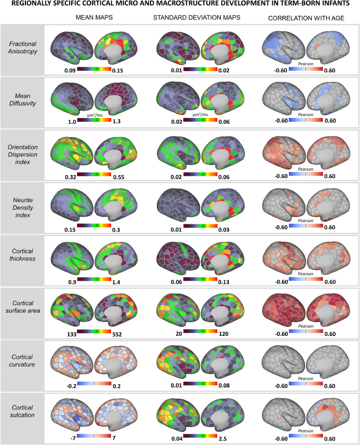 Figure 1
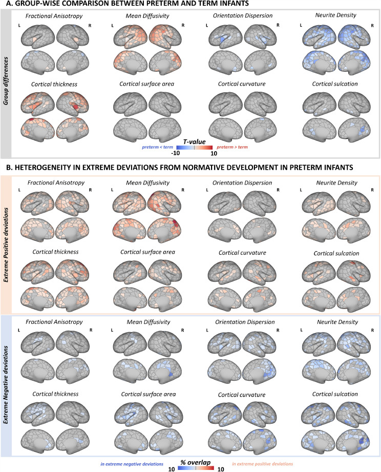 Figure 2
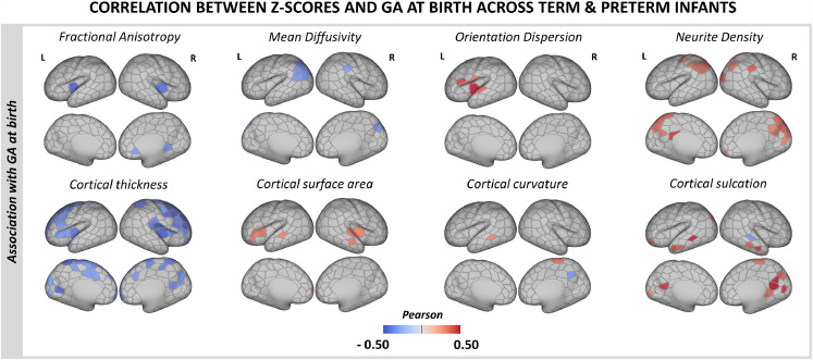 Figure 3
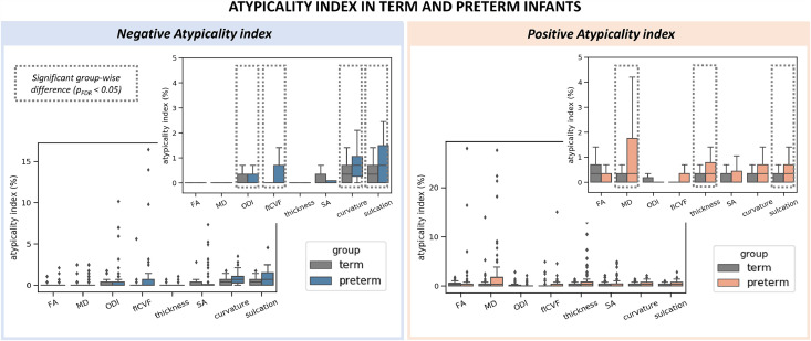 Figure 4
Peer Reviews
No public reviews on file for this paper yet. If you reviewed it on a platform where reviews are public (OpenReview, ICLR, NeurIPS, ICML), you can paste yours below so the community can read it here.
Videos
No videos yet. Explain this paper in a talk, walkthrough, or lecture? Add one.
Taxonomy
TopicsNeonatal and fetal brain pathology · Advanced Neuroimaging Techniques and Applications · Functional Brain Connectivity Studies