DeepMIB: User-friendly and open-source software for training of deep learning network for biological image segmentation
Ilya Belevich, Eija Jokitalo
TL;DR
DeepMIB is a user-friendly, open-source tool that simplifies training deep learning models for segmenting biological images in 2D and 3D.
Contribution
DeepMIB introduces a multi-platform, standalone application for training deep learning models without programming knowledge.
Findings
DeepMIB successfully segments 2D and 3D electron and multicolor light microscopy datasets.
The software is distributed as open-source code and a standalone application for multiple operating systems.
It enables non-experts to use deep learning for image segmentation workflows.
Abstract
We present DeepMIB, a new software package that is capable of training convolutional neural networks for segmentation of multidimensional microscopy datasets on any workstation. We demonstrate its successful application for segmentation of 2D and 3D electron and multicolor light microscopy datasets with isotropic and anisotropic voxels. We distribute DeepMIB as both an open-source multi-platform Matlab code and as compiled standalone application for Windows, MacOS and Linux. It comes in a single package that is simple to install and use as it does not require knowledge of programming. DeepMIB is suitable for everyone interested of bringing a power of deep learning into own image segmentation workflows. Deep learning approaches are highly sought after solutions for coping with large amounts of collected datasets and are expected to become an essential part of imaging workflows. However,…
Genes, proteins, chemicals, diseases, species, mutations and cell lines named across the full text — each resolved to its canonical identifier and authoritative record.
Click any figure to enlarge with its caption.
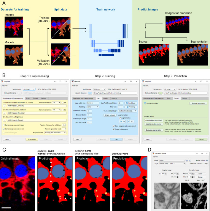 Fig 1
Fig 1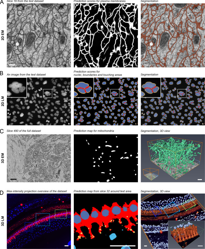 Fig 2
Fig 2- —Biocenter of Finland
- —Academy of Finland
- —Academy of Finland
Peer Reviews
No public reviews on file for this paper yet. If you reviewed it on a platform where reviews are public (OpenReview, ICLR, NeurIPS, ICML), you can paste yours below so the community can read it here.
Videos
No videos yet. Explain this paper in a talk, walkthrough, or lecture? Add one.
Taxonomy
TopicsCell Image Analysis Techniques · Advanced Electron Microscopy Techniques and Applications · Advanced Fluorescence Microscopy Techniques
Introduction
During recent years, improved availability of high-performance computing resources, and especially graphics processing units (GPUs), has boosted applications of deep learning techniques into many aspects of our lives. The biological imaging is not an exception, and methods based on deep learning techniques [1] are continually emerging to deal with various tasks such as image classification [2,3], restoration [4], segmentation [5–8], and tracking [9]. Unfortunately, in many cases, the application of these methods is not easy and requires significant knowledge in computer sciences, making it difficult to adapt by many researchers. Software developers have already started to address this challenge by developing user-friendly deep learning tools, such as Cell Profiler [10], Ilastik [11], ImageJ plug-ins DeepImageJ [12] and U-net [13], CDeep3M [5], and Uni-EM [14] that are especially suitable for biological projects. However, the overall usability is limited because they either rely on pre-trained networks without the possibility of training on new data [10–12], are limited to electron microscopy (EM) datasets [14], or have specialized computing requirements [5,13].
In our opinion, in addition to providing good results, ideal deep learning solution should fulfill the following criteria: a) is capable of training on new data; b) has a user-friendly interface; c) is easy to install and would work straight out of the box; d) is free of charge and e) has open-source code for future development. Preferably, it also would be compatible with 2D and 3D, EM and light microscopy (LM) datasets. To address all these points, we are presenting DeepMIB as a free open-source software tool for image segmentation using deep learning. DeepMIB can be used to train 2D and 3D convolutional neural networks (CNN) on user’s isotropic and anisotropic EM or LM multicolor datasets. DeepMIB comes bundled with Microscopy Image Browser (MIB) [15], forming a powerful combination to address all aspects of an imaging pipeline starting from basic processing of images (e.g., filtering, normalization, alignment) to manual, semi-automatic and fully automatic segmentation, proofreading of segmentations, their quantitation and visualization.
Design and implementation
Deep learning workflow
DeepMIB deep learning workflow comprises three main steps: preprocessing, training and prediction (Fig 1A). Preprocessing requires images supplemented with the ground truth, which can be generated directly in MIB or using external tools [10,11,16,17]. Typically, the application of deep learning to image segmentation requires large training sets. However, DeepMIB utilizes sets of 2D and 3D CNN architectures (U-Net [6,8], SegNet [18]) that can provide good results already with only few training datasets (starting from 2 to 10 images [13]), making it useful for small-scale projects too. To prevent overfitting of the network during training, the segmented images are randomly split into two sets, where the larger set (80–90%) is used for training and the smaller set for validation of the training process. DeepMIB is splitting the images automatically using configurable settings. When only one 3D dataset is available, it can be split into multiple subvolumes using the Chopping tool of MIB to dedicate one subvolume for validation. Alternatively, training can be done without validation. A modern GPU is essential for efficient training, but small datasets can still be trained using a central processing unit (CPU) only. Finally, in the prediction step, the trained networks are used to process unannotated datasets to generate prediction maps and segmentations.
Schematic presentation of DeepMIB deep learning workflow.(A) A scheme of a typical deep learning workflow. (B) Stepwise workflow of DeepMIB user interface for training or/and prediction. (C) Comparison of different padding types and overlapping strategies during prediction. Description for used images is given in Fig 1D. Arrowheads denote artefacts that may appear during prediction of datasets trained with the “same” padding parameter. (D) A snapshot of the GUI of the Activations explorer tool used for visualization of network features. Scale bar, 5 μm.
User interface and training process
The user interface of DeepMIB (available from MIB Menu->Tools->Deep learning segmentation) consists of a single window with a top panel and several tabs arranged in a pipeline order and color-coded to help navigation (Fig 1B). The process starts from choosing the network architecture from four options, 2D or 3D U-Net [6,8], 3D U-Net designed for anisotropic datasets (S1 Fig) or 2D SegNet [18], as preprocessing of datasets is determined according to the selected CNN. To improve usability and minimize data conversion, DeepMIB accepts both standard (e.g., TIF, PNG) and microscopy (Bio-Formats [19]) image formats. The network training is done using the Train tab. DeepMIB automatically generates a network layout based on the most critical parameters that the user has to specify: the input patch size, whether to use convolutional padding, the number of classes and the depth of the network. With these parameters, the network layout can be tuned for specific datasets and available computational power. To improve end-results, data augmentation is a powerful method to expand the training set by using various transformations such as reflection, rotation, scaling, and shear. DeepMIB has separate configurable augmentation options for 2D and 3D networks. The training set can also be extended by filtering training images and their corresponding ground truth models using Elastic Distortion [20] filter of MIB (available from MIB Menu->Image->Image filters). For final fine-tuning, the normalization settings of the input layer and thorough tweaking of training parameters can be done. It is possible to store network checkpoints after each iteration and continue training from any of those steps.
Prediction of new datasets
The prediction process is rather simple and requires only loading of a network file and preprocessing of images for prediction. Overlapping tiles option can be used to reduce edge artefacts during prediction (Fig 1C). The results can be instantly checked in MIB and their quality can be evaluated against the ground truth with various metrics (e.g., Jaccard similarity coefficient, F1 Score), providing that the ground truth model for the prediction dataset is available. In addition, Activations Explorer provides means to follow image perturbations inside the network and explore the network features (Fig 1D).
Results
We have tested DeepMIB on several 2D and 3D datasets from both LM and EM, and here present examples from each of the four cases (Fig 2, S1–S4 Movies). These results should not be considered as winners of a segmentation challenge but rather as examples of what inexperienced users can achieve on their own with only basic knowledge of CNNs. The first example demonstrates the segmentation of membranes from a 2D-EM image, which can be individual micrographs of thin TEM sections or slices of 3D-datasets. Here, the plasma membrane from a slice of a serial section TEM dataset of first instar larva ventral nerve cord [21] of the Drosophila melanogaster was segmented (Fig 2A, S1 Movie). The second example demonstrates the segmentation and separation of objects from 2D-LM images. Here, we segmented nuclei, their boundaries, and detected interfaces between adjacent nuclei from a high-throughput screen on cultured cells [22] (Fig 2B, S2 Movie). The third and fourth examples demonstrate the segmentation of 3D EM and multicolor 3D LM datasets, where we segmented mitochondria from the mouse CA1 hippocampus [23] (Fig 2C, S3 Movie) and inner hear cell cytoplasm, nuclei and ribbon synapses from mouse inner ear cochlea [24] (Fig 2D, S4 Movie), respectively. In all cases, DeepMIB was able to achieve satisfactory results with modest time investment. Typically, the image segmentation is the slowest part of the imaging pipeline. Here, with DeepMIB, we were able to boost this part and decrease manual labor significantly, as the generation of ground truth was the only step requiring manual input. For example, in the fourth case, it took over 2 hours to manually and semi-automatically segment around 5% of the dataset that was used for training. In return, prediction of the whole dataset was completed in less than ten minutes, which was about 20 times faster compared to manual segmentation. The exact times required to train and apply the network for prediction of each four examples are given in S1 Supplementary Material. When necessary, the generated models can be further manually or semi-automatically proofread using MIB for quantitative analysis.
Test datasets and acquired segmentation results.(A) A middle slice of a serial section transmission electron microscopy dataset of the Drosophila melanogaster first instar larva ventral nerve cord supplemented with membrane prediction map and final segmentation of plasma membranes. (B) A random image of DNA channel from a high-throughput screen on human cultured osteocarcinoma U2OS cells (BBBC022 dataset, Broad BioImage Benchmark Collection) supplemented with prediction maps and final segmentation of nuclei, their boundaries (depicted in yellow), and interfaces between adjacent nuclei (depicted in red). The inset highlights a cluster of three nuclei. (C) A slice from a focus ion bean scanning electron microscopy dataset of the CA1 hippocampus region supplemented with prediction maps and 3D visualization of segmented mitochondria. In the right image, the blue box depicts area used for training and green box for testing and evaluation of the network performance. (D) A maximum intensity projection of 3D LM dataset supplemented with predictions and segmentation of inner hair cell located in the cochlea of the mouse inner ear. The inner hair cell cytoplasm depicted in vermillion, their nuclei in dark blue, and ribbon synapses in yellow. Nuclei of the surrounding cells are depicted in light blue. The light blue box indicates the area used for training, dark blue box for validation and green box (magnified in the inset) for testing and evaluation. The dataset was segmented using 3D U-net Anisotropic architecture, which was specially designed for anisotropic datasets. Scale bars, (A) 200 nm, (C) 1 μm, (D) 20 μm. (Scale bar for (B) not known). All presented examples are supplemented with movies (S1–S4 Movies) and DeepMIB projects including datasets and trained network (S1 Supplementary Material).
Availability and future directions
The software is distributed either as an open-source Matlab code or as a compiled standalone application for Windows, MacOS, and Linux. DeepMIB is included into MIB distribution [15] (version 2.70 or newer), which is easy to install on any workstation or virtual machine preferably equipped with GPU. The Matlab version requires license for Matlab and Deep Learning, Computer Vision, Image Processing, Parallel Computing toolboxes. The compiled versions do not require Matlab license and are easily installed using the provided installer. All distributions and installation instructions are available directly from MIB website (http://mib.helsinki.fi) or from GitHub (https://github.com/Ajaxels/MIB2). For a better understanding of the procedure and reducing the learning curve, DeepMIB has a detailed help section, online tutorials, and we provide workflows for all presented examples (S1 Supplementary Material). At the moment, DeepMIB offers four CNNs, but as a future perspective to fulfill the needs of more experienced users, we are aiming to increase the list, add new augmentation options and provide a configuration tool for designing of own networks or import networks trained elsewhere. As the software is distributed as an open-source code, it can be easily extended (for coding tutorials see MIB website) and future development on import/export of the generated networks would allow better interconnection with other deep learning software packages.
Supporting information
S1 FigSchematic representation of the 3D U-Net Anisotropic architecture.The architecture is based on a standard 3D U-net, where the 3D convolutions and the Max Pooling layer of the 1^st^ encoding level are replaced with the corresponding 2D operations (marked using “2D” label). To compensate, the similar swap is done for the last level of the decoding pathway. The scheme shows one of possible cases with patch size of 128x128x64x2 (height x width x depth x color channels), 2 depth levels, 32 first level filters and using “same” padding. This network architecture can be tweaked by modifying configurable parameters and it works best for anisotropic voxels with 1 x 1 x 2 (x, y, z) aspect ratio.(TIF)Click here for additional data file.
S1 Supplementary MaterialDetails of installation details, online tutorials and example datasets.(DOCX)Click here for additional data file.
S1 MovieFinal segmentation results of the test dataset of serial section transmission electron microscopy of the Drosophila melanogaster first instar larva ventral nerve cord.The video is accompanying Fig 2A.(MP4)Click here for additional data file.
S2 MovieFinal segmentation of nuclei (blue), their boundaries (yellow) and interfaces between adjacent nuclei (red) for random images from a high-throughput screen on human cultured osteocarcinoma U2OS cells (BBBC022 dataset, Broad BioImage Benchmark Collection).The video is accompanying Fig 2B.(MP4)Click here for additional data file.
S3 MovieSegmentation of mitochondria from the full focused ion beam scanning electron microscopy dataset of the CA1 hippocampus region.The video is accompanying Fig 2C.(MP4)Click here for additional data file.
S4 MovieA 3D LM dataset from mouse inner ear cochlea.The shown dataset has two color channels: blue, CtBP2 staining of nuclei and ribbon synapses, and red, myosin 7a staining, highlighting inner and outer hair cells. The bottom slice, shown with the model represents the maximum intensity projection through the z-stack. The focus of the study was to segment the inner hair cells and their synapses thus the training and the validation sets were made around those cells omitting other cell type. The video is accompanying Fig 2D.(MP4)Click here for additional data file.
The reference list from the paper itself. Each links out to its DOI / PubMed record.
- 1Moen E, Bannon D, Kudo T, Graf W, Covert M, Van Valen D. Deep learning for cellular image analysis. Nat Methods. 2019;16(12):1233–46. 10.1038/s 41592-019-0403-1 WOS:000499653100023. 31133758 PMC 8759575 · doi ↗ · pubmed ↗
- 2Sullivan DP, Winsnes CF, Akesson L, Hjelmare M, Wiking M, Schutten R, et al. Deep learning is combined with massive-scale citizen science to improve large-scale image classification. Nat Biotechnol. 2018;36(9):820–8. 10.1038/nbt.4225 WOS:000443986000023. 30125267 · doi ↗ · pubmed ↗
- 3Nitta N, Sugimura T, Isozaki A, Mikami H, Hiraki K, Sakuma S, et al. Intelligent Image-Activated Cell Sorting. Cell. 2018;175(1):266–76. 10.1016/j.cell.2018.08.028 WOS:000445120000029. 30166209 · doi ↗ · pubmed ↗
- 4Weigert M, Schmidt U, Boothe T, Muller A, Dibrov A, Jain A, et al. Content-aware image restoration: pushing the limits of fluorescence microscopy. Nat Methods. 2018;15(12):1090–7. 10.1038/s 41592-018-0216-7 WOS:000451826200036. 30478326 · doi ↗ · pubmed ↗
- 5Haberl MG, Churas C, Tindall L, Boassa D, Phan S, Bushong EA, et al. C Deep 3M-Plug-and-Play cloud-based deep learning for image segmentation. Nat Methods. 2018;15(9):677–80. 10.1038/s 41592-018-0106-z WOS:000443439700021. 30171236 PMC 6548193 · doi ↗ · pubmed ↗
- 6Ronneberger O, Fischer P, Brox T. U-Net: Convolutional Networks for Biomedical Image Segmentation. Lect Notes Comput Sc. 2015;9351:234–41. 10.1007/978-3-319-24574-4_28 WOS:000365963800028. · doi ↗
- 7Helmstaedter M, Briggman KL, Turaga SC, Jain V, Seung HS, Denk W. Connectomic reconstruction of the inner plexiform layer in the mouse retina. Nature. 2013;500(7461):168–74. 10.1038/nature 12346 WOS:000322825500027. 23925239 · doi ↗ · pubmed ↗
- 8ÇiçekÖ, Abdulkadir A, Lienkamp SS, Brox T, Ronneberger O, editors. 3D U-Net: Learning Dense Volumetric Segmentation from Sparse Annotation. Medical Image Computing and Computer-Assisted Intervention–MICCAI 2016; 2016; Cham: Springer International Publishing.