Diameters and Fluorescence Calibration for Extracellular Vesicle Analyses by Flow Cytometry
Pasquale Simeone, Christian Celia, Giuseppina Bologna, Eva Ercolino, Laura Pierdomenico, Felisa Cilurzo, Rossella Grande, Francesca Diomede, Simone Vespa, Barbara Canonico, Michele Guescini, Vilberto Stocchi, Lavinia Vittoria Lotti, Maria Teresa Guagnano, Luisa Stellin
TL;DR
This paper introduces a new flow cytometry method for more accurately measuring extracellular vesicles, which could improve their use in therapies.
Contribution
A novel fluorescence-based flow cytometry method is proposed for more reliable and reproducible extracellular vesicle analysis.
Findings
Using fluorescence triggering significantly increased detected extracellular vesicle counts compared to traditional scatter-based methods.
The proposed method achieved high reproducibility across three different flow cytometers with a coefficient of variation under 20%.
Abstract
Extracellular vesicles (EVs) play a crucial role in the intercellular crosstalk. Mesenchymal stem cell-derived EVs (MSC-EVs), displaying promising therapeutic roles, contribute to the strong rationale for developing EVs as an alternative therapeutic option. EV analysis still represents one of the major issues to be solved in order to translate the use of MSC-EV detection in clinical settings. Even if flow cytometry (FC) has been largely applied for EV studies, the lack of consensus on protocols for FC detection of EVs generated controversy. Standard FC procedures, based on scatter measurements, only allows the detection of the “tip of the iceberg” of all EVs. We applied an alternative FC approach based on the use of a trigger threshold on a fluorescence channel. The EV numbers obtained by the application of the fluorescence triggering resulted significantly higher in respect to them…
Genes, proteins, chemicals, diseases, species, mutations and cell lines named across the full text — each resolved to its canonical identifier and authoritative record.
Click any figure to enlarge with its caption.
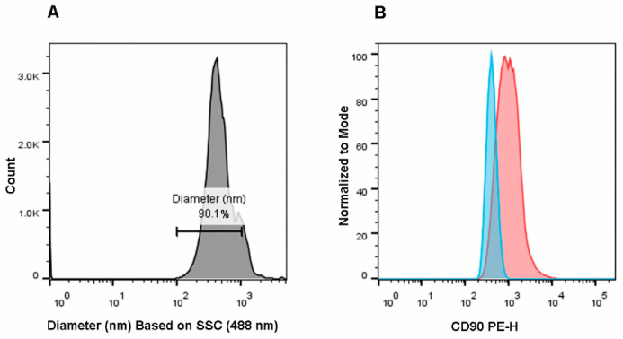 Figure 1
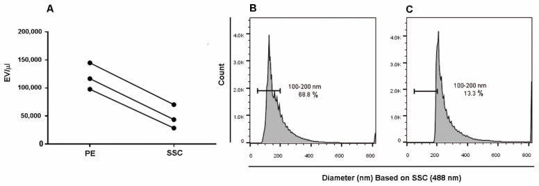 Figure 2
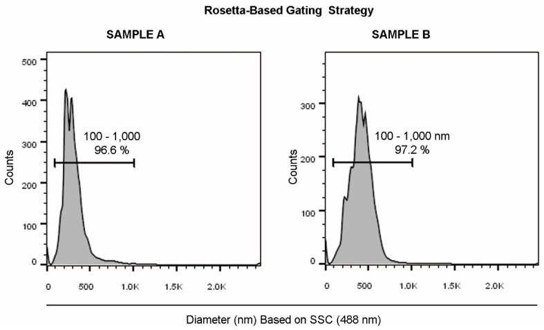 Figure 3
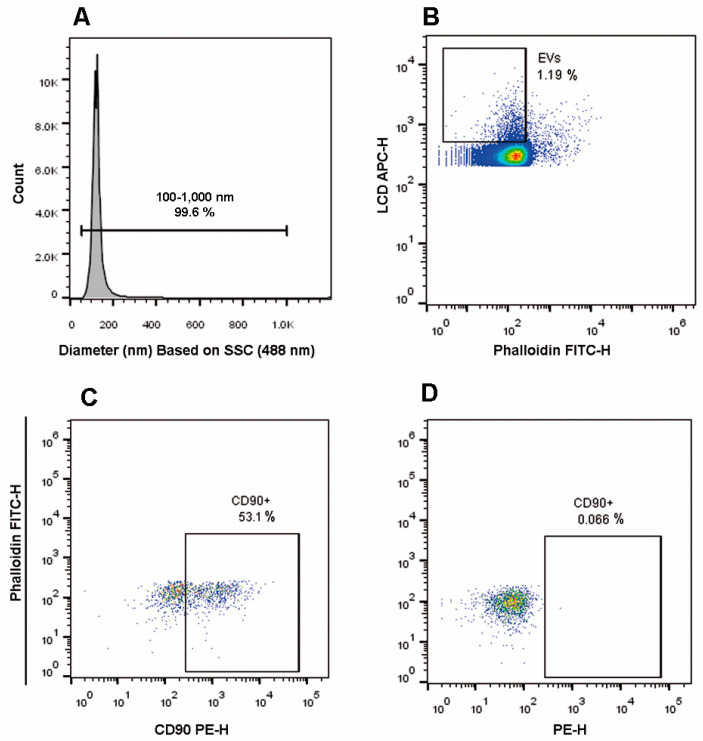 Figure 4
Peer Reviews
No public reviews on file for this paper yet. If you reviewed it on a platform where reviews are public (OpenReview, ICLR, NeurIPS, ICML), you can paste yours below so the community can read it here.
Videos
No videos yet. Explain this paper in a talk, walkthrough, or lecture? Add one.
Taxonomy
TopicsExtracellular vesicles in disease · MicroRNA in disease regulation · Cell Adhesion Molecules Research