Inhibition of autophagy curtails visual loss in a model of autosomal dominant optic atrophy
Marta Zaninello, Konstantinos Palikaras, Deborah Naon, Keiko Iwata, Stephanie Herkenne, Ruben Quintana-Cabrera, Martina Semenzato, Francesca Grespi, Fred N. Ross-Cisneros, Valerio Carelli, Alfredo A. Sadun, Nektarios Tavernarakis, Luca Scorrano
TL;DR
This study shows that blocking autophagy can prevent vision loss in a mouse model of a genetic eye disorder caused by OPA1 mutations.
Contribution
The study identifies AMPK-induced autophagy as a novel therapeutic target for autosomal dominant optic atrophy.
Findings
AMPK and ULK1 accumulate at axonal hillocks in retinal ganglion cells with mutated Opa1, triggering autophagy.
Genetic inhibition of AMPK and autophagy restores mitochondrial content in axons affected by Opa1 mutations.
Depletion of Atg7 in mice reverses visual defects caused by Opa1 deficiency, highlighting autophagy as a targetable pathway.
Abstract
In autosomal dominant optic atrophy (ADOA), caused by mutations in the mitochondrial cristae biogenesis and fusion protein optic atrophy 1 (Opa1), retinal ganglion cell (RGC) dysfunction and visual loss occur by unknown mechanisms. Here, we show a role for autophagy in ADOA pathogenesis. In RGCs expressing mutated Opa1, active 5’ AMP-activated protein kinase (AMPK) and its autophagy effector ULK1 accumulate at axonal hillocks. This AMPK activation triggers localized hillock autophagosome accumulation and mitophagy, ultimately resulting in reduced axonal mitochondrial content that is restored by genetic inhibition of AMPK and autophagy. In C. elegans, deletion of AMPK or of key autophagy and mitophagy genes normalizes the axonal mitochondrial content that is reduced upon mitochondrial dysfunction. In conditional, RGC specific Opa1-deficient mice, depletion of the essential autophagy gene…
Genes, proteins, chemicals, diseases, species, mutations and cell lines named across the full text — each resolved to its canonical identifier and authoritative record.
Click any figure to enlarge with its caption.
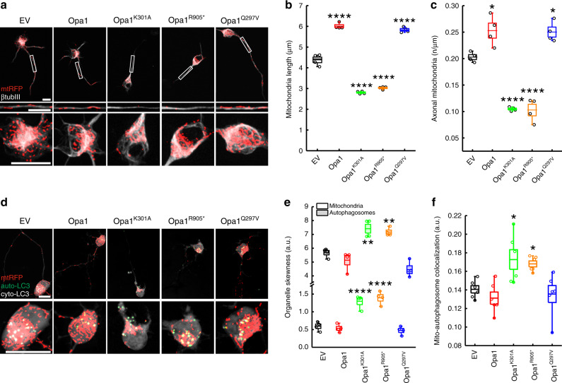 Figure 1
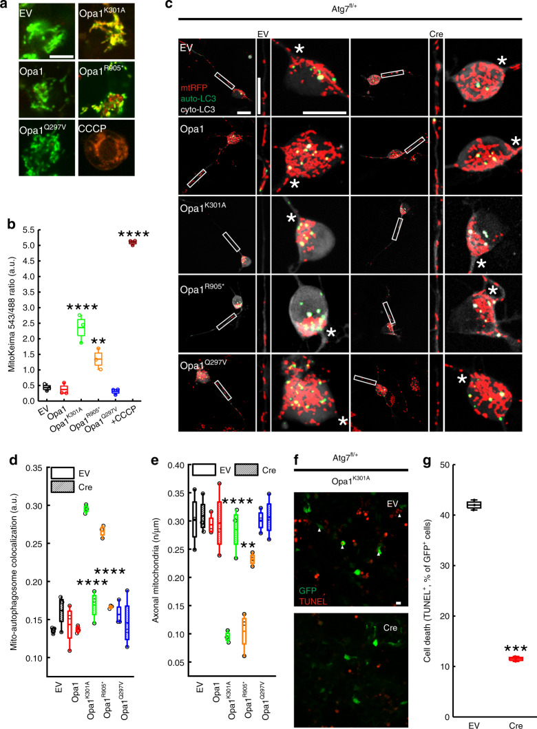 Figure 2
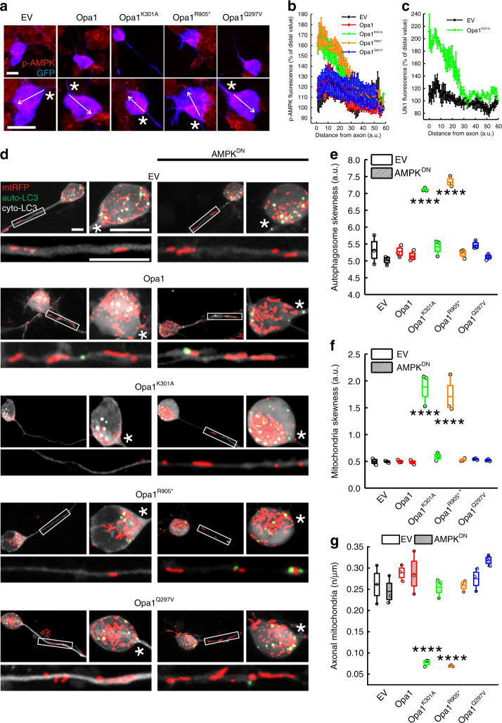 Figure 3
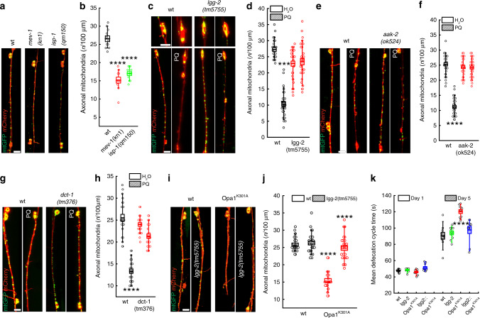 Figure 4
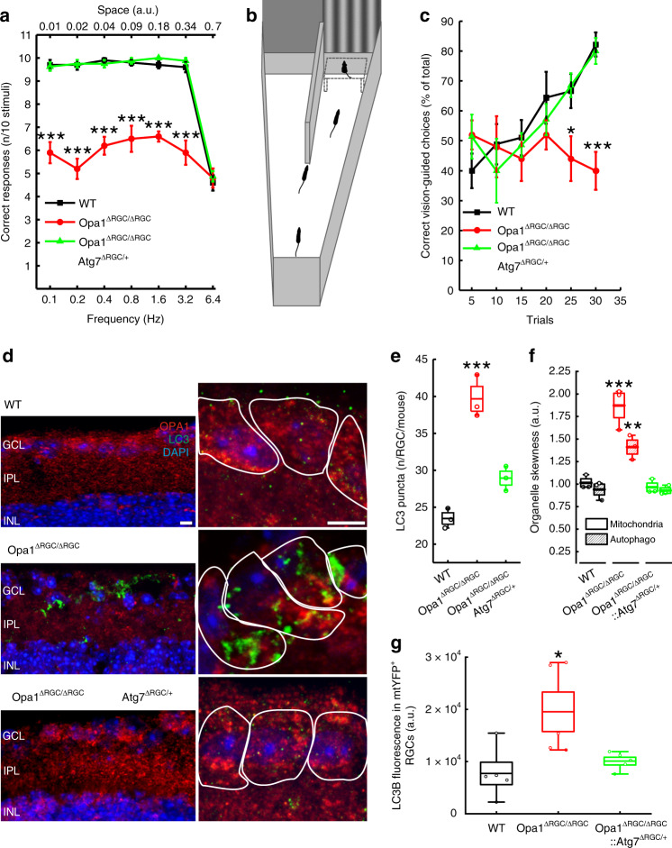 Figure 5
Peer Reviews
No public reviews on file for this paper yet. If you reviewed it on a platform where reviews are public (OpenReview, ICLR, NeurIPS, ICML), you can paste yours below so the community can read it here.
Videos
No videos yet. Explain this paper in a talk, walkthrough, or lecture? Add one.
Taxonomy
TopicsMitochondrial Function and Pathology · Autophagy in Disease and Therapy · Endoplasmic Reticulum Stress and Disease