Arteriovenous and Intercoronary Fistulae Presenting as Heart Failure in an Adult
Emmanouil Petrou, Chrysafios Girasis

Abstract
Genes, proteins, chemicals, diseases, species, mutations and cell lines named across the full text — each resolved to its canonical identifier and authoritative record.
Click any figure to enlarge with its caption.
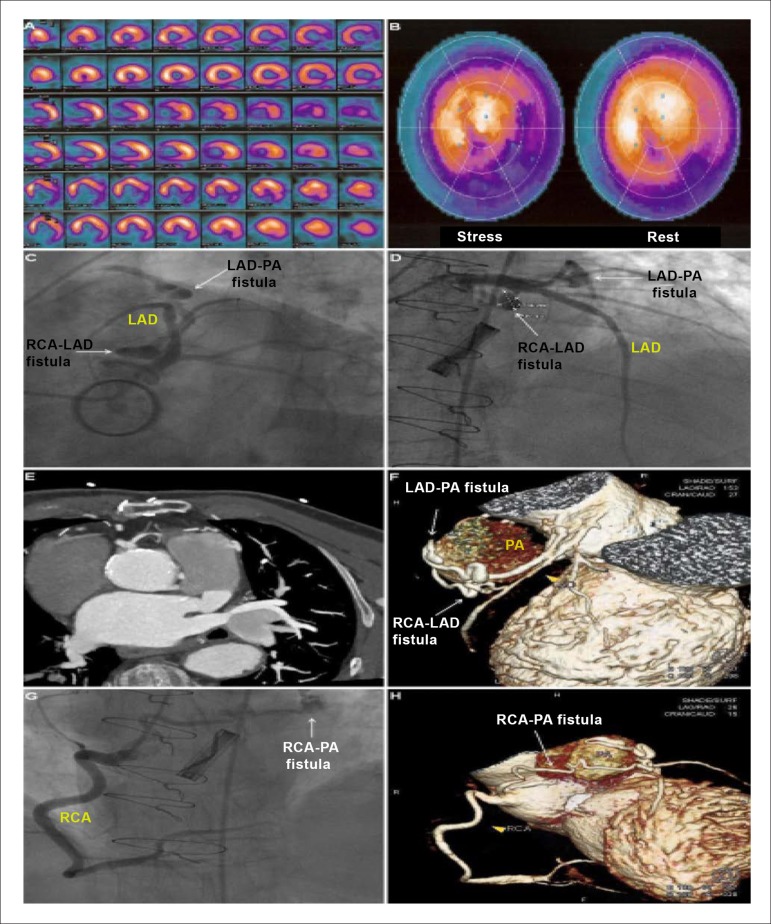 Figure 1
Peer Reviews
No public reviews on file for this paper yet. If you reviewed it on a platform where reviews are public (OpenReview, ICLR, NeurIPS, ICML), you can paste yours below so the community can read it here.
Videos
No videos yet. Explain this paper in a talk, walkthrough, or lecture? Add one.
Taxonomy
TopicsCoronary Artery Anomalies · Vascular anomalies and interventions · Congenital Heart Disease Studies
A 79-year-old man with prior aortic valve replacement was admitted to our hospital due to exertional angina. Single-photon emission computed tomography revealed ischemia at the territory of the left anterior descending artery (LAD) and the right coronary artery (RCA) (Figures 1 A and B). Coronary arteriography (Figures 1 C and D) and cardiac computed tomographic angiography (Figure 1 E), including volume rendering reconstruction (Figure 1 F), revealed an arteriovenous fistula between the proximal LAD and the pulmonary artery (PA). There was another fistula between the ostium of the RCA and the PA (Figures 1 G and H). This vessel gave origin to a small branch to the middle part of the LAD, before reaching the PA, thus forming a third, intercoronary fistula (Figures C, D and F).