Elderly Woman with Abdominal Pain: Bedside Ultrasound Diagnosis of Diverticulitis
Jason D. Heiner

Abstract
Genes, proteins, chemicals, diseases, species, mutations and cell lines named across the full text — each resolved to its canonical identifier and authoritative record.
Click any figure to enlarge with its caption.
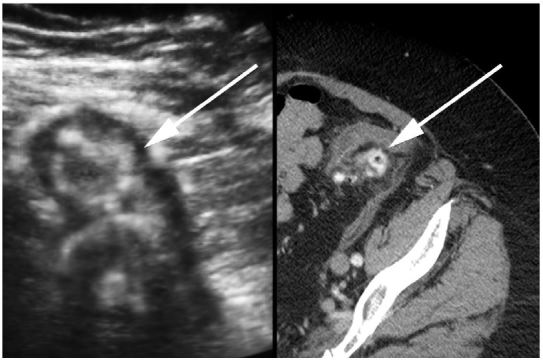 Figure 1
Peer Reviews
No public reviews on file for this paper yet. If you reviewed it on a platform where reviews are public (OpenReview, ICLR, NeurIPS, ICML), you can paste yours below so the community can read it here.
Videos
No videos yet. Explain this paper in a talk, walkthrough, or lecture? Add one.
Taxonomy
TopicsDiverticular Disease and Complications · Gastrointestinal disorders and treatments · Appendicitis Diagnosis and Management
A 72-year-old otherwise healthy female presented to the emergency department with two weeks of worsening abdominal pain. She was afebrile with normal vital signs. Her physical examination was notable for moderate abdominal tenderness without rebound to the left and suprapubic regions of the abdomen. Laboratory studies were remarkable for a white blood cell count of 13,000/mm^3^. A focused bedside ultrasound over the patient’s region of maximal discomfort revealed a thickened bowel wall and several small contiguous hypoechoic projections surrounding a hyperechoic center, suggestive of diverticulitis (Figure). She was given metronidazole and ciprofloxacin and her diagnosis of uncomplicated colonic diverticulitis was confirmed by computed tomography (CT) (Figure).
Acute diverticulitis resulting from inflammation of colonic diverticulum affects over half the population greater 65 years of age.1,2 While an estimated 85% of cases resolve with nonoperative care, complications such as large abscesses, fistula formation, perforation, and peritonitis do occur.1 CT is typically employed to diagnose presumed diverticulitis and recognize the presence of complicated disease, but for several decades ultrasound has been increasingly described as a similarly useful imaging modality.1–5
Particularly in cases of suspected uncomplicated diverticulitis, abdominal ultrasound may reach the diagnostic reliability of CT.1,2,4,5 Ultrasound may detect edema leading to loss of normal bowel architecture, identify inflamed diverticula, and expose mesenteric or omental fat.2–4 Key described songraphic findings include the following: edematous diverticula with thickened hypoechoic walls and hyperechoic centers, air containing diverticula with subsequent hyperechoic acoustic shadowing artifact, enlarged colonic walls greater then 5mm, and surrounding hyperechoic zones representing inflamed fat.1–4 Focused bedside imaging of the area of pain and tenderness may aid in initiation of early antibiotic treatment pending any additional confirmatory studies, but imaging can be hindered by neighboring bowel gas and CT or other complementary imaging may be warranted to search for complications or reveal alternative diagnoses.1,2
The reference list from the paper itself. Each links out to its DOI / PubMed record.
- 1Mazzei MA Cioffi Squitieri N Guerrini S Sigmoid diverticulitis: US findings Crit Ultrasound J 2013155 Suppl 1S 52390279110.1186/2036-7902-5-S 1-S 5PMC 3711739 · doi ↗ · pubmed ↗
- 2Helou N Abdalkader M Abu-Rustum RS Sonography: first-line modality in the diagnosis of acute colonic diverticulitis?J Ultrasound Med 201332101689942406524810.7863/ultra.32.10.1689 · doi ↗ · pubmed ↗
- 3Maturen KE Wasnik AP Kamaya A Ultrasound imaging of bowel pathology: technique and keys to diagnosis in the acute abdomen AJR Am J Roentgenol 20111976 W 1067752210932110.2214/AJR.11.6594 · doi ↗ · pubmed ↗
- 4Vijayaraghavan SB High-resolution sonographic spectrum of diverticulosis, diverticulitis, and their complications J Ultrasound Med 200625175851637155710.7863/jum.2006.25.1.75 · doi ↗ · pubmed ↗
- 5King WC Shuaib W Vijayasarathi A Benefits of sonography in diagnosing suspected uncomplicated acute diverticulitis J Ultrasound Med 20153415382554293910.7863/ultra.34.1.53 · doi ↗ · pubmed ↗