Open lunate enucleation
Monsef El Abdi, Adil Lamkhanter

Abstract
Genes, proteins, chemicals, diseases, species, mutations and cell lines named across the full text — each resolved to its canonical identifier and authoritative record.
Click any figure to enlarge with its caption.
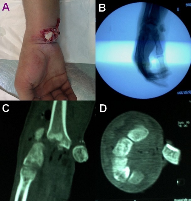 Figure 1
Peer Reviews
No public reviews on file for this paper yet. If you reviewed it on a platform where reviews are public (OpenReview, ICLR, NeurIPS, ICML), you can paste yours below so the community can read it here.
Videos
No videos yet. Explain this paper in a talk, walkthrough, or lecture? Add one.
Taxonomy
TopicsOrthopedic Surgery and Rehabilitation · Elbow and Forearm Trauma Treatment · Congenital limb and hand anomalies
Image in medicine
A 32 year old man, right-handed soldier, was admitted to Emergency Department two hours after undergoing right wrist injury following a 2 meter fall onto the hand, in hyperextension. Clinical examination revealed a deformity of the wrist with an obvious open lunate enucleation (A). The neurovascular status was intact. Standard radiograph of the wrist demonstrated a completely enucleated lunate, associated with scaphoid and radial styloid fracture (B). Computed tomography (CT) scan confirmed diagnosis of trans-radial styloid, trans-scaphoid, perilunate dislocation (C and D). The lunate enucleation was treated by open reduction through a volar approach and internal fixation of associated injuries. Wrist reduction was maintained using K-wires placed through the scapholunate and scaphocapitate. The material was removed after 3 months. The control during one year postoperatively, there was no evidence of complications. The patient had a comfortable range of motion in his right wrist.
(A) clinical aspect at admission revealing an open enucleation of a carpal bone, (B) X-ray of the left wrist shows a lunate dislocation, (C and D) computed tomography scan (transverse and sagittal cut) shows lunate enucleation with scaphoid and radial styloid fracture