Vitamin D Can Ameliorate Chlorhexidine Gluconate-Induced Peritoneal Fibrosis and Functional Deterioration through the Inhibition of Epithelial-to-Mesenchymal Transition of Mesothelial Cells
Yi-Che Lee, Shih-Yuan Hung, Hung-Hsiang Liou, Tsun-Mei Lin, Chu-Hung Tsai, Sheng-Hsiang Lin, Yau-Sheng Tsai, Min-Yu Chang, Hsi-Hao Wang, Li-Chun Ho, Yi-Ting Chen, Ching-Fang Wu, Ho-Ching Chen, Hsin-Pao Chen, Kuang-Wen Liu, Chih-I. Chen, Kuan Min She, Hao-Kuang Wang, Chi-Wei Lin

TL;DR
Vitamin D helps reduce peritoneal fibrosis and damage caused by a dialysis solution by blocking cell transformation in rats and human cells.
Contribution
The study reveals a novel mechanism by which vitamin D inhibits epithelial-to-mesenchymal transition in mesothelial cells, preventing peritoneal fibrosis.
Findings
1α,25(OH)2D3 reduces chlorhexidine gluconate-induced fibrosis and functional deterioration in rat peritoneum.
Vitamin D inhibits TGF-β1-induced epithelial-to-mesenchymal transition markers and migration in human mesothelial cells.
Vitamin D treatment prevents cytoskeleton changes and E-cadherin downregulation in peritoneal cells.
Abstract
Background. Peritoneal dialysis (PD) can induce fibrosis and functional alterations in PD patients' peritoneal membranes, due to long-term unphysiological dialysate exposure, partially occurring via triggering of epithelial-to-mesenchymal transition (EMT) in peritoneal mesothelial cells (MCs). Vitamin D can ameliorate these negative effects; however, the mechanism remains unexplored. Therefore, we investigated its possible links to MCs EMT inhibition. Methods. Peritoneal fibrosis was established in Sprague-Dawley rats by chlorhexidine gluconate (CG) intraperitoneal injection for 21 days, with and without 1α,25(OH)2D3 treatment. Morphological and functional evaluation and western blot analysis of EMT marker were performed upon peritoneum tissue. In vitro study was also performed in a primary human peritoneal MC culture system; MCs were incubated with transforming growth factor-β1…
Genes, proteins, chemicals, diseases, species, mutations and cell lines named across the full text — each resolved to its canonical identifier and authoritative record.
Click any figure to enlarge with its caption.
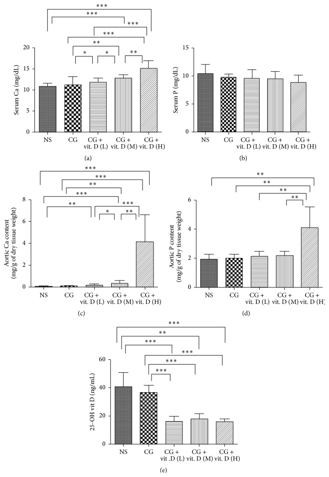 Figure 1
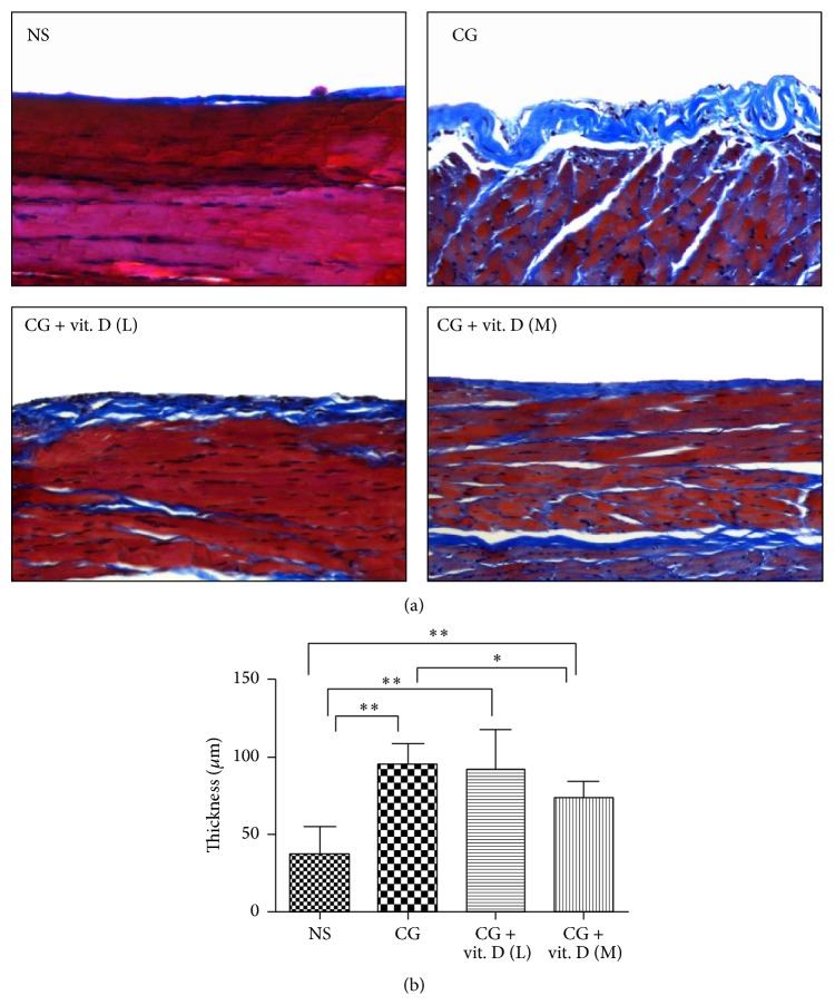 Figure 2
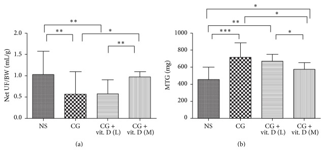 Figure 3
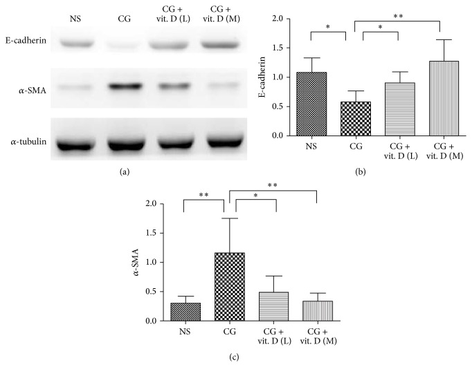 Figure 4
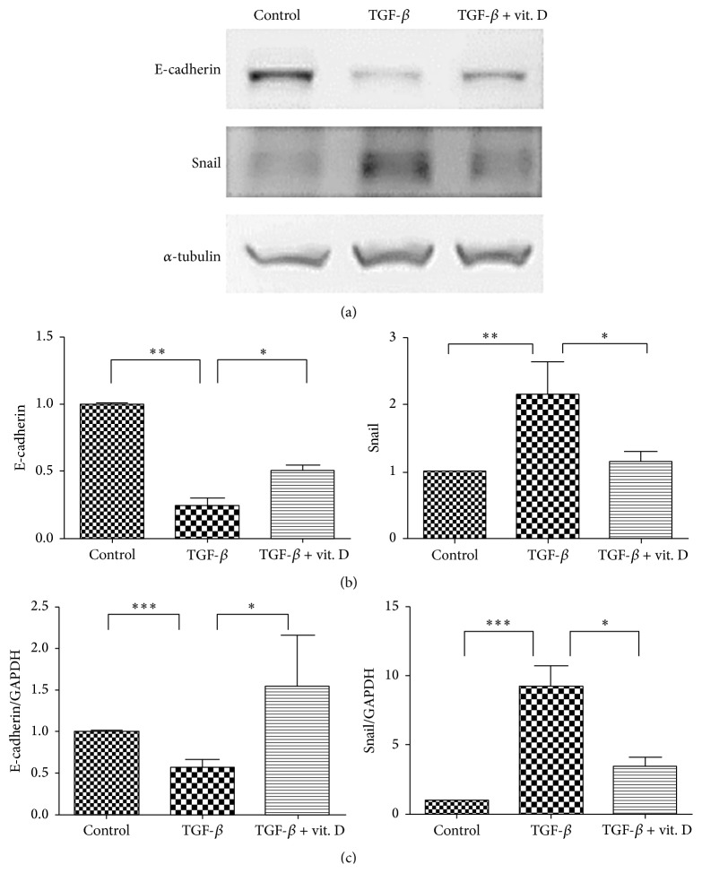 Figure 5
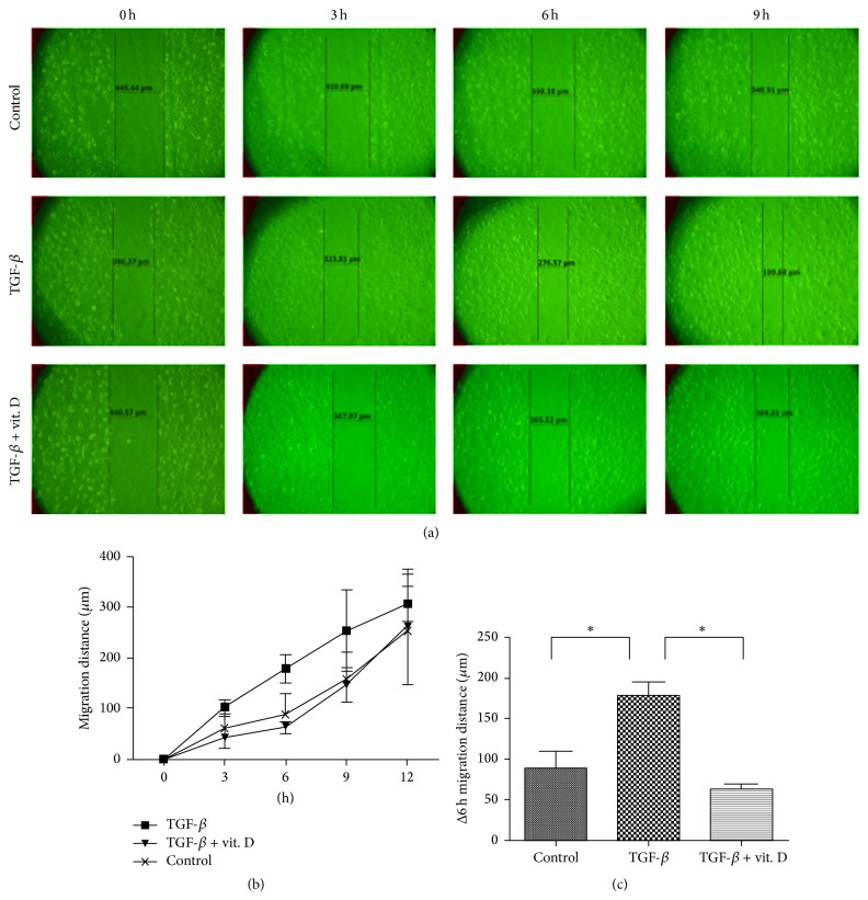 Figure 6
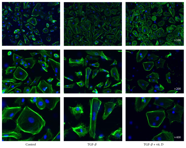 Figure 7
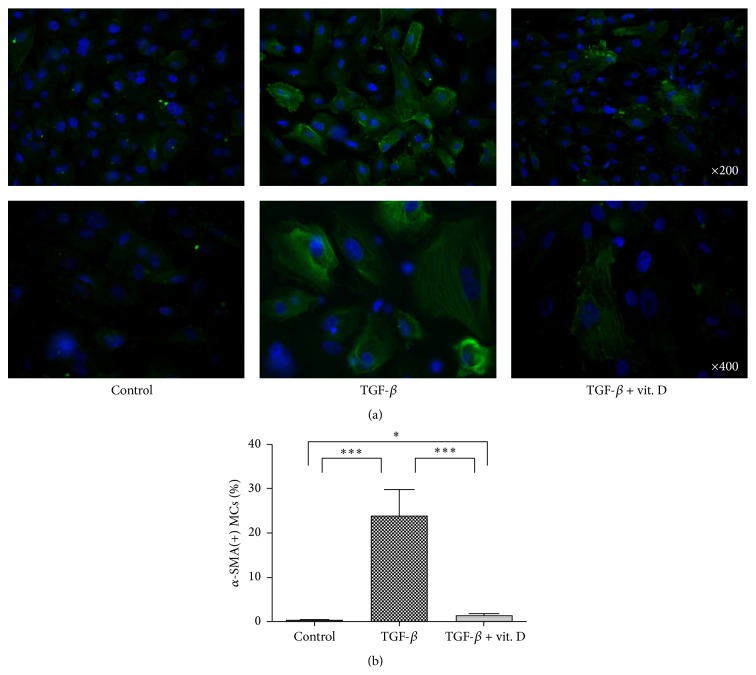 Figure 8
Peer Reviews
No public reviews on file for this paper yet. If you reviewed it on a platform where reviews are public (OpenReview, ICLR, NeurIPS, ICML), you can paste yours below so the community can read it here.
Videos
No videos yet. Explain this paper in a talk, walkthrough, or lecture? Add one.
Taxonomy
TopicsNeuropeptides and Animal Physiology · Thyroid Disorders and Treatments