Left Ventricular Pseudoaneurysm Secondary to Mitral Valve Endocarditis
Bruno Bochard-Villanueva, Jordi Estornell-Erill

Abstract
Genes, proteins, chemicals, diseases, species, mutations and cell lines named across the full text — each resolved to its canonical identifier and authoritative record.
Click any figure to enlarge with its caption.
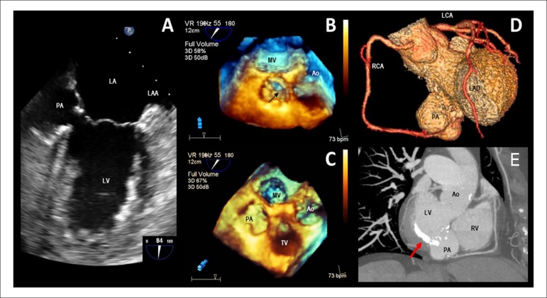 Figure 1
Peer Reviews
No public reviews on file for this paper yet. If you reviewed it on a platform where reviews are public (OpenReview, ICLR, NeurIPS, ICML), you can paste yours below so the community can read it here.
Videos
No videos yet. Explain this paper in a talk, walkthrough, or lecture? Add one.
Taxonomy
TopicsCardiac Structural Anomalies and Repair · Infective Endocarditis Diagnosis and Management · Cardiac Valve Diseases and Treatments
A follow-up transesophageal echocardiography (TEE) was performed on a 76-year-old woman with a recent history of mitral valve endocarditis after 4 weeks of antibiotic treatment. TEE showed a pulsatile perivalvular echo-free space of 32 × 23 mm with a narrow orifice which communicated to the left ventricle at the posterior mitral subannular position consistent with the pseudoaneurysm (Panel A). The real-time three-dimensional TEE allowed us to see its relationship with the neighboring structures (Panels B and C). Subsequently, a coronary CT angiogram confirmed these findings and revealed no significant coronary stenosis (Panels D and E). Therefore, surgery was indicated and a bovine pericardium patch was implanted with good results.