Using PET/CT Bone Scan Dynamic Data to Evaluate Tibia Remodeling When a Taylor Spatial Frame Is Used: Short and Longer Term Differences
Henrik Lundblad, Gerald Q. Maguire, Charlotte Karlsson-Thur, Cathrine Jonsson, Marilyn E. Noz, Michael P. Zeleznik, Hans Jacobsson, Lars Weidenhielm

TL;DR
This study uses PET/CT scans to assess tibia bone remodeling in patients treated with a Taylor Spatial Frame, comparing dynamic data with static methods.
Contribution
A Patlak-like analysis is introduced to approximate blood activity without blood samples, enabling dynamic evaluation of bone remodeling.
Findings
Patlak-like and SUVm slopes showed strong agreement, suggesting static scans can replace dynamic studies.
Positive Patlak-like slope differences of 0.1 min−1 or greater and SUVmax differences of ~5 indicate good remodeling progress.
Negative Patlak-like slope differences of −0.06 min−1 suggest poor remodeling progress in the studied cohort.
Abstract
Eighteen consecutive patients, treated with a Taylor Spatial Frame for complex tibia conditions, gave their informed consent to undergo Na18F− PET/CT bone scans. We present a Patlak-like analysis utilizing an approximated blood time-activity curve eliminating the need for blood aliquots. Additionally, standardized uptake values (SUV) derived from dynamic acquisitions were compared to this Patlak-like approach. Spherical volumes of interest (VOIs) were drawn to include broken bone, other (normal) bone, and muscle. The SUVm(t) (m = max, mean) and a series of slopes were computed as (SUVm(t i) − SUVm(t j))/(t i − t j), for pairs of time values t i and t j. A Patlak-like analysis was performed for the same time values by computing ((VOIp(t i)/VOIe(t i))−(VOIp(t j)/VOIe(t j)))/(t i − t j), where p = broken bone, other bone, and muscle and e = expected activity in a VOI. Paired comparisons…
Genes, proteins, chemicals, diseases, species, mutations and cell lines named across the full text — each resolved to its canonical identifier and authoritative record.
Click any figure to enlarge with its caption.
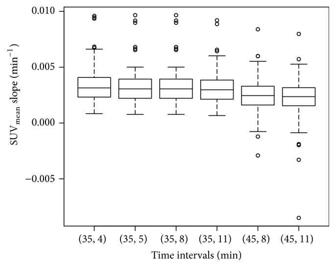 Figure 1
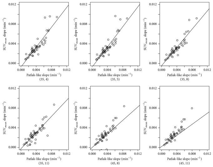 Figure 2
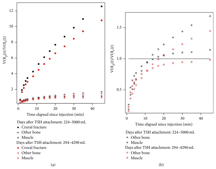 Figure 3
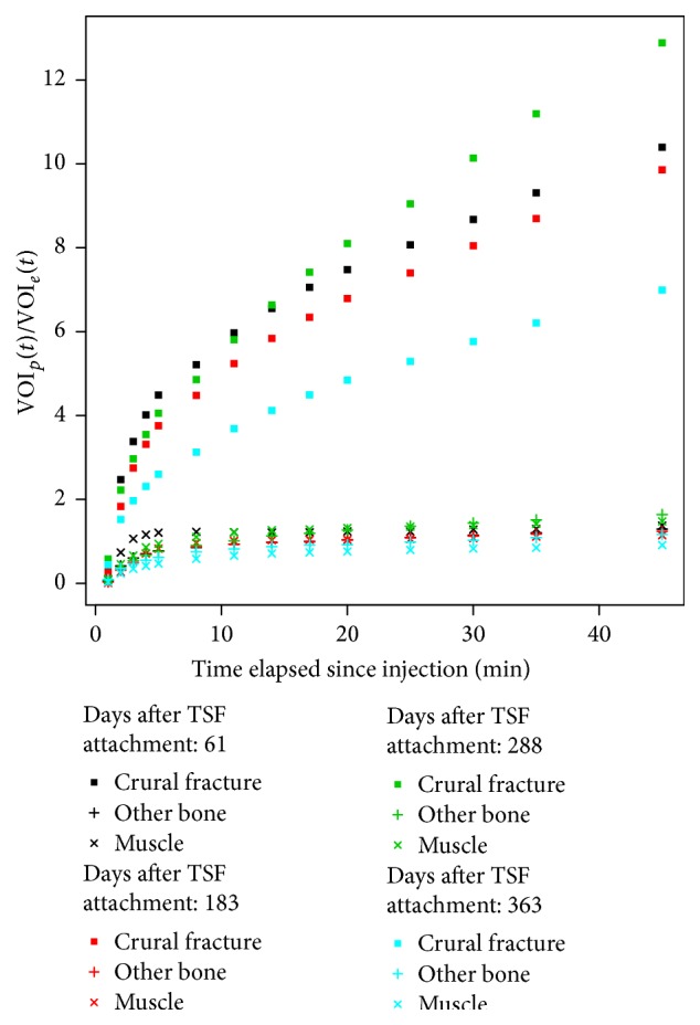 Figure 4
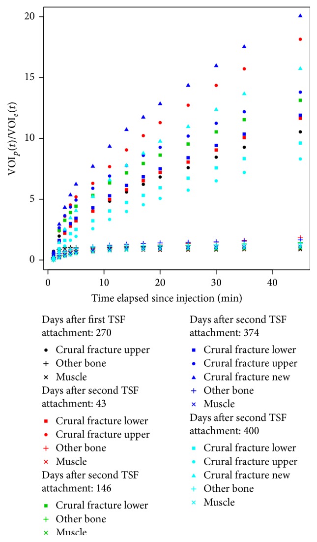 Figure 5
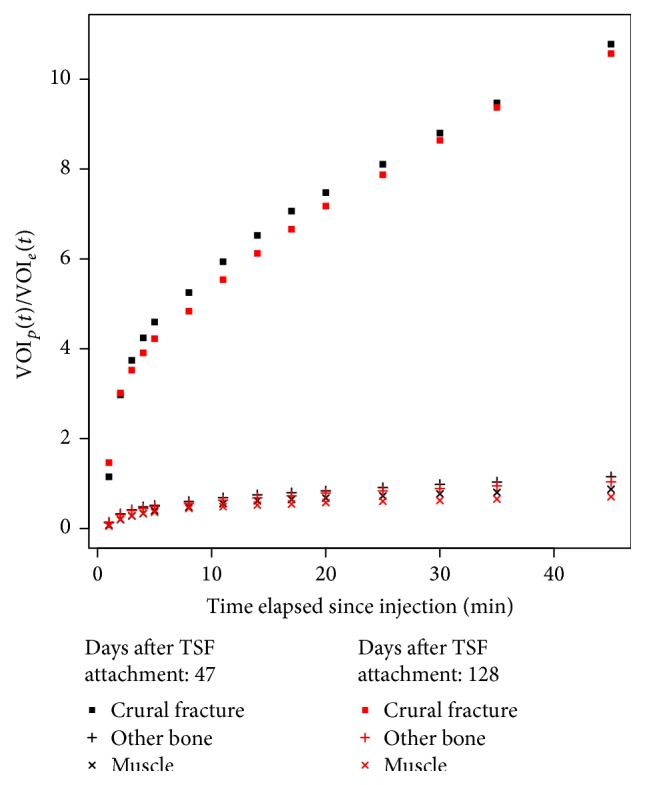 Figure 6
Peer Reviews
No public reviews on file for this paper yet. If you reviewed it on a platform where reviews are public (OpenReview, ICLR, NeurIPS, ICML), you can paste yours below so the community can read it here.
Videos
No videos yet. Explain this paper in a talk, walkthrough, or lecture? Add one.
Taxonomy
TopicsTotal Knee Arthroplasty Outcomes · Medical Imaging Techniques and Applications · Bone fractures and treatments