Giant Right Atrial Mass Following Surgical Aortic Valve Replacement
Teresa Bastante, Fernando Alfonso

Abstract
Genes, proteins, chemicals, diseases, species, mutations and cell lines named across the full text — each resolved to its canonical identifier and authoritative record.
Click any figure to enlarge with its caption.
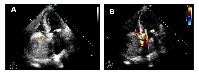 Figure 1
Peer Reviews
No public reviews on file for this paper yet. If you reviewed it on a platform where reviews are public (OpenReview, ICLR, NeurIPS, ICML), you can paste yours below so the community can read it here.
Videos
No videos yet. Explain this paper in a talk, walkthrough, or lecture? Add one.
Taxonomy
TopicsCardiac Structural Anomalies and Repair · Pericarditis and Cardiac Tamponade · Cardiac tumors and thrombi
A 75-year-old man underwent elective biological aortic valve replacement. Two weeks after surgery, transthoracic echocardiography (TTE) showed a small pericardial effusion. On the following day, the patient suffered from syncope. A repeated TTE revealed a "giant" echo-dense mass (90x80 mm) occupying the entire right atrium and severely limiting tricuspid valve inflow. Although the initial differential diagnosis included the development of an intracavitary process, the rapidly-growing mass with a characteristic echo‑lucent layer at its atrial aspect (consistent with the atrial wall and visceral pericardium) (arrows) led to the final diagnosis of a pericardial hematoma mimicking a huge atrial mass. Emergency surgical exploration confirmed the diagnosis.