Anthropomorphic Phantoms for Confirmation of Linear Accelerator-Based Small Animal Irradiation
Julian R Perks, Steven Lucero, Arta M Monjazeb, Jian Jian Li

TL;DR
This paper describes using 3D-printed mouse models to improve the accuracy of radiation dosing for small animal experiments using a human-based linear accelerator.
Contribution
The novel use of anthropomorphic phantoms to refine small animal irradiation accuracy to within 1% of the prescribed dose.
Findings
3D scanning and printing technology was used to create accurate mouse phantom models.
Phantom models enabled dose delivery refinement to within 1% of the prescribed dose.
The phantoms can simulate various tumor types for immunotherapy research.
Abstract
Three dimensional (3D) scanning and printing technology is utilized to create phantom models of mice in order to assess the accuracy of ionizing radiation dosing from a clinical, human-based linear accelerator. Phantoms are designed to simulate a range of research questions, including irradiation of lung tumors and primary subcutaneous or orthotopic tumors for immunotherapy experimentation. The phantoms are used to measure the accuracy of dose delivery and then refine it to within 1% of the prescribed dose.
Genes, proteins, chemicals, diseases, species, mutations and cell lines named across the full text — each resolved to its canonical identifier and authoritative record.
Click any figure to enlarge with its caption.
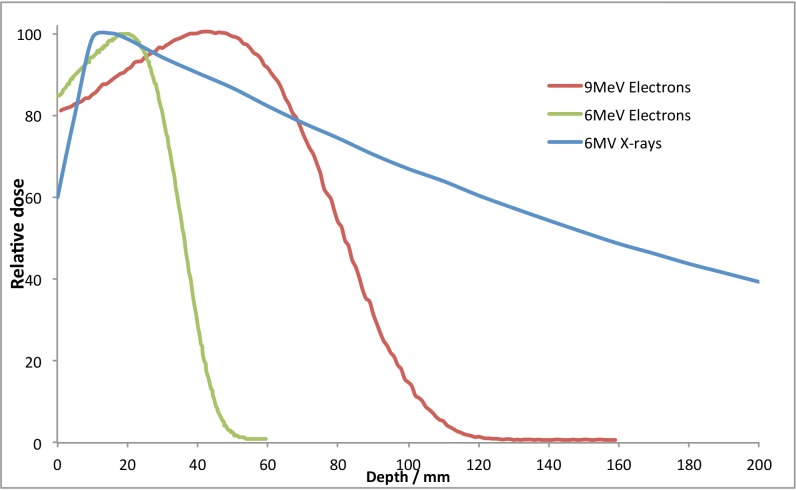 Figure 1
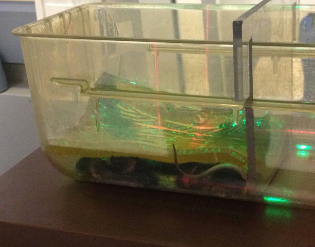 Figure 2
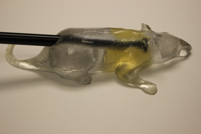 Figure 3
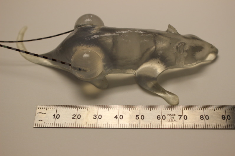 Figure 4
Peer Reviews
No public reviews on file for this paper yet. If you reviewed it on a platform where reviews are public (OpenReview, ICLR, NeurIPS, ICML), you can paste yours below so the community can read it here.
Videos
No videos yet. Explain this paper in a talk, walkthrough, or lecture? Add one.
Taxonomy
TopicsEffects of Radiation Exposure · Effects of Radiation Exposure · Advanced Radiotherapy Techniques