Abscessed Lumbar Hypodermatitis Revealing Pyonephrosis: A Diagnosis to Keep in Mind
Abdennasser Lakrabti, Ali Akjay, Jihad Anzaoui

TL;DR
A rare case shows how a kidney infection can appear as a skin abscess, highlighting the importance of recognizing unusual symptoms.
Contribution
Highlights a rare clinical presentation of pyonephrosis mimicking an abdominal wall abscess.
Findings
Pyonephrosis can present as an abscessed lumbar hypodermis, leading to diagnostic confusion.
Delayed diagnosis due to misinterpretation can have fatal consequences.
The case emphasizes the need for awareness among physicians about this rare manifestation.
Abstract
Pyonephrosis resulting from an obstructing calculus commonly presents with symptoms such as loin pain, fever, and signs indicative of a urinary tract infection. In some cases, significant thinning of the renal parenchyma in pyonephrosis may lead to direct rupture into the retroperitoneum, and exceptionally rarely, into the lumbar abdominal wall, potentially mimicking an isolated abdominal wall abscess, which can be mistaken for a complication of hypodermatitis. Our case is a good illustration of a condition that is not well known among practitioners, particularly family physicians and dermatologists. This lack of awareness can explain the diagnostic delay, which may sometimes result in the patient's death.
Genes, proteins, chemicals, diseases, species, mutations and cell lines named across the full text — each resolved to its canonical identifier and authoritative record.
Click any figure to enlarge with its caption.
 Figure 1
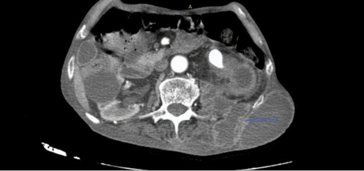 Figure 2
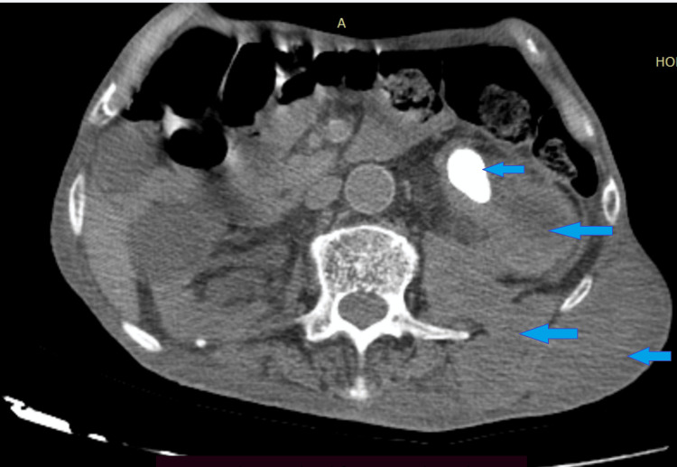 Figure 3
| Biological parameters | Before drainage | After drainage |
| White blood cell, cells/mL | 22000 | 8000 |
| C-reactive protein (CRP), mg/l | 260 | 30 |
| Creatinine, mg/l | 13 | 12 |
| Hg, g/l | 12 | 12 |
Peer Reviews
No public reviews on file for this paper yet. If you reviewed it on a platform where reviews are public (OpenReview, ICLR, NeurIPS, ICML), you can paste yours below so the community can read it here.
Videos
No videos yet. Explain this paper in a talk, walkthrough, or lecture? Add one.
Taxonomy
TopicsInfectious Diseases and Tuberculosis · Orthopedic Infections and Treatments · Omental and Epiploic Conditions
Introduction
Pyonephrosis is an infection of the upper renal-urinary system, which, over time, leads to suppurative destruction of the renal parenchyma [1]. Lithiasis is a common cause of this infection. Kidney stones are a common and urgent problem in urology, usually manifesting as acute colic. In some cases, urolithiasis remains asymptomatic for a long time and may manifest as complications such as lumbar wall abscess secondary to fistulization of pyonephrosis, which is a very rare complication reported in the literature.
Case presentation
An 86-year-old man, with no notable pathological history, was admitted to the emergency department with swelling of the left lumbar abdominal wall (Figure 1).
Left lumbar mass with overlying inflammatory skin
The patient was treated for hypodermatitis of the lumbar region, but with no improvement under antibiotic therapy. In view of the increasing size of the lesion, he consulted the emergency department.
Ultrasound and uroscanner revealed a renal pelvis calculus with pyonephrosis, a perineal abscess, and a psoas abscess extending to the left lumbar abdominal wall (Figures 2-3).
Axial image from the excretory phase of a contrast-enhanced abdominopelvic CT scanThe image demonstrates left pyonephrosis caused by an obstructive stone in the renal pelvis, complicated by a retroperitoneal abscess with fistulization, leading to a subcutaneous lumbar collection in the same region indicated by the blue arrow.
Axial image of an abdominopelvic CT scanThe image reveals left pyonephrosis caused by an obstructive renal pelvic stone, complicated by a retroperitoneal abscess with fistulization, leading to a subcutaneous lumbar collection in the affected region, indicated by blue arrows.
After percutaneous drainage, an Xpert® Mycobacterium tuberculosis/resistance to rifampin MTB/RIF test was carried out for tuberculosis, which proved negative. Biological parameters normalized and general condition improved, with recovery of autonomy after two months' treatment. A nephrectomy is planned to eliminate this undestroyed infection (Table 1).
Discussion
Nephrocutaneous fistula results from the spontaneous formation of an abnormal communication between the kidney and the skin. This fistula traverses the retroperitoneum and the structures of the abdominal wall following pathways of least resistance, such as Petit's triangle and the Grynfeld quadrilateral.
A retroperitoneal abscess, particularly a psoas abscess secondary to calyceal rupture in calculus-induced pyonephrosis, is exceedingly rare [2]. The detection of such a psoas abscess with computed tomography is nearly 100% [3].
In some instances, the abscess may extend into the muscles of the posterior abdominal wall, notably the quadratus lumborum, presenting as a lumbar abscess, as seen in our case. Management typically involves broad-spectrum antibiotics along with percutaneous or surgical drainage of pus via nephrostomy and ureteral stent placement [2]. Surgical removal of the affected kidney is often necessary, particularly when it is non-functioning or affects both the affected and normal-functioning contralateral kidney [4,5].
A thorough review of the existing French and English medical literatures identified fewer than seven reported cases of spontaneous rupture of pyonephrosis secondary to urolithiasis with psoas abscess formation [2,6,7]. In our case, an additional finding was the presence of a posterior abdominal wall abscess in the lumbar region, which has not been reported previously. However, there has been a reported case of lumbar panniculitis with a subcutaneous abscess secondary to pyonephrosis [6]. Additionally, literature describes rare occurrences of peritoneal rupture of pyonephrosis leading to peritonitis and splenic abscess [8,9].
Our case is a good illustration of a condition that is not well known among practitioners, particularly family physicians and dermatologists. This lack of awareness can explain the diagnostic delay, which may sometimes result in the patient's death. Our case is a good example of a pathology that should be considered by practitioners, whether general practitioners, dermatologists, or other specialists, to avoid any diagnostic delay.
Conclusions
Given the increased variability in the symptoms of pyonephrosis, it becomes evident that early diagnosis is crucial. Spontaneous rupture of pyonephrosis is a rare occurrence. Abscesses of the lumbar abdominal wall are a rare complication of pyonephrosis. Accurate diagnosis and thorough examination to identify the source of the abscess are essential prior to intervention. Inclusion of a CT scan of the abdomen in the standard protocol for evaluation of all lumbar abscesses is necessary to exclude a renal origin of the abdominal abscess.
The reference list from the paper itself. Each links out to its DOI / PubMed record.
- 1Pyonephrosis drained by double-J catheter Clin Case Rep Chang CW Huang CN 358535868202010.1002/ccr 3.3204 PMC 775260433363990 · doi ↗ · pubmed ↗
- 2Spontaneous rupture of a calculus pyonephrotic kidney into the retroperitoneal cavity presenting as psoas abscess - a case report Int J Biomed Adv Res Patil V Ichalakaranji R Patil SB Pattar R Ashrith IM 26226352014 https://digitallibrary.bldedu.ac.in/handle/123456789/1591
- 3Psoas abscess: imaging diagnosis of a rare entity Ann Int Med Den Res Kulkarni A Aafreen M Shetkar S Tinmaswala MA 222542018 https://aimdrjournal.com/wp-content/uploads/2021/09/RD 4_CR_PM-edit.pdf
- 4Acute pyelonephritis and pyelonephrosis Turk Klin J Surg Med Sci Eroğlu M KandıralıE 242832007 https://www.turkiyeklinikleri.com/article/en-akut-pyelonefrit-ve-pyonefroz-49715.html
- 5Pyonephrosis: diagnosis and treatment: a review of 14 cases (in French)Ann Urol Rabii R Joual A Rais H 161164342000 https://pubmed.ncbi.nlm.nih.gov/10953791/10953791 · pubmed ↗
- 6Lumbar panniculitis with subcutaneous abscess revealing pyonephrosis (in French)Ann Dermatol Venereol Pauwels C Bulai-Livideanu C Chiavassa H 72772913620091980125910.1016/j.annder.2009.02.006 · doi ↗ · pubmed ↗
- 7Spontaneous rupture of pyonephrosis presenting as anterior abdominal wall abscess: a rare case report Afr J Urol Singh KH Vyas A Rochlani T Patwardhan SK 117272021
- 8A rare complication of renal lithiasis: peritonitis and splenic abscess caused by rupture of pyonephrosis (in French)J Urol Hendaoui MS Abed A M'Saad W Chelli H Hendaoui L 1301331021996 https://pubmed.ncbi.nlm.nih.gov/9091559/9091559 · pubmed ↗