Traumatic brain injury exacerbates mitochondrial dysfunction in APP/PS1 knock-in mice through time-dependent pathways
Elika Z. Moallem, Hemendra J. Vekaria, Teresa Macheda, Margaret R. Hawkins, Kelly N. Roberts, Samir P. Patel, Patrick G. Sullivan, Adam D. Bachstetter

TL;DR
Traumatic brain injury temporarily worsens mitochondrial problems in mice with Alzheimer's disease traits, with males being more affected than females.
Contribution
The study reveals a time-dependent exacerbation of mitochondrial dysfunction in TBI and early amyloidosis, emphasizing sex differences.
Findings
At 1 month post-injury, KI mice showed greater mitochondrial dysfunction than either TBI or AD alone.
Males were more vulnerable to mitochondrial dysfunction than females at 1 month post-injury.
At 4–8 months post-injury, amyloid effects dominated, with TBI-specific changes no longer evident.
Abstract
Cerebral hypometabolism occurs in both traumatic brain injury (TBI) and Alzheimer’s disease (AD), but whether these conditions act through distinct or overlapping mechanisms is unclear. TBI disrupts cerebral metabolism via blood–brain barrier damage, altered glucose transporter expression, calcium buffering abnormalities, and oxidative damage to metabolic enzymes. AD-related hypometabolism is linked to amyloid-β (Aβ) effects on mitochondria, including impaired respiration, oxidative stress, and altered mitophagy, fusion, and fission. We tested whether TBI-induced mitochondrial dysfunction exacerbates Aβ-mediated impairment using a closed-head injury (CHI) model in APP/PS1 knock-in (KI) mice. Injuries were delivered at 4–5 months of age, before plaque formation and mitochondrial deficits in KI mice. Bioenergetics were measured at 1, 4, and 8 months post-injury in hippocampus and cortex…
Genes, proteins, chemicals, diseases, species, mutations and cell lines named across the full text — each resolved to its canonical identifier and authoritative record.
Click any figure to enlarge with its caption.
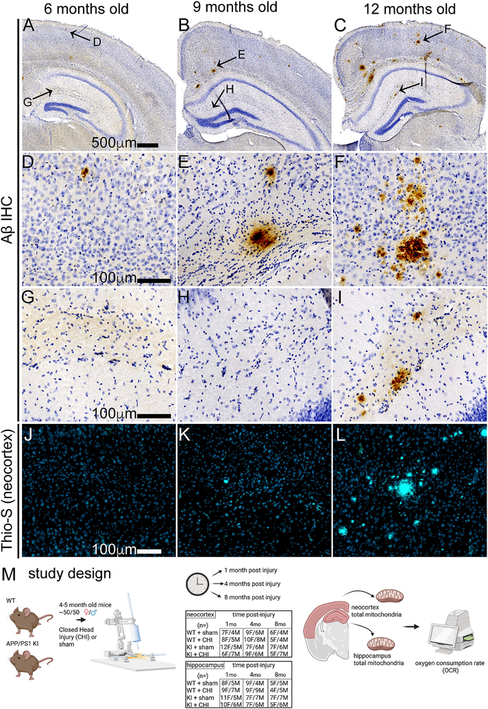 Figure 1
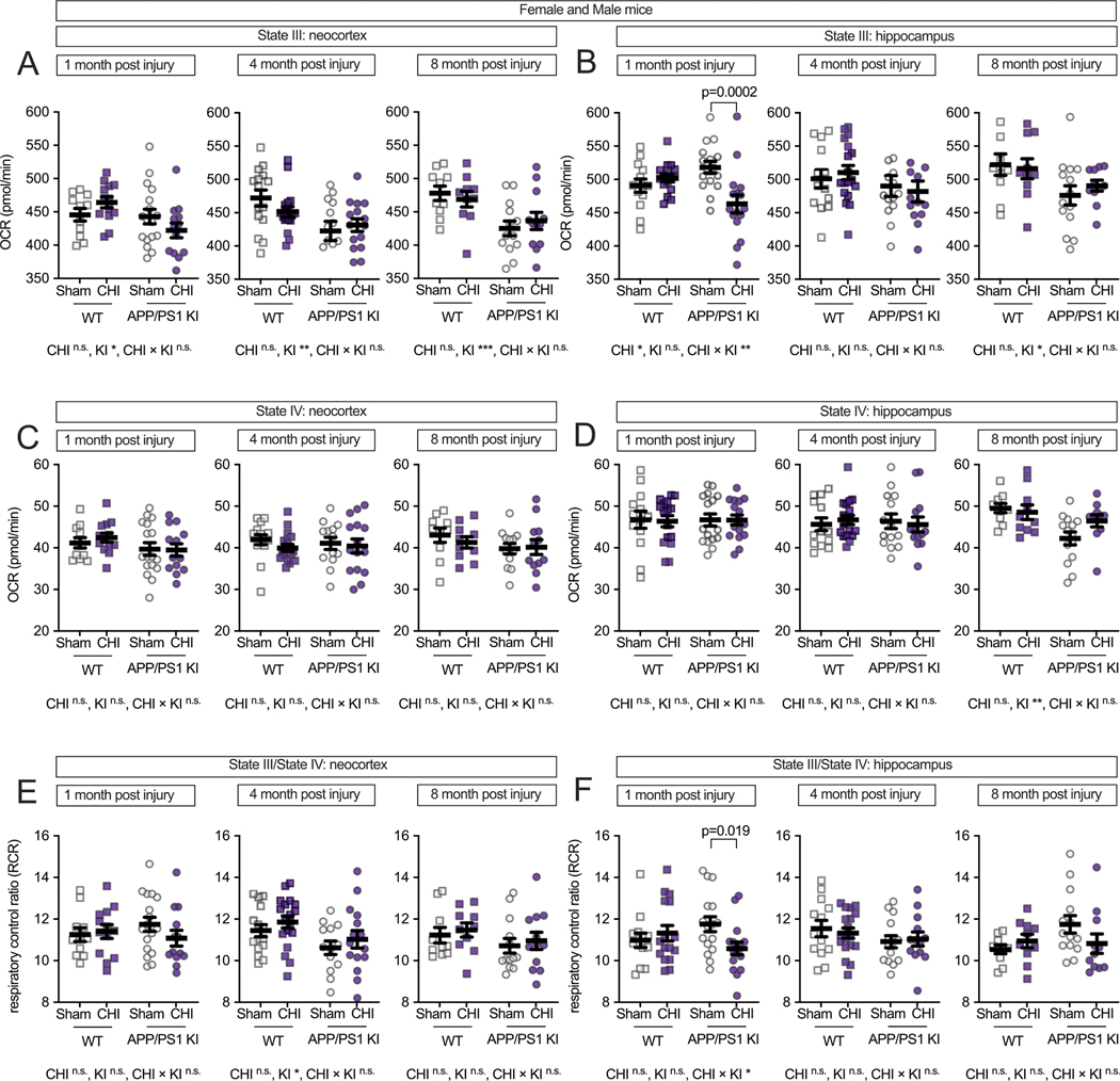 Figure 2
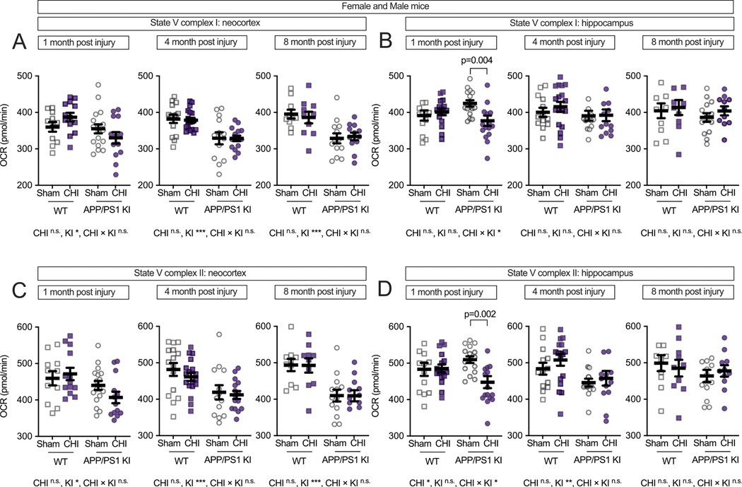 Figure 3
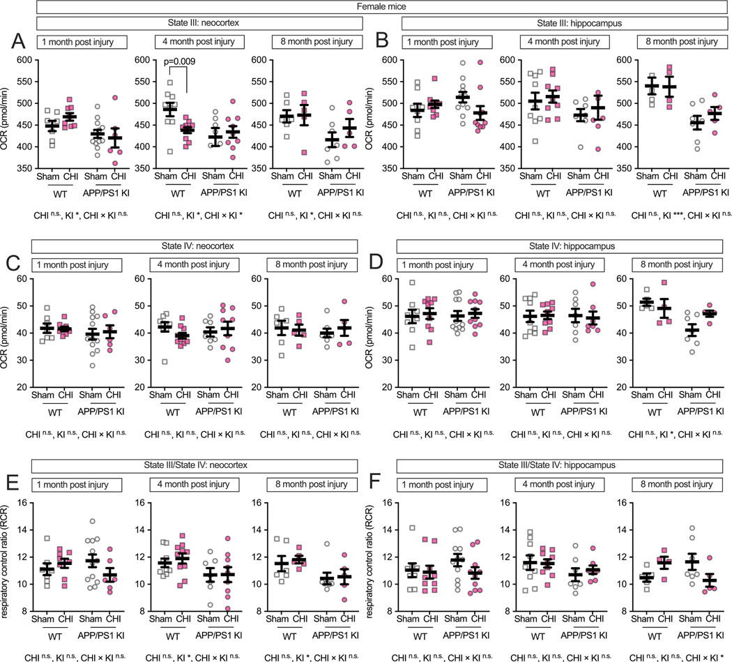 Figure 4
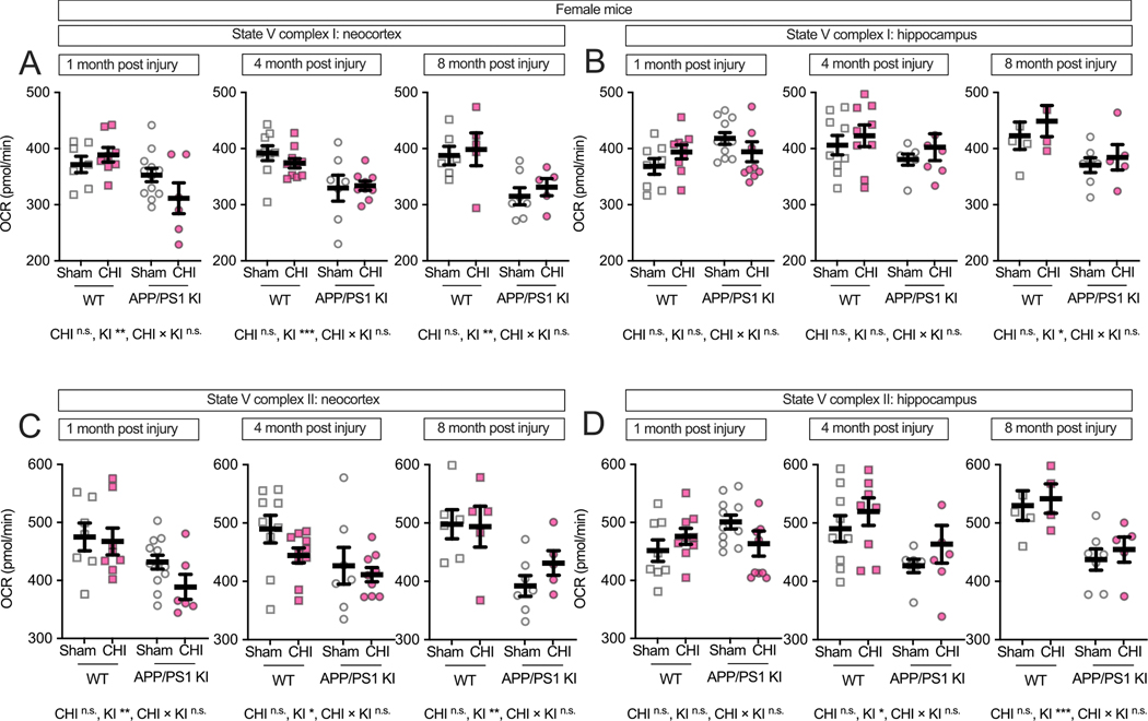 Figure 5
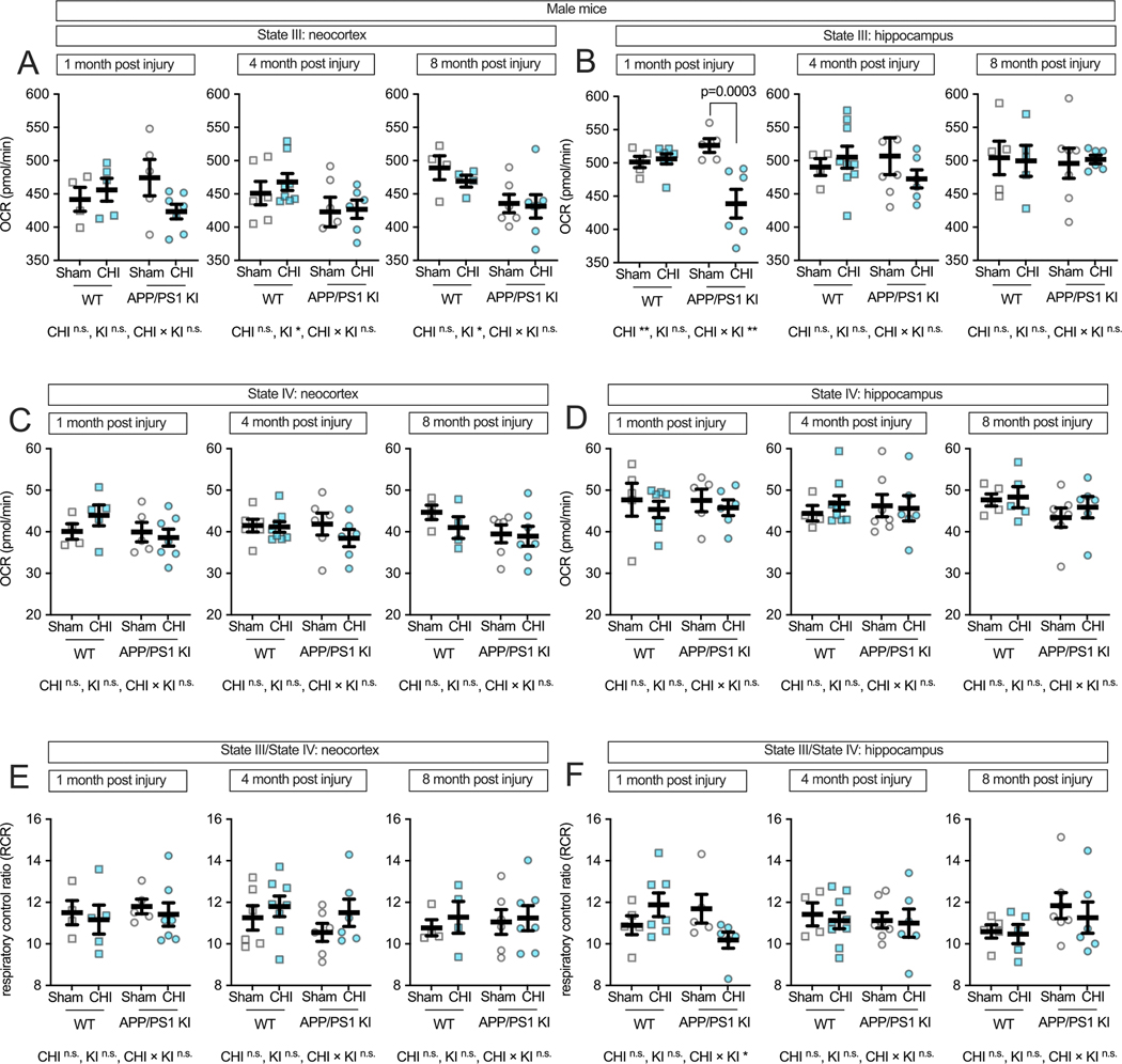 Figure 6
 Figure 7
Peer Reviews
No public reviews on file for this paper yet. If you reviewed it on a platform where reviews are public (OpenReview, ICLR, NeurIPS, ICML), you can paste yours below so the community can read it here.
Videos
No videos yet. Explain this paper in a talk, walkthrough, or lecture? Add one.
Taxonomy
TopicsTraumatic Brain Injury and Neurovascular Disturbances · Traumatic Brain Injury Research · Alzheimer's disease research and treatments
Introduction
Traumatic brain injury (TBI) is an environmental risk factor for Alzheimer’s disease (AD), with human population-level data showing a strong association between TBI and subsequent dementia development (Chiasseu et al., 2020; Cognacq et al., 2025; Dams-O’Connor et al., 2016; Shively et al., 2012). Mitochondrial dysfunction plays a central role in both conditions, contributing to cerebral hypometabolism, neuronal energy deficits, and oxidative stress in people and experimental models (Chandrasekaran et al., 2006; Cheng et al., 2012; Wang et al., 2020). In experimental TBI models, an initial compensatory hyperglycolysis and heightened oxygen utilization near the lesion site is typically followed by prolonged hypometabolism, with metabolic shifts including increased flux through the pentose phosphate pathway (PPP) to support DNA repair processes (Bartnik et al., 2005; Gribnau et al., 2024). These compensatory mechanisms are often insufficient to prevent long-term bioenergetic deficits, as evidenced by hyperglycemia and increased plasma glucose levels in severe TBI patients (Shi et al., 2016; Yuan et al., 2022). Studies using Seahorse and spectrophotometric assay measurements in experimental models have demonstrated that TBI and aging disrupts mitochondrial respiration, reducing adenosine triphosphate (ATP) production and impairing electron transport chain (ETC) function, particularly Complexes I, II and IV (Hubbard et al., 2019; Navarro and Boveris, 2007; Thapak et al., 2024; Vekaria et al., 2024). Mitochondria isolated from injured brains in controlled cortical impact (CCI) models display significant declines in oxidative phosphorylation capacity following CCI (Gilmer et al., 2010; Gilmer et al., 2009; Hill et al., 2017). Similarly, AD exhibits reduced brain glucose metabolism, particularly in temporal and parietal regions in people (Kantarci et al., 2010; Kyrtata et al., 2021), with animal models demonstrating decreased glucose utilization in the hippocampus and cortex (Griffith et al., 2019; Huang et al., 2020; Minhas et al., 2024; Raut et al., 2023). Several studies in experimental models have demonstrated that amyloid precursor protein (APP) and amyloid beta (Aβ) can localize to mitochondria, influencing mitochondrial dynamics and respiratory function (Hansson Petersen et al., 2008; Pinho et al., 2014; Strope and Wilkins, 2023; Wilkins, 2023). Furthermore, experimental model studies show that γ-secretase complexes within mitochondria have been shown to generate Aβ locally, suggesting mitochondrial dysfunction may directly contribute to intramitochondrial Aβ production (Hansson et al., 2004; Wilkins, 2023). Mouse models of cerebral amyloidosis exhibit early mitochondrial abnormalities, including increased mitophagy, reduced mitochondrial biogenesis, and decreased ATP production (Chen et al., 2019; de la Cueva et al., 2022; Zhang et al., 2015). Given these parallel metabolic deficits, TBI may potentially accelerate mitochondrial failure in those susceptible to Aβ pathology.
The aim of this study was to investigate cerebral metabolism during the early and late stages of cerebral amyloid pathology in the APP / presenilin-1 (PS1) knock-in (APP/PS1 KI) mouse model of AD following a closed head injury (CHI). These mice develop both neuritic and non-neuritic amyloid plaque deposition starting at 6 months of age that increases linearly over time, accompanied by increased oxidative stress and metabolic disturbances as early as 1 to 2 months, with cognitive deficits manifesting at 11 months (Webster et al., 2013). In this study, APP/PS1 KI mice 4–5 months of age (representing a stage just before the onset of amyloid pathology) were subjected to CHI and assessed at 1, 4 and 8-months post-injury. We hypothesized that TBI would exacerbate mitochondrial dysfunction in APP/PS1 KI mice, given that injury-related metabolic impairment and Aβ-associated bioenergetic decline both converge on mitochondrial pathways. Our findings reveal time-dependent interactions between injury and genotype, with pronounced mitochondrial dysfunction in injured KI mice at early time points, while amyloid-mediated effects predominate at later stages, providing insights into how these initially distinct insults may converge over time. Clarifying whether these processes interact additively or through overlapping mitochondrial pathways remains an important direction for future work.
Methods
Animals
2.1.
All experimental procedures involving animals were approved by the University of Kentucky’s Institutional Animal Care and Use Committee, following the guidelines set by the Guide for the Care and Use of Laboratory Animals and ARRIVE. Young adult (4-month-old) APP^NLh/NLh^ × PS1^P264L/P264L^ mice (RRID:MGI:3611210), initially developed by Cephalon (Flood et al., 2002), were used in this investigation. This mouse strain was crafted using gene-targeted knock-in (KI) technique to implant the Swedish FAD K670N/M671L mutations, convert the mouse β-amyloid sequence to human equivalent (NLh), and insert a proline to leucine (P264L) mutation into the mouse PS-1 gene (Reaume et al., 1996; Siman et al., 2000). Mice were kept on a CD-1/129 genetic background (RRID:IMSR_CRL:022 x RRID:IMSR_JAX:002448). Wildtype (WT) mice were derived from heterozygous APP-PS1 mating pairs and preserved as a distinct lineage for over 20 inbreeding generations. The WT and APP/PS1 KI mice were backcrossed on average every 5 generations. Mice were grouped and kept under a 12/12 h light/dark cycle with an unrestricted supply of conventional chow diet. The research was carried out with multiple batches of mice using a block design encompassing all experimental categories. Each cage housed several experimental groups, with every mouse receiving a unique identifier that masked its experimental group. Mice were randomly assigned to four experimental groups: WT + sham, WT + CHI, KI + sham, and KI + CHI, and were evaluated at 1, 4, and 8 months post-injury. At the time of surgery, mice were approximately 4.8 months old (group means ranging from 4.3 to 4.9 months, standard deviation (SD) 0.3–1.8 months). A total of 207 mice were included in the final analysis. One female APP/PS1 KI mouse died acutely during surgery due to anesthesia-related apnea and was excluded. Additionally, eight mice died from unknown causes during the study period: one KI + CHI mouse prior to 1 month, one KI + CHI and one WT + sham mouse prior to 4 months, and two WT + CHI, one WT + sham and KI + CHI mice prior to 8 months. No other animals were excluded due to surgical complications or mitochondrial isolation failure. However, four additional mice were excluded following data quality control due to excessive technical variability in mitochondrial respiration measurements (coefficient of variation >25 %). Final sample sizes per group ranged from 9 to 18 animals per condition and time point, as reported in figure legends.
Closed head injury (CHI)
2.2.
As previous described (Bachstetter et al., 2015; Lyons et al., 2018; Macheda et al., 2022; Webster et al., 2015), on the day of surgery the order in which mice underwent procedures was randomized with respect to genotype and assigned treatment (sham or CHI surgery), ensuring that mice from different experimental groups were interspersed throughout the surgical session rather than processed in blocks. This approach minimized potential time-of-day effects. All groups were included at the time of surgery, and the surgeon was blinded to the genotype of the mice. Mice were anesthetized using isoflurane (2.5–4%), and their heads were securely fastened in a stereotaxic apparatus. A midline sagittal cut was made before placing a 1 mL water-filled latex pipette bulb (ThermoFisher Scientific, Waltham, USA) beneath the mouse’s head. A stereotaxic electromagnetic impactor with a 5.0 mm flat steel end (Leica Biosystems, Wetzlar, Germany) was deployed to deliver a single controlled midline impact to the closed skull at coordinates: ML = 0.0 mm; AP = − 1.6 mm, with an impact depth (1.2 mm), a controlled speed (5.0 ± 0.2 m/s), and dwell time (100 ms). Sham-operated mice underwent identical anesthesia and incision without impact. Mice were monitored during recovery and returned to group housing upon regaining full mobility.
Mitochondria isolation
2.3.
Mitochondria were extracted using a multi-step method involving differential centrifugation and nitrogen disruption, as previously established (Hubbard et al., 2019; Sauerbeck et al., 2011). Animal euthanasia was achieved through carbon dioxide (CO_2_) administration, immediately followed by decapitation. Dissection and isolation of the hippocampus and neocortex were performed on a cold block, and tissues were homogenized in a cooled Dounce homogenizer filled with isolation buffer (215 mM mannitol, 75 mM sucrose, 0.1 % bovine serum albumin (BSA), 1 mM ethylene glycol-bis(β-aminoethyl ether)-N,N,N′,N′-tetra-acetic acid (EGTA), and 20 mM 4-(2-hydroxyethyl)-1-piperazineethanesulfonic acid (HEPES) adjusted to pH 7.2 with potassium hydroxide (KOH)). The homogenate was transferred to a 2 mL microcentrifuge tube and centrifuged at 1300 ×g for 3 min. The resulting supernatant was transferred to a new microcentrifuge tube and centrifuged again at 13,000 ×g for 10 min. Following this, the supernatant was removed, and the resultant mitochondrial pellet was resuspended in 400 μL of isolation buffer. This suspension was then subjected to a nitrogen cell disruptor (Parr Instrument Company) pressurized at 1200 lb/in.^2^ (psi) for 10 min at 4 °C, facilitating the release of synaptic mitochondria from synaptosomes. The total mitochondrial suspension (synaptic and non-synaptic mitochondria) was transferred to 1.5 mL centrifuge tubes and centrifuged at 13,000 ×g for 10 min. The supernatants were discarded, and the resulting mitochondrial pellets were resuspended in 50–100 μL of EGTA-free isolation buffer to achieve a final concentration of ≥10 mg/mL. Mitochondrial protein concentration was estimated using a bicinchoninic acid (BCA) protein assay kit (Pierce, Rockford, IL).
Mitochondrial respiration measurements
2.4.
Mitochondrial bioenergetics were measured on isolated cortical and hippocampal mitochondria following established methods (Hubbard et al., 2023). Oxygen consumption rates (OCRs) were measured using a Seahorse XFe96 Flux Analyzer (Agilent Technologies, Santa Clara, CA, USA), in the presence of various mitochondrial substrates, inhibitors, and uncouplers. All reagents were diluted in a respiration buffer containing 125 mM potassium chloride (KCl), 0.1 % BSA, 20 mM HEPES, magnesium chloride (MgCl_2_), and 2.5 mM potassium dihydrogen phosphate (KH_2_PO_4_), adjusted to pH 7.2. Final concentrations were: 5 mM pyruvate, 2.5 mM malate, and 1 mM adenosine diphosphate (ADP) (Port A), 2.5 μM oligomycin A (Port B), 4 μM carbonyl cyanide-p-trifluoromethoxyphenylhydrazone (FCCP) (Port C), and 1 μM rotenone with 10 mM succinate (Port D). Each well was plated with 6 μg of total mitochondria in 30 μL, followed by centrifugation, then respiration buffer was gently added to reach 175 μL total volume. State III respiration (ADP-stimulated maximal respiration) was achieved with pyruvate, malate, and ADP (Port A), followed by State IV (basal respiration) with oligomycin (Port B). Uncoupled respiration representing State V Complex I was achieved with FCCP (Port C), and State V Complex II was achieved with rotenone and succinate (Port D). Each mouse was run in 4–8 technical replicates, and the average of these replicates was used for each animal.
Histology and immunohistochemistry
2.5.
To confirm the presence and progression of amyloid pathology, an independent cohort of APP/PS1 KI mice was used to match the ages of animals at the three experimental post-injury time points: 5–6 months, 8–9 months, and 12–13 months of age. Mice were deeply anesthetized with 5 % isoflurane in 100 % oxygen and transcardially perfused with ice-cold 1× phosphate-buffered saline (PBS) for 5 min. Brains were rapidly extracted, and the left hemibrain was immersion-fixed in 4 % paraformaldehyde (PFA) for 24 h at 4 °C, followed by cryoprotection in 30 % sucrose until fully sunk. Tissue was coronally sectioned at 30 μm using a sliding microtome and stored at − 20 °C in cryoprotectant solution until staining. Immunohistochemical staining for amyloid-β was performed on free-floating sections using standard protocols. Sections were incubated with mouse biotinylated anti-β-amyloid 1–16 antibody (6E10; Covance/BioLegend, cat#39340–200, 1:3000 dilution, RRID: AB_662801). Antibody labeling was visualized using the avidin-biotin complex (ABC) method and 3,3′-diaminobenzidine (DAB) chromogen according to manufacturer instructions. To confirm the presence and progression of amyloid pathology, we analyzed an independent cohort of uninjured APP/PS1 KI mice at ages corresponding to the three experimental post-injury time points: 5–6 months, 8–9 months, and 12–13 months. Brain sections from these mice were stained with 1 % filtered Thioflavin-S (cat# T1892–25G) for 10 min, rinsed in ethanol and distilled water, and then incubated in 1× TrueBlack solution followed by PBS washes. Sections were coverslipped using Vectashield Vibrance Antifade Mounting Media (H-1800). Stained sections were scanned using a Zeiss Axio Scan Z.1 at 20× magnification and analyzed using HALO image analysis software (Indica Labs).
Statistics
2.6.
A median absolute deviation (MAD)-based outlier detection approach implemented in R (version 4.3.0, tidyverse 2.0.0) was used for quality control, removing outliers greater than three MAD units from the group median. After MAD-based quality control, samples with a coefficient of variation exceeding 25 % across technical replicates were excluded from further analysis (n = 4). Remaining replicate data were averaged to generate final subject-level measures. Batch effects introduced by assay day were corrected using ComBat (sva package, 3.48.0), stratified by brain region. For small batch sizes (<3 samples), mean-only correction was applied. The success of batch correction was evaluated using principal component analysis (PCA) and analysis of variance (ANOVA) to confirm elimination of batch effects without loss of biological variability. The effect of treatment groups was analyzed in JMP PRO (version 17.2.0) using two-way ANOVA (genotype x injury) separately for each brain region and time point. When a main effect of injury or a genotype x injury interaction was detected, post hoc t-tests were conducted to compare CHI versus sham within each genotype. This approach reflects our focus on injury effects rather than genotype effects; no post hoc tests were performed for main effects of genotype. Because these contrasts were hypothesis-driven and predefined in the analysis plan, adjustments such as Tukey’s test or false discovery rate (FDR) correction were not applied. As a secondary analysis to evaluate sex as a biological variable (SABV), we performed three-way ANOVAs (genotype × injury × sex) to test for sex interactions. Additionally, all data were stratified by sex and analyzed separately for males and females using two-way ANOVAs (genotype × injury) to ensure transparency in reporting potential sex-specific effects. All statistical tests used batch-corrected data, and significance was set at p < 0.05, and complete ANOVA results, including all F values, are provided in Supplementary Table 1.
Results
Righting reflex time was recorded immediately following CHI as a measure of injury severity and acute neurological function. The latency to return to a prone position (righting reflex) was measured in seconds from the moment of impact until the animal successfully righted itself. Righting reflex latency was prolonged in CHI mice compared to sham controls. In WT + sham mice, females (F) righted in 87.8 ± 28.2 s and males (M) in 76.8 ± 19.2 s; following CHI, WT mice required 237.6 ± 94.6 s (F) and 272.3 ± 83.5 s (M). In APP/PS1 KI + sham mice, females righted in 77.1 ± 47.4 s and males in 122.4 ± 45.5 s; after CHI, KI mice required 297.1 ± 112.9 s (F) and 363.8 ± 158.1 s (M). One-way ANOVA demonstrated a significant effect of group on righting time (F = 48.15, p <0.0001), and post hoc comparisons confirmed that both WT + CHI and KI + CHI mice took significantly longer to right than their respective sham controls (p < 0.0001), with APP/PS1 KI + CHI mice also slower than WT + CHI mice (p < 0.0001).
Traumatic brain injury disrupts mitochondrial respiration in a brain region- and genotype-specific manner
3.1.
We hypothesized that TBI would differentially affect mitochondrial function in wild-type and APP/PS1 KI mice across brain regions and over time. To test this, we measured mitochondrial respiratory function in the neocortex and hippocampus following CHI. These regions were selected for their distinct characteristics: the neocortex receives direct impact during CHI and exhibits early amyloid pathology, while the hippocampus experiences indirect force transmission and shows minimal Aβ plaque formation at 4–5 months of age, as shown in representative staining of the APP/PS1 KI mice without injury to depict the age at which injury was delivered in this study (Fig. 1). In the neocortex (Fig. 2A), we observed a trend toward a genotype-by-injury interaction at 1 month post-injury (F = 1.16, p = 0.069) with the APP/PS1 KI + CHI showing the lowest State III respiration (ADP-stimulated maximal oxygen consumption), with a 4.8 % decrease in APP/PS1 KI + CHI mice (422.25 ± 10.61, least square mean (LSM) ± standard error of the mean (SEM)) compared to APP/PS1 KI + sham mice (442.96 ± 9.28, LSM ± SEM). Overall, the APP/PS1 KI mice showed reduced State III respiration compared to WT mice across all time points (1 month, F(1, 57) = 4.51, p = 0.039, partial η^2^ = 0.083; 4 months, F(1, 57) = 10.35, p = 0.002, partial η^2^ = 0.154; 8 months, F(1, 57) = 12.95, p < 0.001, partial η^2^ = 0.245).
State III respiration in the hippocampus (Fig. 2B) showed a different pattern, with genotype differences appearing later and becoming detectable at 8 months post-injury (F(1, 57) = 6.38, p = 0.016, partial η^2^ = 0.138). Notably, at 1 month post-injury, we observed both a main effect of injury (F(1, 57) = 4.92, p = 0.030, partial η^2^ = 0.080) and a significant injury-by-genotype interaction (F(1, 57) = 10.98, p = 0.002, partial η^2^ = 0.161). APP/PS1 KI mice with CHI showed a 10.6 % reduction in State III respiration compared to APP/PS1 KI + sham controls (p = 0.0002), reflecting reduced mitochondrial oxygen consumption (APP/PS1 KI + CHI: 463.3 ± 53.1 vs. APP/PS1 KI + sham: 518.0 ± 35.4, LSM ± SEM). However, no significant injury or interaction effects were detected at 4 or 8 months post-injury.
State IV respiration (basal oxygen consumption without ADP) remained unchanged across all groups in both brain regions (Fig. 2C-D), indicating that mitochondrial dysfunction primarily affects stimulated rather than basal respiratory capacity. The respiratory control ratio (RCR, State III/State IV), which reflects mitochondrial coupling efficiency, showed a genotype effect in the neocortex at 4 months post-injury (F(1, 57) = 6.00, p = 0.017, partial η^2^ = 0.095; Fig. 2E). In the hippocampus at 1 month post-injury, a significant genotype-by-injury interaction (F(1, 57) = 10.98, p = 0.002, partial η^2^ = 0.161) revealed reduced RCR in injured APP/PS1 KI mice compared to sham controls (p = 0.019; Fig. 2F), suggesting that hippocampal mitochondrial coupling efficiency is particularly vulnerable to CHI in the context of APP/PS1 mutations.
Electron transport chain dysfunction is exacerbated by TBI in APP/PS1 KI mice
3.2.
To further understand the impact on specific components of the ETC, we measured State V respiration for individual complexes. In the neocortex (Fig. 3A), Complex I-driven respiration (NADH-linked oxidative phosphorylation) showed reduced function in APP/PS1 KI mice across all time points (1 month, F(1, 57) = 5.06, p = 0.029, partial η^2^ = 0.092; 4 months, F(1, 57) = 23.88, p < 0.001, partial η^2^ = 0.295; and 8 months, F(1, 57) = 21.07, p < 0.001, partial η^2^ = 0.345.) compared to WT mice. At 1 month post-injury, in the neocortex, the genotype-by-injury interaction did not reach statistical significance (F (1, 57) = 3.60, p = 0.063, partial η^2^ = 0.067, medium effect size). In the hippocampus (Fig. 3B), there was a significant genotype-by-injury interaction (F(1, 57) = 6.10, p = 0.017, partial η^2^ = 0.097), with the APP/PS1 KI mice with CHI showing reduced Complex I-driven respiration compared to APP/PS1 KI + sham controls (p = 0.004).
Complex II-driven respiration (FAD-linked oxidative phosphorylation) showed genotype-associated deficits in both brain regions. In the neocortex (Fig. 3C), APP/PS1 KI mice showed lower respiration than WT mice across all time points (1 month: F(1, 57) = 6.98, p = 0.011, partial η^2^ = 0.123; 4 months: F(1, 57) = 14.32, p ≤ 0.001, partial η^2^ = 0.201; 8 months: F(1, 57) = 25.28, p ≤ 0.001, partial η^2^ = 0.387). In the hippocampus (Fig. 3D), we observed a genotype-by-injury interaction at 1 month post-injury (F(1, 57) = 5.30, p = 0.025, partial η^2^ = 0.085), with APP/PS1 KI mice with CHI showing lower respiration than sham controls (p = 0.002), while genotype differences became detectable at 4 months post-injury (F(1, 57) = 7.47, p = 0.008, partial η^2^ = 0.122).
Female-specific vulnerability to traumatic brain injury in APP/PS1 KI mice
3.3.
We next examined potential sex differences in mitochondrial function following CHI, as previous studies have reported sexually dimorphic effects in both brain injury recovery and amyloid-related pathology (Blaya et al., 2022; Buckley et al., 2019; Ma et al., 2019; Oveisgharan et al., 2018; Park et al., 2023; Starkey et al., 2022; Villapol et al., 2017; Wang et al., 2023; Ye et al., 2024). While we did not detect significant main effects of sex alone (all F < 1.86, all p > 0.17), a three-way ANOVA revealed significant interactions between sex × injury and sex × genotype. Specifically, we observed significant genotype × sex interactions for multiple mitochondrial endpoints (F values: 4.74–12.00, p = 0.001–0.034) and sex × injury type interactions (F = 5.99–6.18, p = 0.016–0.018), with the most robust effects observed in hippocampal tissue at both 1 and 8 months post-injury. Therefore, to assess these sex-dependent patterns, we stratified the data by sex and analyzed males and females separately. In female mice, the neocortex (Fig. 4A) showed a genotype effect at all post-injury time points, with APP/PS1 KI mice exhibiting lower State III respiration than WT controls (1 month: F(1, 29) = 6.96, p = 0.013, partial η^2^ = 0.193; 4 months: F(1,31) = 5.77, p = 0.022, partial η^2^ = 0.157; 8 months: F(1, 19) = 4.99, p = 0.038, partial η^2^ = 0.208). At 4 months post-injury, we observed a genotype-by-injury interaction (F(1, 31) = 4.51, p = 0.042, partial η^2^ = 0.127), with WT + CHI mice showing reduced respiration compared to WT + sham controls (p = 0.009). In the hippocampus of female mice (Fig. 4B), the data visually suggest an interaction between injury and genotype, with KI + CHI mice showing lower State III respiration than KI + sham controls. The interaction had a medium effect size (partial η^2^ = 0.094) but did not reach statistical significance at 1 month post-injury (F(1, 34) = 3.52, p = 0.069). By 8 months post-injury, a genotype effect emerged (F(1, 17) = 16.09, p ≤ 0.001, partial η^2^ = 0.486), with APP/PS1 KI mice exhibiting a 13.5 % reduction in State III respiration compared to WT (APP/PS1 KI = 466.34 ± 11.98, WT = 539.38 ± 13.72, LSM ± SEM).
State IV respiration in female mice showed no differences at 1 and 4 months post-injury. At 8 months in the hippocampus, we observed a genotype effect (F(1, 17) = 8.07, p = 0.011, partial η^2^ = 0.322; Fig. 4C-D). RCR in the neocortex showed genotype effects at 4 and 8 months (F (1, 31) = 5.52, p = 0.025, partial η^2^ = 0.151; F(1, 19) = 5.95, p = 0.025, partial η^2^ = 0.238; Fig. 4E), while the hippocampus exhibited a genotype-by-injury interaction at 8 months (F(1, 17) = 3.79, p = 0.068, partial η^2^ = 0.182; Fig. 4F), with APP/PS1 KI + CHI mice showing an 11.7 % lower RCR than sham controls (LSM ± SEM: KI + CHI = 10.29 ± 0.53 vs. KI + sham = 11.65 ± 0.44).
In female mice, Complex I-driven respiration in the neocortex (Fig. 5A) visually suggests an interaction between genotype and injury at 1 month post-injury, with KI + CHI mice showing an 11.7 % reduction compared to KI + sham controls (KI + CHI = 311.61 ± 18.51 vs. KI + sham = 352.93 ± 13.09, LSM ± SEM). The interaction had a medium effect size (partial η^2^ = 0.100) but did not reach statistical significance (F(1, 29) = 3.23, p = 0.083). A significant genotype effect was present at this time point (F(1, 29) = 8.73, p = 0.006, partial η^2^ = 0.231) and persisted at 4 months (F(1, 31) = 14.87, p < 0.001, partial η^2^ = 0.324) and 8 months (F(1, 19) = 13.15, p = 0.002, partial η^2^ = 0.409). In the hippocampus (Fig. 5B), a genotype effect emerged at 8 months (F(1, 17) = 7.59, p = 0.014, partial η^2^ = 0.309). Complex II-driven respiration in female mice showed genotype effects in the neocortex (Fig. 5C) across all time points (1 month: F(1, 29) = 9.53, p = 0.004, partial η^2^ = 0.247; 4 months: F(1, 31) = 5.77, p = 0.022, partial η^2^ = 0.157; 8 months: F(1, 19) = 11.57, p = 0.003, partial η^2^ = 0.379), with APP/PS1 KI mice showing a 13.0 % reduction at 1 month and a 16.9 % reduction by 8 months compared to WT. In the hippocampus (Fig. 5D), the pattern reversed with APP/PS1 KI mice initially showing slightly higher activity at 1 month (4.0 %), but a 16.8 % reduction by 8 months compared to WT.
Traumatic brain injury-induced hippocampal mitochondrial deficits in male APP/PS1 KI mice
3.4.
Male mice exhibited distinct mitochondrial responses compared to females following CHI. In the neocortex (Fig. 6A), APP/PS1 KI + CHI mice exhibited a 2.1 % reduction in State III respiration compared to KI + sham controls at 1 month post-injury (KI + CHI = 420.69 ± 14.33 vs. KI + sham = 429.82 ± 10.13, LSM ± SEM). The genotype-by-injury interaction showed a large effect size (partial η^2^ = 0.153) but did not reach statistical significance (F(1, 17) = 3.07, p = 0.098). At 4 months post-injury, a genotype effect emerged (F(1,22) = 4.33, p = 0.049, partial η^2^ = 0.164), and by 8 months, this effect strengthened (F(1, 17) = 7.38, p = 0.015, partial η^2^ = 0.303), with APP/PS1 KI mice showing an 8.8 % reduction compared to WT controls (KI = 429.87 ± 13.04 vs. WT = 471.77 ± 13.48, LSM ± SEM). Strikingly, the hippocampus of male mice (Fig. 6B) showed a strong genotype-by-injury interaction at 1 month post-injury (F(1, 19) = 11.45, p = 0.003, partial η^2^ = 0.376), with APP/PS1 KI + CHI mice showing a significant 16.6 % reduction in State III respiration compared to KI + sham controls (p = 0.0003; KI + CHI = 438.71 ± 13.22 vs. KI + sham = 526.38 ± 14.48, LSM ± SEM). State IV respiration remained stable across all groups (Fig. 6C-D). RCR in the neocortex showed no significant changes (Fig. 6E), while the hippocampus showed a genotype-by-injury interaction (F(1, 19) = 5.25, p = 0.034, partial η^2^ = 0.216; Fig. 6F), with APP/PS1 KI + CHI mice exhibiting reduced coupling efficiency at 1 month post-injury.
To examine complex-specific mitochondrial function in male mice, we measured State V respiration for individual ETC complexes. In the neocortex, Complex I-driven respiration (Fig. 7A) showed genotype effects at 4 and 8 months post-injury (4 months: F(1, 22) = 8.24, p = 0.009, partial η^2^ = 0.272; 8 months: F(1, 17) = 7.22, p = 0.016, partial η^2^ = 0.298), with no significant injury effects or interactions at any time point. The hippocampus (Fig. 7B), however, showed vulnerability in male APP/PS1 KI mice 1 month post-injury, with a main effect of injury (F(1, 19) = 9.98, p = 0.005, partial η^2^ = 0.344) and a genotype-by-injury interaction (F(1, 19) = 4.39, p = 0.050, partial η^2^ = 0.188). Male APP/PS1 KI + CHI mice had a 9.5 % reduction in Complex I-driven respiration compared to KI + sham controls (p = 0.0017; KI + CHI = 379.61 ± 11.52 vs. KI + sham = 419.63 ± 12.12, LSM ± SEM). This contrasts with female APP/PS1 KI mice, which showed only a 1.7 % decrease in hippocampal Complex I-driven respiration following CHI (KI + CHI = 394.20 ± 9.82 vs. KI + sham = 406.26 ± 9.33), a 5.5-fold smaller deficit than in males.
Complex II-driven respiration in males showed a similar pattern to Complex I. In the neocortex (Fig. 7C), genotype effects were observed at 4 and 8 months post-injury (4 months: F(1, 22) = 8.05, p = 0.010, partial η^2^ = 0.268; 8 months: F(1, 17) = 11.21, p = 0.004, partial η^2^ = 0.397) without significant injury effects. In the hippocampus (Fig. 7D), we observed a main effect of injury at 1 month post-injury (F(1, 19) = 12.91, p = 0.002, partial η^2^ = 0.405) and a genotype-by-injury interaction of medium effect size (partial η^2^ = 0.141) that did not reach statistical significance (F(1, 19) = 3.12, p = 0.094). Male APP/PS1 KI + CHI mice showed a 20.4 % reduction in Complex II-driven respiration compared to KI + sham controls (p = 0.0014; KI + CHI = 450.27 ± 22.28 vs. KI + sham = 529.43 ± 21.35, LSM ± SEM). At later time points, these injury effects diminished as genotype-associated decline progressed, with injured and non-injured KI mice showing similar levels of impairment by 8 months post-injury.
Discussion
The mechanisms by which TBI increases risk for AD remain incompletely defined. Our mitochondrial bioenergetic analysis of APP/PS1 KI mice following a single CHI revealed significant deficits in mice with both genetic susceptibility and injury that were not observed in other groups. These deficits showed distinct temporal patterns, with the most pronounced genotype-by-injury interactions appearing at early time points, particularly in the hippocampus. The differential vulnerability between brain regions likely reflects both the biomechanics of injury and the spatial pattern of amyloid pathology. Furthermore, biological sex emerged as a critical determinant of vulnerability, with male mice exhibiting substantially greater mitochondrial dysfunction following injury than females. Persistent mitochondrial impairment is increasingly recognized as both a marker and driving force in neurodegeneration, where chronic deficits in oxidative phosphorylation and the resulting oxidative stress may initiate a feed-forward cycle. In this cycle, energy failure promotes protein aggregation and inflammation, which further compromises mitochondrial function. This dynamic interplay between acute injury and chronic pathology may help explain why epidemiological studies in humans consistently identify TBI as a risk factor for dementia.
The temporal patterns we observed, with early injury-induced deficits giving way to genotype-dominated effects at later time points, suggest that initial TBI-associated mitochondrial dysfunction may accelerate or exacerbate processes already compromised by amyloid pathology. For example, within 24 h of mild TBI in mice, reductions in mitochondrial membrane potential are accompanied by increased calcium retention and upregulation of NCLX (Na/Li/Ca exchanger) to mitigate calcium overload (Mira et al., 2023), while Aβ concurrently impairs calcium uptake, compromising buffering capacity, as shown in vitro with oligomeric Aβ1–42 and in transgenic amyloidosis mouse models (Quintana et al., 2020; Santos et al., 2010; Sanz-Blasco et al., 2008). The pronounced deficits we observed in Complexes I and II function, particularly in male mice, align with these calcium dysregulation mechanisms. Similarly, TBI initiates the removal of damaged mitochondria via mitophagy (Chao et al., 2019; Luan et al., 2023; Qian et al., 2024), but Aβ interferes with PINK1 stabilization, leading to inadequate clearance of defective organelles as observed in APPSwInd (J-20) mice in Du et al., 2017 and also reported in human samples, APP/PS1 transgenic mice, and C. elegans (Du et al., 2017; Varte et al., 2023). Moreover, TBI disrupts the balance of mitochondrial dynamics by increasing fission, as evidenced by elevated Drp1 and Fis1 levels and reduced mitochondrial length (Fischer et al., 2016; Sridharan et al., 2024), a process further exacerbated by Aβ’s colocalization with Drp1, demonstrated in human brain tissue and in primary hippocampal neurons from AβPP×PS1 transgenic mice (Manczak et al., 2011). This shift toward excessive fission is compounded by decreased levels of fusion proteins (Mfn1/2 and OPA1) in both TBI and AD (Di Pietro et al., 2017; Varte et al., 2023; Wang et al., 2009), promoting fragmentation, reduced ATP production, and heightened apoptotic vulnerability. Our observation that the hippocampus shows greater vulnerability than the neocortex may reflect the pattern of amyloid pathology, with genotype effects being more pronounced in the hippocampus. This suggests that when genotype effects are stronger, they may act on the same pathways as TBI-induced damage, thus masking or preventing additional injury effects from manifesting. Collectively, these findings are consistent with the hypothesis that TBI-induced compensatory mechanisms may initially clear damaged mitochondria, but that the presence of Aβ impairs these processes, ultimately driving progressive mitochondrial dysfunction that converges with amyloid-mediated pathology over time. However, we cannot exclude the possibility that TBI does not engage compensatory clearance in this context and that the absence of additive TBI effects beyond amyloid pathology in most comparisons reflects a lack of such a mechanism. Distinguishing between these possibilities will require future studies designed to directly test mitochondrial turnover and clearance.
In addition to mitochondrial quality control, both TBI and Aβ directly impact electron transport chain function. These deficits likely stem from multiple mechanisms: Complex I is highly susceptible to oxidative injury and electron leakage, with metabolic damage impairing coenzyme Q10 reduction and ATP synthesis (Dlasková et al., 2008; Hasanloei et al., 2021; Hiebert et al., 2015; Ten and Galkin, 2019). While Aβ specifically inhibits Complex I and exacerbates reactive oxygen species (ROS) production (Bobba et al., 2013), accounting for the robust genotype effects that worsen with increasing Aβ load, TBI-induced oxidative stress, calcium overload, and lipid peroxidation (Thapak and Gomez-Pinilla, 2024) likely contribute to the Complex I respiration decreases we observed. Both insults also impact metabolic enzymes feeding into the ETC. For example, pyruvate dehydrogenase (PDH) is inhibited in both TBI and AD (Sharma et al., 2009; Sorbi et al., 1983), while Aβ accumulating within mitochondria binds to PDH and α-ketoglutarate dehydrogenase, reducing TCA cycle flux and electron donor availability (Pagani and Eckert, 2011). Additionally, both conditions impair mitochondrial membrane potential (Onyango et al., 2016; Thapak and Gomez-Pinilla, 2024; Zorova et al., 2018), which drives Complex V activity, thereby reducing ATP production and increasing ROS (Eckert et al., 2011). The lipid peroxidation product 4-Hydroxynonenal, elevated in both TBI and AD, modifies Complex V in AD brains and accumulates rapidly post-injury (Dodson et al., 2017; Lamade et al., 2020; Siegel et al., 2007).
In WT (CD1/129) mice, we did not detect significant mitochondrial deficits at either 1 or 8 months post-injury, but a clear impairment emerged at 4 months. This delayed pattern contrasts with our previous work in B6 WT mice, where mitochondrial dysfunction was evident at 1 month but not at 3 days post-injury (Lyons et al., 2018). These observations suggest that the temporal profile of mitochondrial vulnerability after CHI is strain-dependent. CD1/129 mice are generally more resilient to long-term behavioral and pathological consequences of TBI than B6 mice, and our prior studies have shown attenuated or absent chronic deficits in this outbred background (Webster et al., 2015). It is also possible that early compensatory responses in WT mice temporarily preserve mitochondrial function after CHI, with these mechanisms diminishing over time and unmasking deficits at later ages. The later onset of mitochondrial dysfunction in CD1/129 mice may therefore reflect delayed degeneration, potentially linked to an accelerated aging–like trajectory of mitochondrial decline, rather than an early and persistent injury effect. Injury severity may further contribute to this attenuated early response. Together, these findings underscore the importance of genetic background and injury parameters in shaping post-traumatic mitochondrial trajectories and support future work directly comparing strains and earlier post-injury time points to delineate the timing and mechanisms of these divergent responses.
Sex differences significantly influence mitochondrial responses to neurodegeneration and injury, with AD models often showing more severe genotype-driven mitochondrial impairments in females, likely due to interactions between Aβ, estrogen, and mitochondrial pathways (Carroll et al., 2010; Li et al., 2016; Roberts et al., 2024; Yang et al., 2018). In contrast, TBI models frequently exhibit greater acute mitochondrial dysfunction in males, particularly in the hippocampus. At the same time, females may benefit from estrogen and progesterone’s neuroprotective effects, including enhanced antioxidant defenses and mitochondrial function (Kalimon and Sullivan, 2021; O’Connor et al., 2006; Robertson and Saraswati, 2015; Stein, 2008). We found the strongest interaction of TBI and Aβ in male mice, suggesting that female mice may show some degree of protection against mitochondrial changes at the 1 month post-injury time point, which we hypothesize could be mediated by hormonal mechanisms. However, at 4 months post-injury, we observed the emergence of mitochondrial deficits in WT female mice, which we hypothesize could be related to perimenopausal-like endocrine changes. These changes typically begin around 9 months of age in mice, though some strains may exhibit irregular cycling as early as 8 months (Diaz, 2012; Herrera-Pérez et al., 2024; Yin et al., 2015). Without assessing reproductive senescence, we cannot determine whether these effects are directly linked to specific stages of endocrine aging in our cohort, and this remains an important area for future study.
Conclusions
This study demonstrates that TBI exacerbates mitochondrial dysfunction in APP/PS1 KI mice in a region- and sex-specific manner, with males showing greater vulnerability to injury-induced deficits in the hippocampus. Our temporal analysis within the APP/PS1 KI mice showed that the early mitochondrial deficits following CHI were detectable at the initial post-injury time points but were no longer distinguishable by 4 months, when the progressive, genotype-driven mitochondrial impairment dominated the phenotype. This pattern suggests that injury-induced and Aβ-driven mechanisms converge onto similar mitochondrial pathways over time, offering a framework for understanding how TBI may accelerate vulnerability to AD-related bioenergetic decline. These findings highlight the need to investigate the specific mechanisms underlying this vulnerability, particularly mitochondrial quality-control pathways (including mitophagy, fusion/fission dynamics, and biogenesis) that may be dysregulated after injury. Given the sex-dependent vulnerability observed in this study, therapeutic strategies may need to account for differential susceptibility and treatment responsiveness in males and females after TBI. Therapeutic strategies aimed at restoring mitochondrial function, including PDH kinase inhibitors (Sakimura et al., 2024) or alternative biofuel substrates such as beta-hydroxybutyrate that bypass damaged metabolic enzymes (Morris et al., 2024), warrant further study. Initiating these interventions before pronounced metabolic deficits develop may provide a critical therapeutic window, allowing for rescue of bioenergetic function and attenuation of later cognitive decline. Targeting these mitochondrial processes early after injury, it may be possible to modify the long-term trajectory of neurodegeneration in vulnerable individuals.
Supplementary Material
MMC1
The reference list from the paper itself. Each links out to its DOI / PubMed record.
- 1Bachstetter AD, Webster SJ, Goulding DS, Morton JE, Watterson DM, Van Eldik LJ, 2015. Attenuation of traumatic brain injury-induced cognitive impairment in mice by targeting increased cytokine levels with a small molecule experimental therapeutic. J. Neuroinflammation 12, 69. 10.1186/s 12974-015-0289-5.25886256 PMC 4396836 · doi ↗ · pubmed ↗
- 2Bartnik BL, Sutton RL, Fukushima M, Harris NG, Hovda DA, Lee SM, 2005. Upregulation of pentose phosphate pathway and preservation of tricarboxylic acid cycle flux after experimental brain injury. J. Neurotrauma 22 (10), 1052–1065. 10.1089/neu.2005.22.1052.16238483 · doi ↗ · pubmed ↗
- 3Blaya MO, Raval AP, Bramlett HM, 2022. Traumatic brain injury in women across lifespan. Neurobiol. Dis 164, 105613. 10.1016/j.nbd.2022.105613.34995753 · doi ↗ · pubmed ↗
- 4Bobba A, Amadoro G, Valenti D, Corsetti V, Lassandro R, Atlante A, 2013. Mitochondrial respiratory chain complexes I and IV are impaired by β-amyloid via direct interaction and through complex I-dependent ROS production, respectively. Mitochondrion 13 (4), 298–311. 10.1016/j.mito.2013.03.008.23562762 · doi ↗ · pubmed ↗
- 5Buckley RF, Mormino EC, Rabin JS, Hohman TJ, Landau S, Hanseeuw BJ, Jacobs HIL, Papp KV, Amariglio RE, Properzi MJ, Schultz AP, Kirn D, Scott MR, Hedden T, Farrell M, Price J, Chhatwal J, Rentz DM, Villemagne VL, Johnson KA, Sperling RA, 2019. Sex differences in the Association of Global Amyloid and Regional tau Deposition Measured by positron emission tomography in clinically Normal older adults. JAMA Neurol. 76 (5), 542–551. 10.1001/jamaneurol.2018.4693.30715078 PMC 6515599 · doi ↗ · pubmed ↗
- 6Carroll JC, Rosario ER, Kreimer S, Villamagna A, Gentzschein E, Stanczyk FZ, Pike CJ, 2010. Sex differences in β-amyloid accumulation in 3x Tg-AD mice: role of neonatal sex steroid hormone exposure. Brain Res. 1366, 233–245. 10.1016/j.brainres.2010.10.009.20934413 PMC 2993873 · doi ↗ · pubmed ↗
- 7Chandrasekaran K, Hazelton JL, Wang Y, Fiskum G, Kristian T, 2006. Neuron-specific conditional expression of a mitochondrially targeted fluorescent protein in mice. J. Neurosci 26 (51), 13123–13127. 10.1523/jneurosci.4191-06.2006.17182763 PMC 2572759 · doi ↗ · pubmed ↗
- 8Chao H, Lin C, Zuo Q, Liu Y, Xiao M, Xu X, Li Z, Bao Z, Chen H, You Y, Kochanek PM, Yin H, Liu N, Kagan VE, Bayır H, Ji J, 2019. Cardiolipin-dependent Mitophagy guides outcome after traumatic brain injury. J. Neurosci 39 (10), 1930–1943. 10.1523/jneurosci.3415-17.2018.30626699 PMC 6407296 · doi ↗ · pubmed ↗