Genome-wide analysis reveals pathways important for the development and maturation of excitatory synaptic connections to GABAergic neurons
Devyn B Oliver, Shankar Ramachandran, Kasturi Biswas, Claire Y Bénard, Maria Doitsidou, Hailey McKillop, Noelia Genao, Michele L Lemons, Michael M Francis

TL;DR
This study identifies genes crucial for forming and maturing synapses connecting excitatory neurons to GABAergic neurons in C. elegans.
Contribution
The research uncovers three novel genes involved in synapse maturation through an unbiased genetic screen in C. elegans.
Findings
Mutation of unc-14 affects GABAergic neuron morphology and dendritic spines.
Mutation of unc-63 disrupts AChR assembly without affecting dendritic spine structure.
Mutation of syd-2 impairs both dendritic spines and AChR localization.
Abstract
A high degree of cell and circuit-specific regulation has presented challenges for efforts to precisely define molecular mechanisms controlling synapse formation and maturation. Here, we pursue an unbiased forward genetic approach to identify Caenorhabditis elegans genes involved in the formation and maturation of cholinergic synaptic connections with GABAergic motor neurons as indicated by the distribution of GFP-tagged postsynaptic acetylcholine receptors (AChR) on GABAergic dendrites. We identified mutations in 3 genes that identify key processes in synapse/circuit maturation. Mutation of the RUN domain (RPIP8, UNC-14, and NESCA) cargo adaptor gene unc-14 dramatically impacts overall GABAergic neuron morphology and dendritic spines. Mutation of the nicotinic acetylcholine alpha subunit gene unc-63 causes a failure in AChR assembly in GABAergic neurons but does not significantly alter…
Genes, proteins, chemicals, diseases, species, mutations and cell lines named across the full text — each resolved to its canonical identifier and authoritative record.
Click any figure to enlarge with its caption.
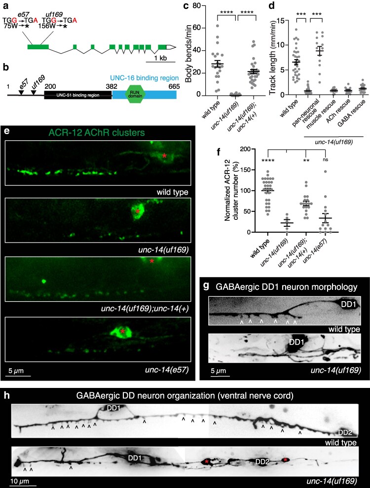 Fig. 1
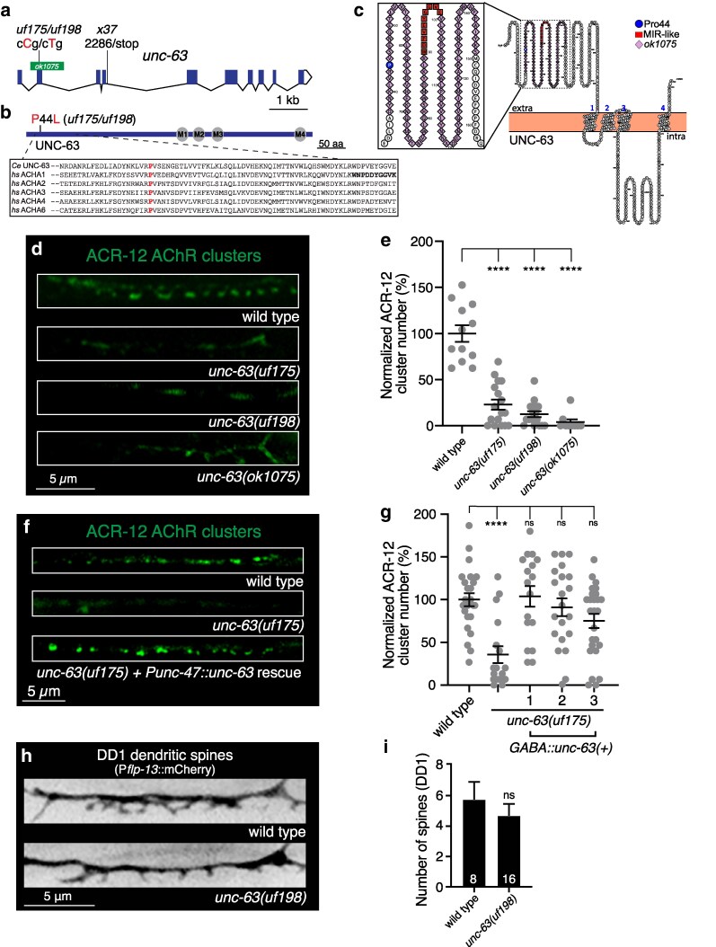 Fig. 2
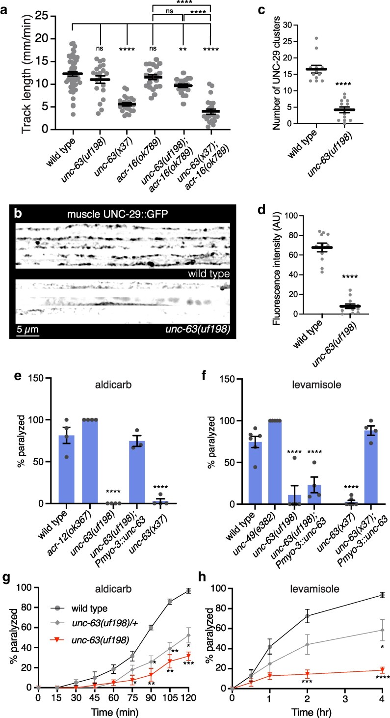 Fig. 3
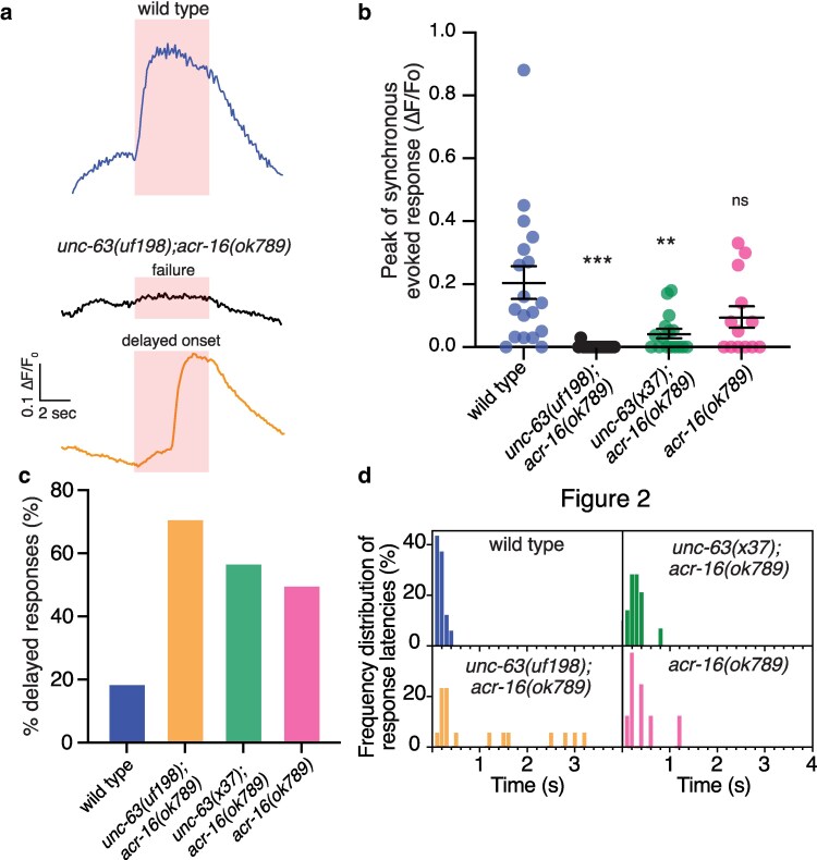 Fig. 4
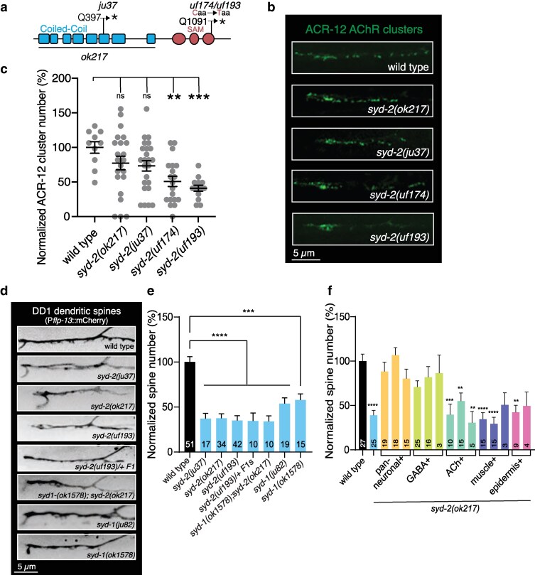 Fig. 5
- —NINDS10.13039/100000065
- —NSF10.13039/100000001
- —NIH10.13039/100000002
Peer Reviews
No public reviews on file for this paper yet. If you reviewed it on a platform where reviews are public (OpenReview, ICLR, NeurIPS, ICML), you can paste yours below so the community can read it here.
Videos
No videos yet. Explain this paper in a talk, walkthrough, or lecture? Add one.
Taxonomy
TopicsGenetics, Aging, and Longevity in Model Organisms · Neurobiology and Insect Physiology Research · Cellular transport and secretion
Introduction
Nervous system performance relies on the precise organization of synaptic connections between neurons. During synapse formation, presynaptic and postsynaptic machinery must be properly trafficked to nascent sites of synaptic contacts and stabilized. Presynaptic protein complexes recruit neurotransmitter-filled synaptic vesicles to specific sites, known as active zones (AZ), and neurotransmitter receptors are clustered at high density in apposition to these AZs. Specific neurotransmitter receptor types are localized in a cell- and synapse-type specific manner, based at least in part on their subunit composition. While we now have generalized knowledge of the various processes that guide these steps in synapse development, the molecular mechanisms involved often show a high degree of cell-type specificity, complicating efforts to gain a comprehensive mechanistic understanding of synapse development processes across neuron classes and neurotransmitter systems.
Genome-wide screening approaches in invertebrates have proven particularly valuable for identifying conserved genetic pathways involved in synapse formation. In Caenorhabditis elegans, these efforts have largely focused on the identification of genes involved in the organization of neuromuscular synapses. For example, screens to identify mutants with mislocalized synaptic vesicle markers at GABAergic neuromuscular synapses identified conserved genes such as syd-2 that encode presynaptic scaffolds important for active zone structural organization (Zhen and Jin 1999). Additionally, screens to identify mutants resistant to the effects of the ionotropic (nicotinic) acetylcholine receptor (AChR) agonist levamisole identified extracellular scaffold genes important for the clustering of muscle AChRs at cholinergic neuromuscular synapses (Gally et al. 2004; Gendrel et al. 2009). By comparison, molecular mechanisms critical for the formation of synapses onto C. elegans neurons are less well defined. Our previous work showed that the presynaptic adhesion protein NRX-1 stabilizes developing postsynaptic spines on GABAergic dendrites (Philbrook et al. 2018; Oliver et al. 2022). However, additional players in the development of spines and their associated postsynaptic sites have remained unidentified.
In this study, we pursued a genome-wide screening approach to obtain mutants in which the organization of postsynaptic cholinergic receptor clusters (AChRs) on GABAergic motor neurons is disrupted. From this screen, we obtained 3 mutants that indicate molecular mechanisms important for the development and maturation of excitatory synaptic contacts located on dendritic spines of GABAergic motor neurons. We isolated a new mutant allele of the unc-14 gene that disrupts both synaptic organization and dendrite outgrowth. unc-14 encodes a RUN domain adaptor protein previously implicated in axonal trafficking and cargo selection (Ogura et al. 1997; Sakamoto et al. 2005; Tsuboi et al. 2005; Ogura and Goshima 2006). Further, we isolated a new mutant allele of the nicotinic receptor alpha subunit unc-63 that disrupts localization of ACR-12 receptor clusters in GABAergic dendrites, providing evidence that UNC-63 is an obligate component of ACR-12-containing receptors. Finally, we isolated a new allele of the synaptic scaffold syd-2/Liprin-α, previously shown to be important for the organization of presynaptic sites (Zhen and Jin 1999; Dai et al. 2006; Chia et al. 2013), which disrupts the outgrowth of dendritic spines and the clustering of postsynaptic receptors in GABAergic neurons. Our evidence linking these genes with synapse formation in C. elegans GABAergic neurons advances a new model for analysis of their functional contributions.
Materials and methods
Strain maintenance
All strains used are derivatives of N2 Bristol and maintained at room temperature. Animals were grown on nematode growth media plates (NGM) seeded with E. coli strain OP50. All experiments were conducted using hermaphrodites. To test for dominance, F1 hermaphroditic cross-progeny were selected using a visible marker. A complete list of all strains used are found in Supplementary Table 1.
EMS mutagenesis, whole-genome sequencing and bioinformatic data analysis
To identify genes important for the development or maintenance of synapses onto GABAergic neurons, we conducted a semi-clonal F2 forward screen (Brenner 1974; Jorgensen and Mango 2002) to obtain mutants where the clustering of GFP-tagged ACR-12 receptors is disrupted. Transgenic animals stably expressing Pflp-13::ACR-12::GFP [ufIs126] were mutagenized for 4 h using 25 µM ethyl methanesulfonate (EMS) following standard protocols (Kutscher and Shaham 2014). Mutagenized L4/young adults were transferred to NGM plates. Three to 4 days later, F1s were transferred (2/plate) to 50 plates (100 F1s). From each of the 50 F1 plates, 8 F2s were transferred to 4 new plates (2/plate) yielding 400 F2s on 200 plates. L4 stage F2 progeny were screened for disruptions in ACR-12::GFP clustering in DD neurons (absent, displaced, ectopic, decrease, or increased number) by spinning disk confocal microscopy. Roughly 3,300 haploid genomes were screened in total.
Candidate mutants were mapped by variant discovery mapping as previously described (Doitsidou et al. 2016; Alexander et al. 2023). Briefly, mutant phenotypes were first confirmed in subsequent generations. Confirmed candidates were then backcrossed to the starting, non-mutagenized strain carrying Pflp-13::ACR-12::GFP together with a visible marker used in the crosses. Homozygous F2 progeny from 16 to 23 backcrossed recombinants were pooled for DNA extraction and whole genome sequencing (Novogene). Homozygous variants within the mapped region were included in the final candidate list. Raw sequence data were processed using Galaxy/Cloudmap mapping, subtracting, and filtering pipelines (Minevich et al. 2012). Sequence reads were aligned to the reference genome (wild type/N2 Bristol WS220) (Sternberg et al. 2024) using the fast-short read Burrows-Wheeler Aligner (BWA). For high coverage and high-quality datasets only reads with a Phred score of >30 (according to FastQC) for each base pair was used. Variants were identified using GATTK Unified Genotyper from the GATK suite. Variations present in the Pflp-13::ACR-12::GFP starting strain were subtracted and filtered (read depth of ≥3) from the mutant sequence results using GATK SelectVariants from the GATK suite. Variants were plotted using CloudMap and annotated using SnpEff.
Imaging and fluorescence quantification
L4 or Day 1 adult animals were immobilized with sodium azide (0.3 M) on 2% or 5% agarose pads. Z-stack confocal volumes were obtained using a Yokogawa CSU-X1 spinning disk confocal mounted on an Olympus BX51WI upright microscope equipped with a Hamamatsu EM-CCD camera and 63 × oil immersion objective. Analysis of synapse number and fluorescence intensity was performed using FIJI ImageJ software (Schindelin et al. 2012) using defined threshold values acquired from control experiments for each fluorescent marker. Statistical comparisons for all synaptic and spine analyses were performed by student's t-test for experiments with 2 groups, or by 1-way or 2-way ANOVA with the appropriate post hoc test for experiments with multiple groups.
Molecular biology
Plasmids were constructed using the 2-slot Gateway Cloning system (Invitrogen) and confirmed by restriction digest and sequencing. All plasmids and primers used in this work are described in Supplementary Tables 2 and 3.
CRISPR/Cas9 design and gene editing
The Bristol N2 strain was used as the wild-type strain for all CRISPR/Cas9 editing. unc-63 and syd-2 gene editing was performed by CRISPR/Cas9 using established protocols (Arribere et al. 2014, Ward 2015). Site-specific sgRNAs and repair template donor ssODNs were manually designed and generated by IDT (Integrated DNA Technologies, Inc.). Mutations in the dpy-10 gene were used as a CRISPR co-conversion marker. Base substitutions were confirmed via Sanger sequencing. All crRNAs, ssODNs, and PCR screening sequences are reported in Supplementary Table 3 and File 1.
Aldicarb and levamisole assays
Assays were performed on synchronous populations of Day 1 adult animals. At least 10 worms of each genotype were transferred to separate plates containing NGM agar and either 1 mM aldicarb or 250 μM levamisole. The number of paralyzed animals was recorded every 15 min and the rate of paralysis was plotted as a function of time using GraphPad Prism. Assays were repeated on at least 3 separate days. Single time point (2 h) and time course drug exposure experiments were conducted independently.
Behavioral assays
Staged Day 1 adult animals were placed on behavioral assay plates coated with a thin OP50 lawn. Following 1 min of habituation, worm movement was recorded using the WormLab Imaging System (MBF Bioscience). Post hoc analysis was performed using WormLab software. The total length of the worm's movement path (track length), calculated by summing the lengths of all forward and backward movements, over 1-min duration from the start of image acquisition was calculated. Statistical comparisons were performed by 1-way ANOVA with Bartlett's correction using GraphPad Prism.
Calcium imaging
Simultaneous optogenetics and calcium imaging was performed as previously described (Philbrook et al. 2018). Imaging was conducted using Yokogawa CSU-X1-A1N spinning disk confocal system equipped with EM-CCD camera (Hammamatsu, C9100-50) and 40X C-Apochromat 1.2 NA water immersion objective. Optogenetic stimulation experiments employed a 625 nm (40 W) LED (Mightex Systems). Optical output through the objective was 0.3 mW/mm^2^ at the focal plane of the specimen. Simultaneous GCaMP excitation (488 nm) and emission (525 nm) acquisition and Chrimson activation were achieved using a 556 nm edge BrightLine single-edge short-pass dichroic beam splitter positioned in the light path (Semrock). Ca^2+^ transients were recorded from muscle cells in transgenic animals co-expressing Pmyo-3::NLSwCherry::SL2::GCaMP6 s (from M. Alkema) along with Pacr-2::Chrimson. Animals were placed on plates seeded with OP50 containing 2.75 mM All-Trans Retinal (ATR) for 24 h prior to experiments. Young adults were immobilized on 5% agarose pads in 2,3-Butanedione monoxime (BDM) (30 mg/mL). For all genotypes, control animals grown in the absence of ATR were imaged. Data were acquired at 10 Hz sampling using Volocity software. Pre-stimulus baseline fluorescence (F0) was calculated as the average of the corrected background-subtracted data points in the first 4 s of the recording (prior to stimulus onset). The corrected fluorescence response data were normalized to prestimulus baseline as ΔF/F0, where ΔF = F–F0. Peak ΔF/F0 was determined by fitting a Gaussian function to the ΔF/F0 time sequence using Multi peak 2.0 (Igor Pro, WaveMetrics). All data collected were analyzed, including failures (no response to stimulation). All responses occurring within the stimulation window (5 s) that could be fitted with a Gaussian function were scored as positive and quantified. Latency was calculated as the time after stimulus onset for the fluorescence intensity to reach 2 times the pre-stimulus baseline standard deviation (Larsch et al. 2015).
Results
A forward screen to identify genes important for neuronal AChR clustering/assembly
To identify genes important for the formation or maintenance of synaptic connections in the C. elegans motor circuit, we performed a forward genetic screen to obtain mutants where the clustering and/or distribution of AChRs in dorsal type D class (DD) GABAergic motor neurons are disrupted. ACR-12-containing AChR clusters are organized into synapses located at the tips of spines in mature DD GABAergic dendrites (Oliver et al. 2018; Philbrook et al. 2018; Cuentas-Condori et al. 2019; Oliver et al. 2022). We screened L4 stage F2 progeny of EMS-mutagenized animals stably expressing ACR-12::GFP in GABAergic neurons (Petrash et al. 2013; Barbagallo et al. 2017) for mutants with either misplaced or altered density of AChR clusters in the dendrites of DD GABAergic neurons. We identified 3 mutant alleles of note: uf169, uf175, and uf174. For each of these mutant alleles, we noted striking decreases in ACR-12::GFP fluorescence in GABAergic dendrites. Using next generation sequencing and variant discovery mapping, we identified the causal mutations in these 3 candidates and describe our analysis of each below.
A mutation in the unc-14/RUN domain cargo adaptor disrupts GABAergic neurite outgrowth and organization
Whole genome sequence analysis and variant discovery mapping of uf169 mutants revealed a point mutation that introduces an early stop in the first exon of the unc-14 gene, presumably resulting in a null allele (Fig. 1a, b). UNC-14 encodes a RUN domain (RPIP8, UNC-14, and NESCA) cargo adaptor protein (Ogura et al. 1997). RUN domain-containing proteins associate with small GTPases, motor proteins, and cytoskeletal networks and are implicated in a variety of cellular signaling pathways and processes, including neuronal development (Callebaut et al. 2001; Yoshida et al. 2011). UNC-14 was previously implicated in the outgrowth of GABAergic axons through interactions with the UNC-51 serine/threonine kinase and Netrin signaling components (Ogura et al. 1997; Lai and Garriga 2004; Tsuboi et al. 2005; Ogura and Goshima 2006; Asakura et al. 2010) and synaptic vesicle transport through interactions with kinesin motors UNC-116 and KLC-2 (Sakamoto et al. 2005), but its importance in the extension of GABAergic dendrites and the structural organization of postsynaptic sites has not been previously investigated.
unc-14 mutation disrupts the outgrowth of GABAergic dendrites. a) Schematic of unc-14 transcript. Reference allele (e57) and screen isolate allele (uf169) mutations are indicated, both resulting in a premature stop () in the first exon. b) UNC-14 protein domains including the predicted binding sites of UNC-51 and UNC-16. Modified from Sakamoto et al. (2005). Reference allele (e57) and screen isolated allele (uf169) mutations are indicated. Numbers, amino acid positions. c) Quantification (mean ± SEM) of the average body bends per minute during exploratory movement for the indicated genotypes. Points indicate measurements from independent animals. ****P < 0.0001, 1-way ANOVA with Dunnett's multiple comparison's test. n = 22,32,30. d) Quantification (mean ± SEM) of track length (mm/min) during exploratory movement for the indicated genotypes. The decreased movement of unc-14(uf169) mutants was rescued by pan-neuronal (Prgef-1) unc-14(+) expression but not by cholinergic (Punc-17β), GABAergic (Punc-47) or muscle (Pmyo-3) unc-14(+) expression in unc-14(uf169) mutants. Points indicate measurements from independent animals. ***P < 0.001, 1-way ANOVA with Dunnett's multiple comparison's test. n = 33,42,13,15,21,25. e) Representative confocal images of ACR-12 AChR localization (Pflp-13::ACR-12::GFP) in ventral GABAergic DD1 dendrites of control, screen isolate unc-14(uf169), unc-14(e57), and unc-14(uf169) rescued by expression of wild type unc-14 coding sequence and promoter region. Red asterisks, DD1 cell body. f) Quantification (mean ± SEM) of the number of ACR-12 AChR clusters in a 30 µm synaptic region of the DD1 dendrite in the ventral nerve cord of wild type, screen isolate unc-14(uf169), unc-14(e57), and unc-14(uf169) rescued by expression of wild-type unc-14 coding sequence and promoter region. Points indicate measurements from independent animals. ****P < 0.0001, *P < 0.01, 1-way ANOVA with Dunnett's multiple comparison's test. ns, not significant. n = 29,5,14,18. g) Representative confocal images of DD1 dendrite in the ventral nerve cord (Pflp13::mCherry) of wild type and unc-14(uf169) mutants. White carets indicate DD dendritic spines. h) Representative images showing expanded views of the ventral nerve cord processes of DD1 and DD2 GABAergic neurons in wild type and unc-14(uf169) mutant. Black carets point to possible DD dendritic spines. Red asterisks indicate varicosities along the ventral nerve cord.
unc-14(uf169) mutants show severely uncoordinated movement (Fig. 1c), similar to previously characterized unc-14(e57) loss-of-function mutants. Expression of the wild-type unc-14 coding sequence and 2.9 kb promoter region upstream of the unc-14 start in unc-14(uf169) mutants robustly restored normal movement (Fig. 1c), supporting that unc-14(uf169) is a loss-of-function mutation. Pan-neuronal, but not muscle, expression of wild-type unc-14 was also sufficient to rescue the locomotor deficits of unc-14(uf169) mutants (Fig. 1d), indicating a neuronal requirement. However, specific expression in either GABAergic or cholinergic neurons was not sufficient for behavioral rescue (Fig. 1d), consistent with a broad neuronal requirement for unc-14 expression.
We next quantified the effects of the uf169 mutation on ACR-12::GFP fluorescence. We observed the number of fluorescent receptor clusters is severely reduced in DD GABAergic dendrites of uf169 mutants (Fig. 1e, f). Dendritic ACR-12::GFP fluorescence is similarly reduced in the DD dendrites of previously characterized unc-14(e57) loss-of-function animals (Fig. 1e, f). The reduction in ACR-12 AChR clusters was rescued by expression of the wild-type unc-14 sequence (2.9 kb promoter) (Fig. 1e, f). We also sometimes noted a qualitative increase in ACR-12::GFP fluorescence in DD neuronal somas of unc-14(uf169) mutants relative to the unmutagenized starting strain, perhaps indicative of impaired trafficking from the soma, though we did not quantify this effect further. To better understand how mutation of unc-14 impacts AChR localization, we examined the morphological features of DD neurons in unc-14 mutants expressing a Pflp-13::mCherry transcriptional reporter. We observed a significant reduction in dendritic spines in unc-14(uf169) mutants (Fig. 1g). However, this reduction was accompanied by severe morphological defects in ventral nerve cord DD neurites, including ectopic branching, large varicosities, and misplaced DD somas in the uf169 mutant (Fig. 1h). Given the severe dendrite morphology defects observed in these animals, the reduction in spine number likely arises from a generalized requirement for UNC-14 in dendrite extension rather than a specific role for UNC-14 in dendritic spine outgrowth. While unc-14 has previously been shown to play a role in neurite outgrowth and extension (Ogura et al. 1997; Lai and Garriga 2004; Tsuboi et al. 2005), our data suggest a potential additional role for UNC-14 in postsynaptic receptor trafficking and localization.
A mutation in the nAChR alpha subunit unc-63 gene disrupts ACR-12 AChR clustering in DD motor neurons
uf175 mutants displayed a striking reduction in AChR clusters in GABAergic dendrites. Sequence analysis and mapping of uf175 mutants revealed a missense mutation in exon 2 of the unc-63 gene (Fig. 2a). unc-63 encodes a nicotinic acetylcholine receptor (nAChR) alpha subunit that is expressed in both the nervous system and muscles and is incorporated into heteromeric nAChRs that vary in subunit composition across cell types (Lewis et al. 1980; Culetto et al. 2004; Boulin et al. 2008; Jospin et al. 2009; Barbagallo et al. 2010; Philbrook et al. 2018). The sensitivity of UNC-63-containing nAChRs at the C. elegans neuromuscular junction to the nicotinic agonist levamisole (L-AChR) has been well-documented (Fleming et al. 1997; Richmond and Jorgensen 1999). UNC-63 also contributes to other heteromeric nAChR classes in both cholinergic and GABAergic motor neurons (Jospin et al. 2009; Barbagallo et al. 2010; Philbrook et al. 2018). The missense mutation carried by unc-63(uf175) mutants introduces a P44L substitution at a highly conserved position in the N-terminal extracellular region of UNC-63 immediately adjacent to amino acids implicated in forming the main immunogenic region (MIR) of the human muscle alpha1 nAChR subunit (Fig. 2b, c). The MIR is a major target for autoantibodies in humans suffering from myasthenia gravis (Tzartos et al. 1991; Lindstrom and Luo 2012). Sequence in this region is thought to be important for mediating the conformational maturation of AChRs during receptor assembly as well as for determining sensitivity to activation by ACh (Luo et al. 2009).
*Substitution of a highly conserved proline residue near the N-terminus of the UNC-63 AChR subunit disrupts ACR-12 AChR localization in GABAergic DD dendrites. a) Transcript of unc-63 with annotated unc-63 alleles. Screen isolated uf175 and CRISPR generated uf198 alleles are indicated. Green bar indicates region unc-63(ok1075) deletion. b) Domain structure of UNC-63 protein. Transmembrane domains M1 through M4 are indicated. Screen isolated uf175 and CRISPR generated unc-63(uf198) P44L mutation are shown. Sequence alignments of UNC-63 across species are detailed in box. The red “P” indicates the unc-63(uf198) P44L mutation, a proline residue at this position is highly conserved from C. elegans (Ce) to humans (hs). The putative main immunogenic region (MIR) is indicated in bold font. c) Schematic of UNC-63 protein topology. P44 is indicated in blue, sequence corresponding to the main immunogenic region (MIR) in red, and the unc-63(ok1075) mutation in purple. Generated using the open-source protein visualization tool Protter (Omasits et al. 2014). d) Confocal images of ACR-12 localization (Pflp-13::ACR-12::GFP) in GABAergic DD1 dendrites of wild type, unc-63(ufI175), unc-63(uf198) and unc-63(ok1075) animals. e) ACR-12 clusters in GABAergic dendrites are decreased by mutation of unc-63. Quantification of receptor clusters, normalized to wild type, for each genotype. Points indicate measurements from independent animals. Mean ± SEM are indicated. ****P < 0.0001, 1-way ANOVA with Dunnett's multiple comparisons test. n = 12,17,17,9. f) Confocal images of DD1 ACR-12 localization (Pflp-13::ACR-12) in GABAergic DD1 dendrites of wild type, unc-63(uf175), and unc-63(uf175) rescued by GABA-specific (Punc-47::unc-63 cDNA) expression of wild type unc-63. g) GABA-specific (Punc-47::unc-63 cDNA) expression of a wild type unc-63 cDNA in unc-63(uf175) mutants restores ACR-12 clustering (3 independent lines). Quantification of receptor clusters, normalized to wild type, for each genotype. Points indicate measurements from independent animals. Mean ± SEM are indicated. ***P < 0.0001, 1-way ANOVA with Dunnett's multiple comparisons test. ns, not significant. n = 24,17,17,21,26. h) Representative confocal images of DD1 spines (Pflp-13::mCherry) in wild type and unc-63 (uf198) animals. i) Quantification of spine number (mean ± SEM) in DD1 dendrites (Pflp-13::mCherry) of wild type and unc-63(uf198) animals are indicated. ns, not significant. Numbers in bars indicate the number of animals quantified.
ACR-12 AChR clusters in the DD neurons of unc-63(uf175) mutants are decreased by ∼65% compared with wild type (Fig. 2d, e). Mutant unc-63(uf198) animals, carrying the same cCg/cTg P44L mutation engineered by CRISPR/Cas9 genome editing, showed a similar decrease in ACR-12 AChR clusters (Fig. 2d, e), providing additional evidence that the P44L substitution is the causal mutation. Similar decreases in ACR-12 clustering were also observed in previously characterized unc-63(ok1075) animals (Fig. 2d, e) that carry a deletion in the unc-63 locus (Philbrook et al. 2018), suggesting the uf175 mutation isolated from the screen and the corresponding CRISPR-generated uf198 mutation cause a severe loss-of-function. Cell-specific expression of wild-type unc-63 in the GABAergic neurons of unc-63(uf175) mutants rescued the decrease in ACR-12 AChR clusters (Fig. 2f, g), demonstrating a cell-autonomous requirement for UNC-63 in AChR assembly and synaptic clustering in DD neurons. In contrast to the severe decrease in AChR clusters, the number of dendritic spines in DD neurons is not appreciably altered by the UNC-63 P44L substitution (Fig. 2h, i), consistent with our prior findings that spine formation and stabilization occur independently of postsynaptic receptor localization (Philbrook et al. 2018; Oliver et al. 2022).
The UNC-63 P44L substitution is semi-dominant and disrupts neuromuscular function
In addition to motor neurons, unc-63 is strongly expressed in body wall muscles and serves an obligate subunit of heteropentameric postsynaptic levamisole-sensitive L-AChRs at neuromuscular synapses (Boulin et al. 2008). We next investigated the function of muscle L-AChRs in unc-63(uf198) mutants to explore how the P44L substitution may impact classes of UNC-63 containing AChRs in other cell types. Our interest in this question was motivated in part by our initial qualitative observation that the movement of unc-63(uf198) animals appeared phenotypically less severely affected compared with the previously characterized unc-63(x37) loss-of-function reference allele. This mutation disrupts a splice junction consensus site (Culetto et al. 2004) and has been shown to eliminate UNC-63 protein at the cell surface (Jones et al. 2011). To define the impacts of unc-63(uf198) on movement, we measured movement velocity by video monitoring and post hoc analysis (Fig. 3a). We found that the unc-63(x37) loss-of-function significantly decreased the movement velocity as previously reported (Culetto et al. 2004). In contrast, the P44L substitution in unc-63(uf198) mutants produced no appreciable decrease in movement velocity. To explore this more deeply, we investigated the effects of the unc-63 mutation in combination with deletion of the acr-16 gene. acr-16 encodes another nAChR alpha subunit that forms homomeric iAChRs (N-AChR) which operate in parallel with L-AChRs at muscle synapses. Combined loss of function of these 2 receptor classes produces severe deficits in both cholinergic neuromuscular transmission and movement (Francis et al. 2005; Touroutine et al. 2005). Consistent with prior work, we found that the movement of unc-63(x37); acr-16(ok789) double mutants is severely decreased compared to either wild type or acr-16(ok789) single mutants. In contrast, the movement of unc-63(uf198); acr-16(ok789) double mutants is not significantly decreased (Fig. 3a). Likewise, the movement of unc-63(x37); acr-16(ok789) double mutants is significantly decreased compared with unc-63(uf198); acr-16(ok789) double mutants. Together, our results suggest there may be sufficient muscle L-AChR function in unc-63(uf198) mutants to support muscle contraction and movement.
*The UNC-63 P44L substitution disrupts neuromuscular synapse organization and presents resistance to aldicarb and levamisole. a) Quantification of track length (mm/min) during exploratory movement. unc-63(uf198) mutants move similarly to wild type while the movement of unc-63(x37) mutants is more severely disrupted. Similar results are obtained in animals that also lack functional ACR-16 N-AChRs. Points indicate measurements from independent animals. Mean ± SEM are indicated. **P < 0.01, ****P < 0.0001, 1-way ANOVA with Tukey's multiple comparisons test. ns, not significant. n = 52,22,20,21,22,19. b) Representative confocal images of muscle regions along the ventral nerve cord from 5 wild type and unc-63(uf198) mutants expressing Pmyo-3::UNC-29::GFP (muscle AChRs). (c, d) UNC-29::GFP AChR clusters (c) and muscle fluorescence intensity (d) are each significantly decreased in unc-63(uf198) animals. Points indicate measurements from independent animals. Mean ± SEM are indicated. ****P < 0.0001, student's t-test. c) n = 12,13. d) n = 11,12. (E, F) Quantification of the percentage of animals paralyzed following 2 hr exposure to aldicarb (1 mM) (E) or levamisole (250 µM) (F). unc-63(uf198) animals are resistant to aldicarb and levamisole. Muscle expression (Pmyo-3::unc-63 cDNA) fails to fully restore levamisole sensitivity of unc-63(uf198) mutants but is sufficient to restore aldicarb sensitivity. The same Pmyo-3::unc-63 rescuing transgene is used for all aldicarb and levamisole experiments. Points indicate independent measurements from trials of at least 10 animals each. Mean ± SEM are indicated. ****P < 0.0001, 1-way ANOVA with Tukey's multiple comparisons test. e) WT n = 4, acr-12 n = 4, unc-63(uf198) n = 4, unc-63(uf198); Pmyo-3::unc-63(+) n = 3, unc-63(x37) n = 4. f) WT n = 6, unc-49 n = 5, unc-63(uf198) n = 5, unc-63(uf198); Pmyo-3::unc-63(+) n = 4, unc-63(x37) n = 5, unc-63(x37); Pmyo-3::unc-63(+) n = 4. (g,h) Percentage of animals paralyzed over time in the presence of aldicarb (1 mM) (g) or levamisole (250 µM) (h). Heterozygous unc-63(uf198/+) animals (gray) treated with either aldicarb or levamisole paralyze more slowly than drug-treated wild type controls (black). Each data point indicates the mean of 4 (g) or 5 (h) independent trials of at least 10 animals each. *P < 0.05, **P < 0.01, ***P < 0.001, ***P < 0.0001, 2-way ANOVA with Tukey's multiple comparisons test.
We next sought to investigate the assembly and clustering of L-AChRs at neuromuscular synapses in unc-63(uf198) animals. We quantified the clustering of L-AChRs in muscles using muscle-specific expression of UNC-29::GFP, an obligate non-alpha subunit of L-AChRs (Fleming et al. 1997; Boulin et al. 2008). The number and fluorescence intensity of muscle L-AChR (UNC-29::GFP) clusters were strikingly decreased in unc-63(uf198) mutants compared with control (Fig. 3b–d). This result suggests that the assembly of muscle L-AChR and GABA neuron ACR-12 AChR complexes are similarly disrupted in unc-63(uf198) mutants. However, our observation that movement is not appreciably affected in these animals supports the idea that a low level of receptor assembly persists in unc-63(uf198) animals and is sufficient to support muscle contraction and movement.
To further investigate the function of muscle L-AChR in unc-63(uf198) animals, we measured the effects of exposure to either the cholinesterase inhibitor aldicarb or the L-AChR agonist levamisole. Wild-type animals are paralyzed in response to prolonged treatment with either chemical. The time course over which paralysis occurs is widely used as a measure of cholinergic function. We found that unc-63(uf198) animals are strongly resistant to the paralyzing effects of either aldicarb or levamisole, similar to animals carrying the severe unc-63(x37) loss-of-function allele (Fig. 3e, f). Muscle-specific expression of wild-type unc-63 in unc-63(uf198) animals restored paralysis in response to aldicarb treatment; however, only weakly altered paralysis in response to levamisole. Expression of the same transgene in unc-63(x37) mutants was sufficient to restore levamisole-induced paralysis (Fig. 3f). Together, our analysis suggests that the P44L substitution in unc-63(uf198) mutants may have dominant negative effects or exhibit haploinsufficiency, particularly for activation by levamisole. Consistent with this interpretation, we found that unc-63(uf198) heterozygous animals had increased resistance to paralysis by aldicarb and levamisole compared with control (Fig. 3g, h), suggesting unc-63(uf198) is a semi-dominant allele.
To investigate muscle responses to cholinergic activation in unc-63(uf198) mutants, we next measured muscle calcium transients evoked by photostimulation of cholinergic motor neurons. To measure muscle calcium responses, we expressed the genetically encoded calcium sensor GCaMP6 s in muscles together with the red-shifted channelrhodopsin Chrimson in cholinergic motor neurons (Philbrook et al. 2018). As the cholinergic synaptic response is mediated through both UNC-63-containing L-AChRs and homomeric ACR-16 AChRs (Richmond and Jorgensen 1999; Francis et al. 2005; Touroutine et al. 2005), we pursued these experiments in animals lacking acr-16, allowing us to focus on L-AChR-mediated muscle calcium responses. Depolarization of cholinergic motor neurons elicited robust calcium responses that were closely synchronized to the onset of the light stimulus in the muscles of wild-type control animals (response within 200 ms of stimulus onset) (Fig. 4a, b). The amplitude of these synchronous calcium responses was dramatically reduced in unc-63(uf198); acr-16(ok789) double mutants compared to wild type (Fig. 4a, b). We observed a similar reduction in these synchronous calcium responses in animals that carried the presumptive null allele unc-63(x37) in combination with the acr-16(ok789) deletion (Fig. 4b). Together, these results suggest that unc-63(uf198) is a severe reduction-of-function allele. However, we noted another intriguing feature of muscle calcium responses in unc-63(uf198); acr-16(ok789) animals. Many muscles in unc-63(uf198); acr-16(ok789) animals displayed large, delayed calcium transients that were not closely synchronized to the light stimulus (Fig. 4a, bottom). These asynchronous responses occurred more frequently in unc-63(uf198); acr-16(ok789) muscles and the delays were more extended compared with either control or unc-63(x37); acr-16(ok789) muscles (Fig. 4c, d), suggesting this is a property conferred by the UNC-63 P44L substitution.
*unc-63(uf198) muscle responses to cholinergic stimulation are severely disrupted. a) Representative evoked muscle calcium responses for wild type and unc-63(uf198); acr-16(ok789) animals. Example traces for the 2 major response classes observed in the unc-63(uf198); acr-16(ok789) animals (response failures and delayed onset responses) are shown. Red shading indicates the period of cholinergic photostimulation. b) Quantification (mean ± SEM) of the peak muscle responses synchronized to cholinergic depolarization for the indicated genotypes. Points indicate measurements from independent animals. Mean ± SEM are indicated. Responses were scored as synchronous if occurring within 200 ms of stimulus onset. ***P < 0.001, *P < 0.01, 1-way ANOVA with Tukey's multiple comparisons test. ns, not significant. n = 18,17,16,13. c) Proportion of delayed onset responses calculated for the indicated genotypes. Note the high proportion of delayed onset responses in unc-63(uf198) animals. Responses were classified as delayed if occurring more than 200 ms from the onset of stimulation. Response failures were excluded from the analysis. n = 16,16,14,8. d) Frequency distribution of response latencies (time from the onset of stimulation to response) for the indicated genotypes for all measurements (including failures). Nearly 90% of wild-type muscle calcium responses occurred within 200 ms of stimulus onset. Note the increased response latencies in unc-63(uf198) animals. Response failures were excluded from the analysis. n = 16,16,14,8.
Mutation of syd-2/Liprin-α disrupts dendritic spine formation
Mapping and sequence analysis of uf174, a third mutant isolated from our screen with reduced AChR clusters in GABAergic dendrites, revealed a novel point mutation in the syd-2 gene (Fig. 5a). syd-2 encodes the sole C. elegans Liprin-α. syd-2/Liprin-α was shown to interact with LAR family receptor protein tyrosine phosphatases (LAR-RPTP) (Serra-Pagès et al. 1998) and implicated as a structural component of active zones based on a series of elegant genetic studies in C. elegans (Zhen and Jin 1999; Dai et al. 2006; Chia et al. 2013). The SYD-2/Liprin-α family proteins contain a N-terminal coiled-coil region followed by 3 C-terminal SAM (sterile-α-motif) domains (Fig. 5a). The coiled-coil domains are implicated in homo- and hetero-multimerization while the SAM domains form the LH (Liprin Homology) region that binds LAR-RPTP (Serra-Pagès et al. 1998). The syd-2(uf174) mutants isolated from the screen carry a C/T missense mutation in exon 18 that produces an early stop (Q1091stop) and truncation of the predicted SAM domain (Fig. 5a).
*Mutation of syd-2 disrupts GABAergic dendritic spines. a) Domain structure of SYD-2. Coiled-coil (blue) and SAM domains (red) are indicated. Deleted region in syd-2(ok217) and approximate positions of mutations in syd-2(ju37), screen isolate syd-2(uf174) and CRISPR-engineered syd-2(uf193) are indicated. b) Confocal images of DD1 ACR-12 AChR localization (Pflp-13::ACR-12:GFP) in wildtype, screen isolate syd-2(uf174), CRISPR engineered syd-2(uf193), syd-2(ju37) and syd-2(ok217) animals. c) Quantification (mean ± SEM) of ACR-12 AChR clusters in DD GABAergic dendrites of the indicated syd-2 and syd-1 alleles. **P < 0.01, ***P < 0.001, 1-way ANOVA with Dunnett's multiple comparison test. ns, not significant. n = 10,22,24,19,13. d) Confocal images of DD1 dendritic spines (Pflp-13::mCherry) for the genotypes indicated. e) Quantification (mean + SEM) of DD1 GABA neuron spine number for the indicated genotypes normalized to wild type. ****P < 0.0001, 1-way ANOVA with Dunnett's multiple comparison test. Numbers in bars indicate the number of animals quantified. f) Quantification (mean ± SEM) of GABAergic DD1 dendritic spine number for the genotypes indicated normalized to wild type. Pan-neuronal, but not muscle or epidermal, expression of wild type syd-2 in syd-2(ok217) mutants is sufficient to restore spine number. GABAergic, but not cholinergic, expression is also sufficient for rescue. ****P < 0.0001, *P < 0.01 compared to wild type, 1-way ANOVA with Dunnett's multiple comparison test. Bars indicate independent rescue lines. Numbers in bars indicate number of animals quantified.
ACR-12 AChR clustering was decreased by roughly 50% in syd-2(uf174) mutants compared to wild type (Fig. 5b, c). To gain support that the mutation in syd-2 was causal for the disruption in AChR clustering, we engineered the same mutation in otherwise wild-type animals by CRISPR/Cas9 genome editing. CRISPR/Cas9-engineered syd-2(uf193) animals showed similar deficits in AChR clustering to those observed in DD neurons of syd-2(uf174) isolated from the screen (Fig. 5b, c). We found that AChR clustering is also disrupted in previously characterized loss-of-function, nonsense syd-2(ju37) and deletion syd-2(ok217) alleles that each produce a premature stop (Fig. 5b, c) (Zhen and Jin 1999). However, the disruptions in these animals appeared less severe and more variable, perhaps suggesting the uf174/uf193 alleles have additional impacts, such as disrupting the function of another protein. We next analyzed GABAergic DD dendritic spines (Pflp-13::mCherry) in syd-2(uf193) L4 animals. The number of DD1 dendritic spines in syd-2(uf193) animals is decreased by ∼65% compared to wild type animals (Fig. 5d, e). To further investigate the impact of the predicted SYD-2 truncation, we analyzed the number of GABAergic DD dendritic spines in syd-2(uf193/+) heterozygotes. Animals heterozygous for syd-2(uf193) had decreased spines compared to wild type, similar to homozygous syd-2(uf193) mutants (Fig. 5e), suggesting either haploinsufficiency or that uf193 may represent a dominant allele of syd-2. Notably, we found that dendritic spine number is also significantly decreased compared to wild type in homozygous syd-2(ju37) and syd-2(ok217) animals. Pan-neuronal expression of wild type syd-2 cDNA in syd-2(ok217) mutants restored spines, while expression in either muscles or epidermis did not, indicating a neuronal requirement (Fig. 5f). Interestingly, specific expression of wild type syd-2 cDNA in GABAergic, but not cholinergic, neurons of syd-2(ok217) mutants also restored spines. These results suggest a primary involvement of postsynaptic SYD-2 in the formation or stabilization of spines; however, additional experiments will be required to confirm this finding. As the RhoGAP-like protein SYD-1 is implicated to act upstream of SYD-2 in presynaptic assembly (Patel et al. 2006; Taru and Jin 2011), we also examined the effects of syd-1 mutation on spines. We found a significant decrease in spines on DD dendrites of syd-1 mutants, though this effect was less severe compared with syd-2 mutation (Fig. 5d, e). Our results indicate that, similar to the synaptic adhesion protein NRX-1 (Philbrook et al. 2018; Oliver et al. 2022), SYD-2 is important for the formation or stabilization of both dendritic spines and ACR-12 AChR clusters at postsynaptic sites.
SYD-2 has been broadly implicated in both presynaptic structural organization and cargo trafficking (Zhen and Jin 1999; Dai et al. 2006; Wagner et al. 2009). Our findings newly implicate SYD-2 in the formation or stabilization of postsynaptic receptor clusters and dendritic spines, and provide initial evidence that a postsynaptic SYD-2 pool may be sufficient to direct these processes.
Discussion
We pursued an unbiased, genome-wide strategy to elucidate mechanisms directing structural organization of dendrites and postsynaptic specializations localized to dendritic spines in C. elegans GABAergic neurons. We identified 3 genes that, when mutated, disrupt the organization of postsynaptic receptor clusters on GABAergic dendrites. These genes define distinct functional pathways required for establishing synaptic connections: (1) the RUN domain cargo adaptor UNC-14 is required for dendrite outgrowth, (2) the nAChR subunit UNC-63 is an obligate subunit of postsynaptic receptors and required for receptor assembly, and (3) the synaptic scaffold SYD-2 is required for spine outgrowth or maintenance.
The unc-14 allele we obtained from our screen has an early stop codon and is a predicted null. Prior studies of unc-14 null mutants demonstrated an interaction between UNC-14 and the S/T kinase UNC-51 and implicated UNC-14 in axonal elongation as well as axonal delivery of a variety of cargoes including synaptic vesicles, the axon guidance cue UNC-6/Netrin, and the Netrin receptor UNC-5 (Ogura et al. 1997; Sakamoto et al. 2005; Ogura and Goshima 2006; Asakura et al. 2010). Our analysis of dendritic structure revealed striking morphological abnormalities in GABAergic DD dendrites in addition to the disruption of ACR-12/AChR clustering identified from our initial screen. We are therefore unable to distinguish whether the defects in postsynaptic organization we observed reflect a requirement for UNC-14 in trafficking post-synaptic components, arise secondarily to a failure in the extension and fasciculation of dendritic processes within the ventral nerve cord, or a combination of both. Similar to previous findings for unc-14(e57) mutants, we observed bulbous regions in the dendrites of DD GABAergic neurons in unc-14(uf198) animals that may represent foci where dendritic material, including post-synaptic proteins, has accumulated due to a failure in trafficking. unc-14 is known to be broadly expressed in neurons (Ogura et al. 1997). It is therefore possible that some of the phenotypes we observed in GABAergic DD dendrites reflect a parallel requirement for unc-14 in the organization of presynaptic cholinergic axons. Consistent with this hypothesis, we found that unc-14 expression using either an ∼2.9 kb unc-14 promoter fragment or a pan-neuronal promoter was sufficient to rescue the uncoordinated movement of unc-14 mutants. In contrast, specific expression in either cholinergic or GABAergic motor neurons was not sufficient to rescue, most consistent with a requirement for unc-14 expression in both neuron classes for proper motor function.
The P/L substitution mutation in unc-63 that we isolated disrupts cholinergic receptor clustering in both GABAergic dendrites (Pflp-13::ACR- 12::GFP) and muscles (Pmyo-3::UNC-29::GFP). The mutation, however, does not affect the formation or maintenance of GABAergic dendritic spines, consistent with prior work demonstrating spine outgrowth proceeds independently of receptor clustering (Philbrook et al. 2018; Oliver et al. 2022). Prior structural studies of acetylcholine binding proteins from the freshwater snail, Lymnaea stagnalis (L-AChBP), saltwater mollusk Aplysia californica, and the extracellular ligand binding domain of the mouse muscle alpha1 subunit place this residue adjacent to the N-terminal α-helical region at the apical surface of the receptor (Brejc et al. 2001; Hansen et al. 2005; Dellisanti et al. 2007). Prior mutagenesis studies of chimeric mouse AChRs have implicated this region in receptor assembly and ligand sensitivity (Luo et al. 2009), consistent with our findings for the P/L substitution in UNC-63. In addition to available null alleles, C. elegans mutations that alter UNC-63 subunit functional properties have been characterized previously. For instance, unc-63(x26) (C151Y) disrupts the Cys-loop region, a motif important for forming the agonist binding site, and causes a decrease in channel opening frequency (Jones et al. 2011). However, unc-63(x26) is thought to exclusively alter channel function, not expression. Our analysis suggests that the conserved proline which is altered in unc-63(uf174) and unc-63(uf198) animals is important for proper assembly and function of UNC-63-containing AChRs in both muscles and GABAergic dendrites. Although the P44L substitution is not at the ligand binding Cys-loop motif, the proline to a leucine change may alter the structure of the extracellular ligand binding domain, impacting the both assembly/folding of UNC-63 subunit and ligand binding.
Given the severe disruption in nAChR clustering we observed and the resistance to pharmacological activation by aldicarb or levamisole, it is surprising that the movement of unc-63(uf198) animals is not more severely disrupted. Our studies of evoked calcium responses in muscles indicate that calcium responses do sometimes occur in muscles of unc-63(uf198) animals, but these responses are not strictly time-locked to the stimulus. The most parsimonious interpretation of our findings is that the allele is a strong reduction-of-function but allows sufficient residual receptor activity to support movement. One possibility is that inefficient activation of the receptor may over time produce muscle depolarization. Alternatively, the mutation may indirectly lead to increased spontaneous muscle activity, though a mechanism to account for such an effect remains unclear. Mammalian ionotropic AChRs are linked with myasthenic syndromes and nicotine addiction (Albuquerque et al. 2009) while nematode AChRs are common targets for anthelmintic compounds (Choudhary et al. 2022). Our identification and characterization of unc-63(uf198) animals therefore offer additional tools for comparative structure-function studies of this conserved receptor family.
The isolation of a novel syd-2 allele from our screen newly highlights the importance of this scaffold protein for maintaining postsynaptic organization. Evidence from prior studies across a variety of models implicates SYD-2/Liprin-α as one of several core scaffold proteins responsible for orchestrating presynaptic active zone organization (Hallam et al. 2002; Dai et al. 2006; Patel et al. 2006; Fouquet et al. 2009; Owald et al. 2010; Stigloher et al. 2011; Wentzel et al. 2013; Emperador-Melero and Kaeser 2020; McDonald et al. 2020; Frankel and Kurshan 2025). Though less well-characterized compared with its presynaptic roles, immuno-EM studies of rodent cortical neurons have demonstrated that Liprin-α is also localized to postsynaptic sites (Wyszynski et al. 2002). Postsynaptic Liprin-α has been implicated in AMPAR clustering and in shaping morphological features of dendritic spines through interactions with GRIP1 (glutamate receptor-interacting protein 1) and LAR family of receptor protein tyrosine phosphatases (Wyszynski et al. 2002; Dunah et al. 2005; Spangler and Hoogenraad 2007) (Ko et al. 2003). Similarly, postsynaptic Liprin-α organizes AChRs at the mouse neuromuscular junction (Bernadzki et al. 2017). In C. elegans, current evidence supports predominant presynaptic SYD-2 localization (Zhen and Jin 1999; Dai et al. 2006). While our cell-specific rescue experiments may point toward a primarily postsynaptic role for SYD-2 in supporting dendritic spines, additional studies investigating the subcellular localization of SYD-2 in GABAergic dendrites will be required to further solidify this observation.
At presynaptic sites, SYD-2/Liprin-α and the RhoGAP domain and PDZ domain-containing scaffold protein, SYD-1, are suggested to localize to nascent active zones in the early stages of synapse development, and to subsequently recruit additional presynaptic proteins for presynaptic assembly (Hallam et al. 2002; Dai et al. 2006; Patel et al. 2006; Fouquet et al. 2009; Owald et al. 2010; Stigloher et al. 2011; Wentzel et al. 2013; Emperador-Melero and Kaeser 2020; McDonald et al. 2020; Frankel and Kurshan 2025). In particular, SYD-1 is suggested to function upstream of SYD-2 and to promote SYD-2 localization and activity at presynaptic sites (Dai et al. 2006; Frankel et al. 2025). Interestingly, Drosophila dSyd1 and neurexin (dnrx) mutants have similar active zone defects and form an in vivo complex where dSyd1 stabilizes neurexin at active zones through PDZ domain interactions (Dai et al. 2006; Owald et al. 2012; Frankel et al. 2025). Similarly, in mammalian neurons, Liprin-α forms a complex with Neurexin1 and another presynaptic scaffold protein Mint1 to regulate the stability of Neurexin1 (LaConte et al. 2016). We showed previously that presynaptic NRX-1/neurexin is required for the maintenance of developing spines (Philbrook et al. 2018; Oliver et al. 2022), raising the alternate possibility that the postsynaptic alterations we observe in syd-2 and syd-1 mutants arise secondarily to a mislocalization of NRX-1 at presynaptic sites. Additional studies employing strategies for cell-specific inactivation or deletion of syd-2 will be required to cleanly dissect potential pre- vs postsynaptic roles at synapses onto GABAergic neurons.
Regardless of the precise mechanism of action, the syd-2(uf174)/syd-2(uf193) alleles offer additional tools for functional analysis of SYD-2 at synapses. The mutation we isolated generates an early stop codon in the region encoding the predicted SYD-2 SAM domain and has similar impacts on spines in syd-2(uf193) homozygotes and syd-2(uf193/+) heterozygotes, indicating the mutation may have dominant effects. Prior work identified a putative syd-2 gain-of-function mutation, syd-2(ju487), which also has dominant effects. syd-2(ju487) carries a missense mutation that produces an R/C substitution at position 184 (R184C) of the conserved Liprin Homology 1 (LH1) domain (Dai et al. 2006). This mutant has synaptic phenotypes opposite to those associated with syd-2 loss-of-function alleles, including elongated dense core projections and hypersensitivity to the acetylcholine esterase inhibitor aldicarb (Dai et al. 2006; Kittelmann et al. 2013). In contrast, our analysis of syd-2(uf193) demonstrates these mutation phenocopies known loss-of-function mutants. Our findings suggest the possibility that a dominant-negative, truncated SYD-2 protein is produced in which the SAM domain is disrupted, perhaps locking SYD-2 in an inactive conformation or interfering with interactions with activating proteins (Taru and Jin 2011; Chia et al. 2013). Our observation that the reduction in ACR-12 AChR clusters in the syd-2 mutant isolated from our screen is more severe compared with other available loss-of-function alleles may also point toward a dominant negative role.
Collectively, our studies of unc-14, unc-63, and syd-2 offer new avenues for understanding synapse development and organization. Moreover, the novel alleles we isolated from our screen provide new tools for gaining additional insights into the functions of these conserved proteins at synapses.
Supplementary Material
jkag028_Supplementary_Data
The reference list from the paper itself. Each links out to its DOI / PubMed record.
- 1Albuquerque EX, Pereira EFR, Alkondon M, Rogers SW. 2009. Mammalian nicotinic acetylcholine receptors: from structure to function. Physiol Rev. 89:73–120. 10.1152/physrev.00015.2008.19126755 PMC 2713585 · doi ↗ · pubmed ↗
- 2Alexander KD et al 2023. The homeodomain transcriptional regulator DVE-1 directs a program for synapse elimination during circuit remodeling. Nat Commun. 14:7520. 10.1038/s 41467-023-43281-4.37980357 PMC 10657367 · doi ↗ · pubmed ↗
- 3Arribere JA et al 2014. Efficient marker-free recovery of custom genetic modifications with CRISPR/Cas 9 in Caenorhabditis elegans. Genetics. 198:837–846. 10.1534/genetics.114.169730.25161212 PMC 4224173 · doi ↗ · pubmed ↗
- 4Asakura T, Waga N, Ogura K, Goshima Y. 2010. Genes required for cellular UNC-6/netrin localization in Caenorhabditis elegans. Genetics. 185:573–585. 10.1534/genetics.110.116293.20382828 PMC 2881138 · doi ↗ · pubmed ↗
- 5Barbagallo B, Prescott HA, Boyle P, Climer J, Francis MM. 2010. A dominant mutation in a neuronal acetylcholine receptor subunit leads to motor neuron degeneration in Caenorhabditis elegans. J Neurosci. 30:13932–13942. 10.1523/JNEUROSCI.1515-10.2010.20962215 PMC 2965043 · doi ↗ · pubmed ↗
- 6Barbagallo B et al 2017. Excitatory neurons sculpt GAB Aergic neuronal connectivity in the C. elegans motor circuit. Development. 144:1807–1819. 10.1242/dev.141911.28420711 PMC 5450832 · doi ↗ · pubmed ↗
- 7Bernadzki KM et al 2017. Liprin-α-1 is a novel component of the murine neuromuscular junction and is involved in the organization of the postsynaptic machinery. Sci Rep. 7:9116. 10.1038/s 41598-017-09590-7.28831123 PMC 5567263 · doi ↗ · pubmed ↗
- 8Boulin T et al 2008. Eight genes are required for functional reconstitution of the Caenorhabditis elegans levamisole-sensitive acetylcholine receptor. Proc Natl Acad Sci U S A. 105:18590–18595. 10.1073/pnas.0806933105.19020092 PMC 2587545 · doi ↗ · pubmed ↗